Trauma in Pregnancy


Trauma in Pregnancy


Victor J. Hassid

Miren A. Schinco


Trauma is the primary cause of death in women of reproductive age and is the leading nonobstetric cause of both fetal and maternal death during pregnancy (1,2,3,4). The potential for pregnancy must be considered in any girl or woman between the ages of 10 and 50 years. Accidental injury is estimated to occur in 6% to 7% of all pregnancies, and its incidence increases as pregnancy progresses until, by the end of the third trimester, minor trauma occurs more frequently than at any other time during female adulthood (1,2,5,6,7,8).

The majority of blunt trauma during pregnancy results from motor vehicle crashes, with the remainder being relatively evenly distributed between falls and assaults (3,4,9,10,11,12,13). Some authors report that up to 11% of women are victims of physical abuse during pregnancy (14,15,16,17) and up to 31.5% of pregnant women admitted following trauma are victims of intentional injury (11). During the third trimester, an altered center of gravity (caused by the enlarged uterus) combined with pelvic ligamentous laxity produces a degree of gait instability that makes the pregnant woman particularly susceptible to injury by falls (1,9). This represents the second most common mechanism of injury during pregnancy (13).

In a recent review of over 77,000 women of childbearing age in the American College of Surgeon’s National Trauma Data Bank, pregnant patients were significantly younger, more likely to be underinsured, and of African American or Hispanic descent than their nonpregnant counterparts. While they were less likely to have used alcohol or illicit drugs, still 13% had been drinking and 20% had used drugs prior to their trauma. Although pregnant trauma patients were more likely to use seat belts, still only two thirds were restrained. After admission for a trauma, 5.1% of patients went on to delivery, three quarters within 24 hours and by cesarean section.

The foundation of trauma management in pregnancy is that “the best chance for fetal survival is to assure maternal survival” (5). The physician caring for a pregnant trauma patient should always remember that there are two patients. Initial treatment priorities for an injured pregnant patient remain the same as for the nonpregnant patient. The use of imaging studies should not be withheld because of pregnancy. Because the anatomic and physiologic alterations of pregnancy can alter the gravid woman’s response to injury, an understanding of these changes is critical when approaching trauma management. Early communication between the trauma surgeon and the obstetrician is very important in order to optimize the management of the pregnant trauma patient.


OUTCOMES

Regardless of severity, trauma during pregnancy has been associated with spontaneous abortion, premature labor, uterine injury, placental abruption, maternal death, and low birth weight fetal loss. When comparing traumatized gravid women with nontrauma controls, for any injury, both maternal and fetal outcomes are worse, with progressively worse outcomes with higher injury severity score (ISS). Injuries associated with the highest maternal mortality are thoracoabdominal and pelvic followed by brain injuries. Women who sustain
trauma, but do not deliver at that time, continue with significant risk of increased morbidity and mortality throughout their pregnancy and therefore require close monitoring (13).

Maternal predictors of fetal death consist of maternal factors (ISS >15, severe head injury, pelvic fractures, shock, hypoxemia, and absent fetal heart tones) and mechanisms of injury (auto-pedestrian collisions, maternal ejection, motorcycle collision, and lack of restraints) (18,19,20,21). Infants under 28 weeks’ gestational age appear to have the highest potential for complications, possibly due to their intolerance of maternal stress (13).


ANATOMIC AND PHYSIOLOGIC ALTERATIONS OF PREGNANCY


Anatomic Changes

Until approximately the 12th gestational week, the uterus remains in the pelvis, and by 20 weeks and 34 to 36 weeks, it reaches the umbilicus and the costal margin, respectively. During the last 2 weeks of gestation, the fundus frequently descends as the fetal head engages the pelvis. The progressive enlargement of the uterus causes the bowel to be displaced cephalad and therefore keeps it relatively protected from blunt abdominal trauma, whereas the uterus and its contents become more vulnerable (Fig. 4.1).

During the second trimester, the uterus enlarges beyond its protected intrapelvic location, but the fetus remains mobile and protected by a generous amount of amniotic fluid; the amniotic fluid can be a source of embolism and disseminated intravascular coagulation following trauma if it gains access to the intravascular space.

By the third trimester, secondary to the location of the fetal head within the pelvis, pelvic fractures may result in fetal skull fracture or significant intracranial injury. Unlike the elastic myometrium, the placenta has little elasticity, which predisposes it to shear forces at the uteroplacental interface andplacental abruption.

While interpreting pelvic x-rays, it is important to remember that the pubic symphysis widens to 4 to 8 mm and the sacroiliac joint spaces increase by the 7th month of gestation.


Cardiovascular System

Maternal cardiac output begins to increase during the first 10 weeks of pregnancy, reaching a peak of 30% to 50% above nonpregnant levels by the latter part of the second trimester (22). After the 10th week of pregnancy, the cardiac output can be increased by 1.0 to 1.5 L per minute, due to the increase in plasma volume and decrease in vascular resistance of the uterus and placenta, which during the third trimester of pregnancy receive 20% of the patient’s cardiac output. During the same period, the cardiac output may be transiently lowered by aortocaval compression by the gravid uterus, resulting in decreased placental perfusion. This phenomenon, known as the supine hypotension syndrome, may be observed clinically in otherwise normal pregnant patients (22,23). It is very important to remember while managing a pregnant trauma patient that changing the maternal position from the supine to the left lateral decubitus position may increase cardiac output by as much as 25% at term (23).

The average maternal heart rate increases 20% to 30% during pregnancy such that by term, the normal maternal pulse rate is 80 to 95 beats per minute, making borderline tachycardia difficult to evaluate (2,9,14,23). Mean arterial blood pressure gradually declines during the first two trimesters of pregnancy, reaching its nadir by approximately 28 weeks’ gestation (22).
Systolic arterial pressure decreases by 0 to 15 mm Hg, whereas diastolic pressure declines by 10 to 20 mm Hg, creating a widened pulse pressure. These changes are the result of diminishing peripheral vascular resistance and should not be mistaken as evidence of hypovolemia during the first two trimesters of pregnancy. During the third trimester, blood pressure gradually increases, returning to pre-pregnancy levels near term (2,7,9,22,23). Hypertension, systolic or diastolic, is never expected during pregnancy and, if present, may either be a response to pain, anxiety, or injury or be the result of a direct complication of pregnancy such as pregnancy-induced hypertension (9).


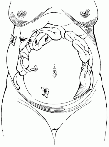
FIGURE 4.1 Compartmentalization of intestines during pregnancy and sites of gunshot wounds above and below the umbilicus.

Associated with the underlying decreased peripheral vascular resistance of the first two trimesters of pregnancy is a paradoxical response to stimuli that
would normally cause vasoconstriction. This altered response causes the skin to be warm and dry, instead of cool and clammy which would be expected during hypovolemic shock (9). In addition, central venous pressure, normally approximately 9 cm H2O in the nonpregnant patient, gradually decreases throughout pregnancy until it reaches 4 to 6 cm H2O during the third trimester (4,7,14,24). Venous hypertension in the lower extremities is present during the third trimester.

Total maternal blood volume increases by as much as 50% at 34 weeks’ gestation, improving maternal response to hemorrhage. A smaller increase in red blood cell (RBC) volume occurs, resulting in a decreased hematocrit (physiologic anemia of pregnancy) (1,22). The white blood cell (WBC) count increases, resulting in counts as high as 15K and 25K/mm3 during pregnancy and labor, respectively (1,5,9). Levels of serum fibrinogen and other clotting factors are mildly elevated, leading to a hypercoagulable state (14,22). During periods of stress, the mother maintains homeostasis at the expense of the fetus. Acute maternal hemorrhage or maternal hypoxia induces uterine artery vasoconstriction, which can reduce uterine perfusion by 10% to 20% before clinical evidence of maternal hypovolemia occurs (1,4,5,9). Consequently, 30% to 35% of maternal blood volume may be lost before clinical signs of hypovolemia develop (1,6,7,9,14,24). This, combined with the sensitivity of the placental vasculature to catecholamines, places the fetus at risk during maternal hemorrhage that may, upon clinical examination of the mother, appear minimally significant. Early and adequate maternal volume replacement and thorough fetal evaluation, including fetal monitoring, are therefore critical in the management of the gravid trauma patient.

Electrocardiographic (ECG) changes in late pregnancy are not specific and reflect the leftward shift of the cardiac axis, by approximately 15 degrees, due to diaphragm elevation. The T waves may be flattened and inverted in lead III, and the Q waves may appear in leads III and aVF. Supraventricular ectopic beats may also be seen (9,14).


Respiratory System

Minute ventilation increases by 40% to 50% over control values at term due to increases in tidal volume and respiratory rate (9,14,22,25). The metabolic effect of this change is a partially compensated respiratory alkalosis, with arterial blood gases showing a decrease in PCO2 to approximately 30 to 34mm Hg and a decrease in serum bicarbonate to 18 to 22mEq/L. The net effect is to leave the pregnant woman with a decreased buffering capacity after trauma (1,9,22). A PaCO2 of 35 to 40 mm Hg may indicate impending respiratory failure during pregnancy. Maternal arterial PO2 increases by 10 mm Hg, but oxygen consumption increases by as much as 20% by term (9,14,25). Coupled with fetal oxygen demand and fetal sensitivity to hypoxia, these alterations place the pregnant trauma patient and her fetus at risk for hypoxic insult. Supplemental oxygen therefore becomes a priority in the pregnant trauma patient (9,25). Anatomic alterations in the thoracic cavity appear to account for the decreased residual volume, associated with diaphragmatic elevation, increased lung markings, and prominence of the pulmonary vessels noticed on chest x-ray. These should be kept in mind when considering chest tube placement in a pregnant trauma patient.


Gastrointestinal System

The major physiologic alterations of the gastrointestinal system are those of increasing compartmentalization of the small intestine into the upper abdomen and a progesterone-induced decrease in gastrointestinal motility. The gravid
uterus may protect the abdominal viscera from injury to the lower abdomen, but penetrating wounds to the upper abdomen may injure many loops of tightly crowded small intestine (Fig. 4.1). In addition, stretching of the abdominal wall alters the normal response to peritoneal irritation, at times masking significant intra-abdominal organ injury. Decreased gastric emptying increases the possibility of aspiration during trauma and intubation, therefore early gastric tube decompression is important in order to prevent aspiration of gastric contents (1,7,9,22,24,25). Position of the patient’s spleen and liver is essentially unchanged by pregnancy.


Urinary System

Progesterone-induced smooth muscle relaxation of the ureters and mechanical compression by the gravid uterus cause dilatation of the ureters and renal pelvises, which may appear as hydronephrosis on radiographic studies (14,22). In addition, ureteropelvic dilatation predisposes the urinary collecting system to stasis and subsequent infection, particularly after catheterization (14,22). Anatomically, the urinary bladder is displaced anteriorly and superiorly by the enlarging uterus, making it increasingly vulnerable to injury after the first trimester (1,2,8,22). Furthermore, the glomerular filtration rate and the renal plasma blood flow increase during pregnancy and the levels of creatinine and serum urea nitrogen drop to approximately one half of normal pre-pregnancy levels.


Endocrine System

The pituitary gland increases in size and weight by 30% to 50% during pregnancy, and any significant drop in blood pressure can result in necrosis of the anterior pituitary gland and subsequent pituitary insufficiency.


Neurologic System

One of the complications of late pregnancy is eclampsia, which can present with the same signs and symptoms as a head injury. It should always be considered in case of seizures associated with hypertension, hyperreflexia, proteinuria, and peripheral edema. It may be necessary to obtain expert neurologic consultation in these cases in order to differentiate between eclampsia and other causes of seizures.

Table 4.1 outlines the physiologic, anatomic, and laboratory changes associated with pregnancy.


PATTERNS OF MATERNAL AND FETAL INJURY


Blunt Abdominal Trauma

Following blunt abdominal trauma during the latter part of pregnancy, the gravid uterus is subject to direct injury, as well as to the shearing forces resulting from sudden deceleration. Most fetal morbidity is a result of catastrophic maternal trauma; however, some serious complications, including preterm delivery, abruptio placentae, fetal injury, fetal death, and massive fetomaternal hemorrhage (FMH), have occurred after seemingly minor injuries (5,11,26,27).

The abdominal wall, uterine myometrium, and amniotic fluid act as buffers to direct fetal injury from blunt trauma. Still, fetal injuries can occur when the abdominal wall strikes the dashboard or steering wheel, or in case, the pregnant patient is struck by a blunt instrument. Indirect fetal injury may take place secondary to rapid deceleration, contrecoup effect, or a shearing force leading to placental abruption.


TABLE 4.1 Physiologic and Anatomic Changes in Pregnancy


System


Physiologic Change


Effect


Cardiovascular


↑ Cardiac output


Tachycardia


↑ Blood volume


↑ Maternal tolerance to hemorrhage


↑ Peripheral vascular resistance


↓ Blood pressure; ↑ skin temperature


↓ Centralvenous pressure Aortocaval compression


4-6cm H2O during third trimester Supine hypotension


↓ Uterine perfusion in response to hemorrhage


↑ Fetal risk during hemorrhage


Pulmonary


↑ Minute ventilation


Partially compensated respiratory alkalosis;


↓ toleranceto acidosis


↑ Oxygen consumption


↓ Tolerance to hypoxia


↑ Fetal oxygen demand


Elevation of diaphragm 4 cm by term


Thoracostomies performed one to two interspaces higher than usual


Gastrointestinal


↓ Motility; ↓ gastric emptying compartmentalization


↑ Risk of aspiration


↑ Risk of injury with upper abdominaltrauma


↓ Risk with lower abdominal trauma


Genitourinary


↓ Motility of collecting system; ↓ bladder emptying


Physiologic abnormality of radiographic studies


↑ Risk of infection with catheterization


Displacement of bladder


↑ Risk of injury


↑ Uterine size and blood flow


↑ Risk of hemorrhage from abdominal wounds


Hematologic


↑ Plasma volume without proportional ↑ in red cell mass


Dilutional anemia


Leukocytosis


Normal WBC count can reach 18,uuu/mm3 in second and third trimester


↑ Factors VII, VIII, IX, and X and fibrinogen


Hypercoagulable state


Note: ↑ Increased; ↓ decreased. WBC, white blood cell.


Abruptio placentae is a significant cause of fetal loss in both catastrophic and noncatastrophic trauma. While the exact mechanism of traumatic abruption is not known, the suggested mechanism is based on the fundamental differences in tissue characteristics between the relatively elastic myometrium of the uterus and the relatively inelastic tissue of the placenta.
When an external deforming force is applied to the abdomen, shearing of the uteroplacental interface occurs. Shearing is further aggravated by the increased intrauterine pressure that results from impact (10,15). Signs and symptoms suggesting abruption include vaginal bleeding, uterine tenderness or contractions, fetal heart rate abnormalities, and fetal death. Although the presence of these symptoms is significant, the absence of symptoms following trauma does not exclude the possibility of placental abruption (3,27,28,29). Most cases of significant abruption can be identified by clinical signs or electronic fetal monitoring within 4 to 6 hours of the traumatic event (15,23,30), however, even in minor abdominal trauma, significant placental abruption can occur without significant symptoms. This supports the importance of fetal monitoring after abdominal trauma (20). Cases of abruptio placentae have been reported to occur up to 5 days following severe trauma (29,31).

Uterine rupture may also result from blunt trauma. Uterine rupture complicates approximately 0.6% of traumatic events during pregnancy and tends to occur only with major blunt abdominal trauma (15,23). The presentation of uterine rupture ranges from subtle findings such as uterine tenderness and worrisome fetal heart rate patterns, without changes in maternal vital signs, to rapid onset of maternal hypovolemic shock associated with fetal and maternal death (15). Fetal mortality rate approaches 100%; maternal mortality is usually due to concurrent injury (23).

Amniotic fluid embolism is a rare complication of pregnancy characterized by poor response to treatment and high mortality. The incidence is between 1 in 8,000 and 1 in 80,000 live births with mortality ranging from 61% to 86%. While it most frequently occurs in the peripartum period, it has also been described after blunt abdominal trauma. It typically presents with sudden onset of hypoxia, altered mental status, hemodynamic compromise, and disseminated intravascular coagulation. The diagnosis remains largely a clinical one. Management is mainly nonspecific supportive therapy aimed at cardiopulmonary resuscitation, correction of coagulopathy, and treatment of hemorrhage. If delivery has not occurred, emergent cesarean section prevents further hypoxic insult to the fetus and facilitates treatment of the mother (32).

Premature uterine contractions are another common sequela of maternal trauma (1,3,30,31,33,34,35,36). Studies have shown that up to two thirds of traumatized gravidas experience frequent contractions during the initial 4 hours of monitoring (3,30,34). Postulated etiologies include placental abruption, uterine contusion, membrane ischemia, and membrane rupture (1). The use of tocolytics to halt premature contractions associated with trauma is controversial. Although some authors report successful cessation of preterm contractions with these agents (36,37), others discourage their use, believing that regular uterine activity after trauma will spontaneously subside during observation. In those cases in which contractions persist, placental abruption must be considered until proven otherwise (14,34). Although the mean abdominal abbreviated injury score (aAIS), a direct indicator of injury severity of intra-abdominal structures, is usually higher among patients who sustained acute termination of pregnancy and/or fetal loss, in some studies, a high aAIS (>3) did not independently predict these complications (38).

Direct fetal injury complicates <1% of all pregnancies in which trauma occurs (15). The most common fetal injuries after blunt trauma are skull fractures and intracranial hemorrhage; these injuries are frequently fatal (5,6). The most commonly described mechanism of fetal head injury is that associated with fracture of the maternal pelvis late in gestation when the fetal head is engaged (39).


When evidence of serious or life-threatening hemorrhage is found, laparotomy should not be delayed because of pregnancy (26,37,40).


Fetomaternal Hemorrhage

FMH, the transplacental passage of fetal blood into the maternal circulation, is reported to occur in 8.8% to 30.6% of victims of blunt abdominal trauma (14,30,34,36,41

Only gold members can continue reading. Log In or Register to continue

Stay updated, free articles. Join our Telegram channel

Jun 17, 2016 | Posted by in OBSTETRICS | Comments Off on Trauma in Pregnancy

Full access? Get Clinical Tree

Get Clinical Tree app for offline access