Principles of Management of Respiratory Problems


Principles of Management of Respiratory Problems


Sergio G. Golombek

Eugenia K. Pallotto

William E. Truog


Although the principles of management of respiratory disorders are constant, the techniques of treatment have become more complex recently because of increasing and increasingly sophisticated technology. This complexity results from the incorporation into routine care of highly technical equipment and the necessity for skilled personnel—dedicated to the smooth functioning of life-sustaining devices. Knowledge of the pathophysiology of lung disorders and of the maturational status of the lung is essential to applying the imperfect evidence derived from clinical trials and from experience to everyday use of assisted ventilation.

The goal of respiratory treatment is to provide tissue oxygenation and carbon dioxide (CO2) removal in a safe and effective manner. Excessive oxygen delivered to the airways or to organs can be harmful. Excessive distention can result in stretching and tearing of the lung. Excessive alveolar ventilation with resultant respiratory alkalosis and hypocarbia can in a detrimental manner alter blood flow distribution and oxygen unloading. In contrast, respiratory and/or metabolic acidosis may constrict pulmonary blood vessels, thus affecting pulmonary blood flow and oxygen uptake. Attention to organ system dysfunction other than the lung must occur in parallel with respiratory management. One of the most challenging aspects of ventilatory treatment is the dynamic way in which the ventilatory needs can change because of either the treatment applied or the progression of the underlying disease. This chapter reviews the indications, methods, and complications of respiratory management and available methods of assessment.


▪ OXYGEN THERAPY


Optimal Level of Oxygenation

Uncertainty persists about what constitutes an acceptable level of oxygen tension or hemoglobin (Hb) saturation, especially in extremely preterm infants (1). This remains true in spite of continually available pulse Doppler oximetry equipment. The debate has focused on the contribution of excessive or of insufficient supplemental oxygen levels and hence levels of arterial pO2 in the etiology of multiple disorders of prematurity. These disorders include central nervous system injuries, especially leukomalacia or hemorrhagic infarction; development or progression of retinopathy of prematurity (ROP); chronic lung disease (CLD) of prematurity; and perhaps other disorders associated with excess level of reactive oxygen species (ROS).

Because of the direct relationship between elevated fraction of inspired oxygen (FiO2) and arterial oxygen partial pressure (PaO2) and the generation of ROS, it is reasonable that the arterial partial pressure of oxygen (PO2) and/or oxyhemoglobin saturation be minimized to a level sufficient to allow adequate oxygen tissue delivery with satisfactory reserves. There are several practical limitations to this seemingly innocent idea. Cardiopulmonary status of extremely preterm babies is inherently unstable. There is intrinsic cyclicity to such physiologic events as cardiac output and to spontaneous respiratory rate and depth and the variable contribution of spontaneously generated respirations in addition to those associated with assisted respiration. This respiration variability affects minute-to-minute pCO2 and therefore potentially the position of the oxygen Hb dissociation curve.

Hb concentration is subject to significant changes, and the relative quantity of the various forms of Hb can change rapidly over the first days and weeks (e.g., decreasing fetal Hb and increasing adult Hb). As this occurs, the relationship between PO2 tension and oxygen saturation can change in somewhat unpredictable ways in short periods of time. Relying on only one measurement exclusively (e.g., PaO2 or pulse oximeter saturation [SpO2]) may either mask or exaggerate changes in tissue oxygen delivery. Hb concentration is measured per mL or 100 mL of blood, but the actual blood volume, and hence total body Hb concentration, can vary sometimes unpredictably.

Key elements of tissue oxygen delivery are not routinely assessed at bedside. For example, arterial blood pressure measurements, either by sphygmometry or by direct arterial measurement, do not measure cardiac output or oxygen tissue delivery. Clinicians have available only summary information, at best, of total body oxygen delivery and consumption. In fact, the body organs might be considered as interdependent with individualized oxygen consumption. Satisfactory levels of oxygen delivery in one or more of these may not be satisfactory for a particularly high-consuming area, and interorgan changes and distribution of blood flow can have a marked impact on local oxygen tissue delivery and consumption and hence on the risk of organ-specific ischemic injury.

For infants treated with assisted ventilation, it is possible to manipulate arterial oxygen tension, perhaps at the cost of other kinds of injury, especially to the lung. The tradeoff between mean airway pressure (MAP) and FiO2 in the treatment of conditions associated with low end-expiratory volume (EEV) (e.g., respiratory distress syndrome or RDS) is one example of a difficult balancing act in clinical medicine. Central venous sampling or selective sampling of organ-specific venous drainage would be crucial to optimizing the MAP FiO2 dichotomy, but obtaining that information is impractical.

Hyperoxemia with excess generation of ROS in one or more organs is associated with increased risk of severe ROP and increased risk of developing bronchopulmonary dysplasia (BPD). Hypoxemia is associated with increased risk of at least episodic elevation of pulmonary vascular resistance (PVR) and subsequent ventilation-perfusion mismatching, and inadequate organ tissue oxygen delivery. The balance between hyperoxemia and hypoxia has proven to be extremely difficult (2).


Recent Trials Testing Optimal Oxygen Saturation Levels

In an effort to determine if reduced saturation levels in the first weeks after birth for infants born less than 28 weeks of gestation would result in improved outcomes for ROP, five international trials were conducted almost simultaneously from approximately 2005 to 2010. Short-term results and some follow-up information of those multicenter, blinded, double-masked, randomized, stratified trials have now been published. Three studies were conducted simultaneously but analyzed together as BOOST 2 (3). The NICHD Neonatal Research Network’s SUPPORT study (4) and the Canadian Oxygenation Trial (COT) (5) were the other two studies. Similar study entry criteria across the studies included gestational age (<28 weeks at birth), initiation of the high versus low oximetry range within hours to 1 to 2 days after birth; blinding of the minute-to-minute oximetry values at the bedside; and a similar high versus low saturation comparison range (85% to 89% and 91% to 95%). Although there were similarities in design, there were potentially important differences in the inclusion/exclusion criteria and in the use of a modified software algorithm for measuring saturations. Based on mortality rates, either up to the time of discharge in the case of the COT study, or for the 18- to 22-month follow-up studies in the case of the BOOST and SUPPORT, the
results do not support maintaining oxygen saturation levels in very preterm infants in the first weeks of life between 85% and 89%. This is still a very controversial area. Although lower saturations are not routinely recommended, high saturations may also have increased risks, so limiting high saturation alarm to 95% is a common recommendation for these preterm infants (1). Additional analyses are being conducted that will combine individual patient data from these studies in an effort to arrive at a more precise and timely recommendation for specific groups of patients based on gestational age, postnatal age, and perhaps other confounding factors.


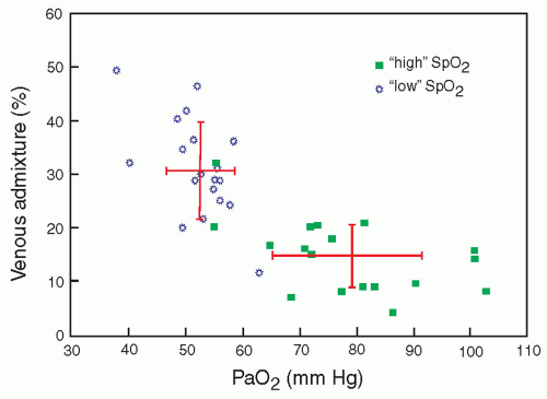
FIGURE 28.1 Relationship between venous admixture and PaO2. From Schulze A, Whyte RK, Way RC, et al. Effect of the arterial oxygenation level on cardiac output, oxygen extraction, and oxygen consumption in low birth weight infants receiving mechanical ventilation. J Pediatr 1995;126(5):777, with permission.

One possibility for why there is a difference in mortality may relate to episodes in which arterial oxygen saturation (SaO2) levels in the lower target group were associated with lower than expected concomitant arterial PO2 tension measurements. An example of this possibility, obtained from a separate set of infants less than 29 weeks of gestational age (6), is demonstrated in Figure 28.1. Low oxygen saturations also have been associated with transient increases in PVR in infants.


Calculations of Oxygen Gradient

The gap between the alveolar partial pressure of oxygen (PAO2) and the PaO2 indicates the magnitude of the arterial O2 gradient across the lungs and provides an indication of the magnitude of right-to-left shunting of blood. A simplification of the alveolar air equation provides an estimate of PAO2, that is, PAO2 = PiO2 – PaCO2.

Because at sea level, the barometric pressure, minus water vapor pressure, is approximately 700 mm Hg, the percent of inspired oxygen multiplied by 7 equals PiO2 in mm Hg (e.g., 21% ≅ 147 mm Hg, 50% ≅ 350 mm Hg). Because PaCO2 approximates PACO2 (because of a usually small arterial-alveolar CO2 gradient [aADCO2]), PaCO2 can be substituted for PACO2, and PAO2 can be derived. For example, if an infant is breathing gas with FiO2 of 0.6, with the measured PaO2 of 70 mm Hg and the PaCO2 of 40 mm Hg, the PAO2 = 420 – 40 = 380 mm Hg, and the alveolar-arterial gradient for O2 (AaDO2) approximates 310 mm Hg. In an infant without either lung disease or a significant right-to-left cardiac shunt, the AaDO2 should not exceed 25 mm Hg while breathing ambient air. Infants with severe RDS may have an AaDO2 in excess of 500 mm Hg while breathing 100% oxygen.


Oxygen Delivery

Each gram of HbF binds 1.37 mL of oxygen. The full-term newborn with a Hb of 17 g/dL binds and transports 23 mL of oxygen per 100 mL of blood. Less than 2% of transported O2 is carried as oxygen dissolved in plasma. Normal tissue oxygen consumption extracts 4 mL O2/100 mL if oxygen consumption and cardiac output are normal. HbF binds oxygen with a greater affinity than does adult HbA. The oxyhemoglobin saturation curve is nonlinear, and the P50, the PaO2 at which Hb is 50% saturated, increases with gestational age (Fig. 28.2). The higher the P50, the greater the driving pressure for oxygen unloading. The curve gradually shifts to the right as HbA increases after birth. Several factors can adversely affect tissue oxygen delivery, including decreased cardiac output, maldistribution of cardiac output, arterial vasoconstriction, and shifts in the O2 dissociation curve. Oxygen unloading in the tissues is increased with a shift to the right of the O2 dissociation curve (i.e., decreased O2 affinity of Hb) facilitated by a local decrease in pH, increase in PaCO2, and increase in temperature. A shift to the right of the O2 dissociation curve can result from transfusion of adult red blood cells. Oxygen uptake depends on adequate alveolar ventilation (VA), an appropriate ventilation-perfusion match in the lungs, and absence of rightto-left shunting. Oxygen uptake or increased O2 affinity of Hb (associated with a shift of the curve to the left) is enhanced by alkalosis, decreased temperature, decreased 2,3-diphosphoglycerate, and increased HbF.


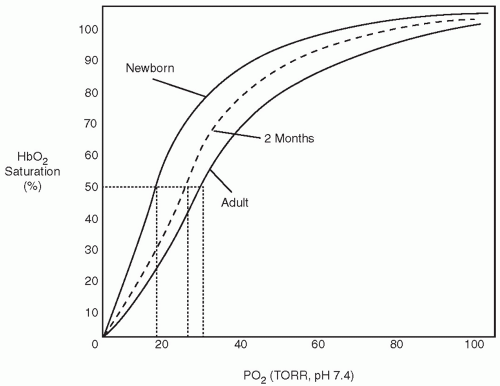
FIGURE 28.2 Oxygen equilibrium curves of hemoglobin at birth, 2 months of age, and adulthood. Note the increase in P50 with age.


Oxygen Administration

There are two methods by which to deliver supplemental oxygen to neonates: an oxygen hood with sufficient gas flow to prevent CO2 retention and a nasal cannula or prongs. The concentration and the rate of flow are varied, and the precise amount of oxygen delivered to the lungs by nasal prongs is difficult to determine. This is because there may be dilution of inspired air through ill-fitting prongs or through an open mouth. Estimates of the effective FiO2 have been based on patient’s weight, gas flow rate, and concentration of oxygen blended in the blender (7). The O2-air mixture should be warmed to the same temperature as the incubator air, which should be in the range of thermal neutrality.


▪ ASSESSMENT OF GAS EXCHANGE


Clinical Assessment

The infant with respiratory problems may present with a wide spectrum of clinical findings. The infant’s response depends on the degree of prematurity, lung and chest wall development, and maturation of respiratory control. The full-term infant may be
able to increase the work of breathing to accomplish adequate gas exchange without treatment, including oxygen administration. The extremely premature infant will have a much weaker respiratory drive and inadequate muscular development and, thus, is less able to compensate for lung abnormalities. The classic clinical signs of respiratory distress are helpful in the assessment of the mature newborn infant. Nasal flaring, grunting respirations, and tachypnea are almost always present. With progression of lung disease and decreased lung compliance, chest wall retractions become more marked. With increased work of breathing, retractions progress from sternal to subcostal, to intercostal, and then to a seesaw pattern of chest and abdominal wall movement. The full-term infant may increase respiratory rate above 100 per minute, with shallow respirations. This pattern is the most efficient way to increase gas exchange, with the least costly work of breathing. Expiratory grunting represents an effort to retard expiratory flow to increase end-expiratory pressure and maintain alveolar patency. It is unsafe to rely on color changes as an indication of oxygenation, as abnormalities in peripheral perfusion as a result of poor cardiac output, hypotension, or hypovolemia may be misleading. Similarly, infants with recurrent apnea will have intermittent deficiency in gas exchange. Auscultation assists in determining the quality of air entry in various parts of the lung and the presence of airway secretions or obstructions.


Transcutaneous PO2 and PCO2

Transcutaneous PO2 monitoring can be used selectively in conjunction with pulse oximetry. Most devices contain oxygen (O2) and CO2 electrodes. Noninvasive PO2 measurements continue to be helpful in certain situations, particularly if transcutaneous PCO2 also is obtained. Calibration before use and correlation with an arterial sample are necessary, but the need for subsequent blood samples should be reduced.

Arterial PO2 and transcutaneous PO2 are not identical. Differences can arise from local O2 consumption by the skin or by the electrode itself, heating of the skin, O2 diffusion time, and response time of the electrode (8). Skin blood flow may be affected by vasopressor medications, hypotension, and shock (9). Use of transcutaneous PO2 can reduce the number of blood sampling procedures, particularly during a period when rapid changes in O2 administration or mechanical ventilatory settings are taking place. Continuous monitoring for several hours allows assessment of changes as a result of position, handling, suctioning, and feeding and for comparison with SaO2 monitoring.


Pulse Oximetry

Pulse oximetry provides a safe, accurate, and noninvasive adjunct to the assessment of tissue oxygenation (10). Oxygen saturation is determined by infrared spectrometry, utilizing two electrodes and a small cuff that can be placed around a hand, foot, or toe without requiring heating or calibration. One electrode contains two diodes that emit light at two wavelengths: red at 660 nm and infrared at 940 nm. The other electrode senses the light from both of these diodes that has not been absorbed by blood or tissue. The relative concentration of hemoglobin-oxygen (HbO2) and deoxyhemoglobin determines the amount of transmitted light, because different forms of Hb have markedly different absorption characteristics. The ratio of the amount of light absorbed at each wavelength is used to calculate a SaO2 value. The pulsed element of the apparatus allows the instrument to differentiate added arterial blood oxygenation and absorption from tissue, and it subtracts the amount contributed by nonpulsatile venous blood flow. With PO2 values greater than 40 mm Hg, the saturation accurately reflects measurements of PO2 obtained by catheter sample or by transcutaneous PO2 (11). The latest monitors, like the Masimo (Masimo Corporation, Irvine, CA), use signal extraction technology (SET), a better method of acquiring, processing, and reporting both the SaO2 and pulse rate. This greatly improves the accuracy of SpO2 monitoring, even with motion and poor peripheral perfusion.

A PaO2 of 60 to 90 mm Hg results in a saturation value of 94% to 98% (see Fig. 28.2), and changes of 1% to 2% usually reflect a PaO2 change of 6 to 12 mm Hg. The point of inflection at which the HbO2 dissociation curve grows steep has considerable variability and depends on proportions of HbA, HbF, PCO2, pH, and temperature. Generally, these variables are not so critical to the interpretation of the percent SaO2 in arterial blood as they are to PaO2. Below 40 mm Hg, the SaO2 falls below 90%. Poor correlation with PaO2 exists when the SaO2 is above 96%, in which case the PaO2 may be well above 100 mm Hg.


Near-Infrared Spectroscopy

Utilization of the unique light-absorbing properties of Hb and HbO2, as used in pulsed oximetry, has led to a sophisticated method of appraising tissue oxygenation by means of near-infrared spectroscopy. Near-infrared light penetrates the skin, bone, and various tissues and can be detected by electrodes placed in two locations, typically the skull and flank to estimate the percentage of Hb saturated with oxygen in the tissue under the sensor, thus monitoring for changes in the circulatory systems by following the trend of changes in the regional Hb oxygen saturation. This permits assessment of cerebral tissue O2 and alterations in cerebral blood volume. Hb and cytochrome a and a3 (cyt a, a3) change their absorption characteristics according to the degree of oxygenation. The wavelength at which maximal absorption occurs is different for HbO2, deoxygenated Hb, total Hb, and reduced and oxygenated cyt a, a3. Preterm and term infants have been the focus of several research projects (12,13,14,15).

The technique is gaining more widespread use in neonatology and in the pediatric and neonatal cardiac intensive care patient, particularly those patients greater than 2.5 kg at risk for reduced flow or no-flow ischemic states.


End-Tidal CO2 Monitoring

The concentration of CO2 at the mouth or nose rises to reach a plateau at the end of each breath. This plateau reflects the alveolar CO2 concentration under normal conditions but may be inaccurate if there is marked ventilation-perfusion inequality or inhomogeneity of lung disease. Recent refinements to end-tidal CO2 detection equipment permit in-line or “mainstream” infrared monitoring just proximal to the endotracheal tube (capnography), with a continuous display of the PCO2 waveform. There is minimal dead space of the apparatus, and sampling accuracy has improved to compensate for the low expiratory flow rates characteristic of small premature infants. End-tidal CO2 has historically been less reliable in extremely small premature infants. Capnography is as accurate as capillary PCO2 but is less precise than is transcutaneous monitoring (16,17).


Hazards of High or Low Arterial CO2

Identifying a safe range of PCO2 has proven as difficult as for PaO2. There are substantial risks to hypocarbia (Fig. 28.3) (18) (Table 28.1). Limiting the exposure time to hypocarbia may be important in presenting the development of cystic periventricular leukomalacia or decreasing the risk of CLD (19,20). There has been an increase in use of permissive hypercarbia, by which PaCO2 levels of 50 to 60 mm Hg are sought and maintained. The rationale is to avoid high peak inspiratory pressure (PIP), or to delay the onset of, or to avoid, assisted ventilation (21).


▪ STRATEGIES FOR RESPIRATORY SUPPORT


Continuous Positive Airway Pressure

The application of end-expiratory pressure is intended to prevent alveoli and/or terminal airways from collapsing to airlessness. Continuous positive airway pressure (CPAP) may be applied during
spontaneous breathing or as positive end-expiratory pressure (PEEP) during mechanical ventilation. This usually requires pressures from at least 5 cm H2O to 8 cm H2O if the lung disease is severe.


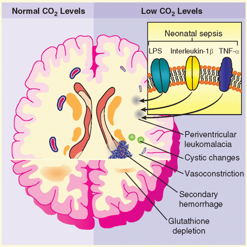
FIGURE 28.3 Effects of hypocapnia on the brain in premature infants. Hypocapnia has been implicated in the pathogenesis of neonatal white matter injuries, including periventricular leukomalacia, resulting in IVH. At normal CO2 levels (left-hand side of figure), cerebral blood flow is determined by local metabolic demand. Prolonged or severe hypocapnia includes severe cerebral vasoconstriction, resulting in brain ischemia, particularly in poorly perfused areas of the brain such as watershed areas (right-hand side of figure). This ischemia may initiate white matter destruction in the brain of premature infants. Additionally, antioxidant depletion (caused by excitatory amino acids), lipopolysaccharide (LPS), and cytokines produced in response to sepsis, such as interleukin-1β and tumor necrosis factor-α (TNF-α), potentiate the process. Finally, restoration of the normal partial pressure of arterial CO2 can result in cerebral vasodilation, which may precipitate or contribute to intraventricular hemorrhage. From Laffey JG, Kavanagh BP. Hypocapnia. N Engl J Med 2002;347(1):43, with permission.

The physiologic effects of CPAP/PEEP may vary depending on the underlying pulmonary pathology, although the primary goal is to prevent alveolar collapse. Grunting respirations in infants with respiratory distress suggest laryngeal narrowing and increased resistance to expiratory flow to increase end-expiratory alveolar pressure. In the surfactant-deficient state, alveoli will collapse at end-expiration unless a minimum distending pressure is maintained. CPAP of 4 to 5 cm H2O will prevent alveolar collapse but will not recruit atelectatic alveoli. Opening pressures of 12 to 15 cm H2O are required to inflate collapsed alveoli. The infant will need to create a large distending airway pressure in the absence of CPAP. The shear forces from opening and closing of small airways may contribute to alveolar epithelial damage. Additionally, resultant abnormal distending forces on terminal or respiratory bronchioles will contribute to small airway injury. Therefore, inflation and deflation may occur on the flatter portion of the pressure-volume curve and increase the work of breathing. CPAP theoretically could stimulate surfactant secretion. Maintenance of alveolar volume will reduce right-to-left shunting of blood through atelectatic alveoli, hence reducing oxygen needs.


TABLE 28.1 Risks of Hypocarbia and Hypercarbia


Hypocarbia


  • Overventilation increases risk for pulmonary injury


  • Decreases cerebral perfusion, associated with ischemic white matter injury


  • Increase in pH, interfering with tissue O2 unloading


Hypercarbia


  • Underventilation may increase areas of lung collapse and increase VA/Q mismatching necessitating higher FiO2


  • May decrease pH elevating PVR and increase VA/Q mismatching


  • Increases cerebral perfusion with associated risk of hemorrhagic infarctions


Recurrent Apnea

CPAP helps some infants with recurrent apnea of prematurity to sustain a more regular respiratory rate. The mechanism of its action is not well understood, although an increase in functional residual capacity (FRC) may alter the Hering-Breuer reflex or stabilize the thoracic cage, minimizing chest wall distortion and possibly altering inhibitory spinal cord reflexes (23). CPAP also helps to overcome obstructive apnea and decreases total respiratory system resistance (24,25).

Earlier methods of applying CPAP utilized an enclosed head box, face masks, and nasopharyngeal tubes. More recently, nasal prongs have been adapted to fit most infants. One device (the ALADDIN Infant Flow System, Hamilton Medical Inc., Reno, NV) appears to be well tolerated by both large and small infants. This apparatus maintains a constant flow of air by incorporating a double fluidic jet system within the apparatus. During inspiration, one jet maintains the flow to match the infant’s inspiratory effort; during expiration, gas flow is reversed by a second jet to assist outflow while maintaining a constant minimum pressure. This system presumably does not add to the work of breathing and reduces the need to use high flow rates to compensate for air leak around the nasal prongs. A second system utilizes a variable depth water seal for the expiratory circuit to sustain continuous airway pressure (26). CPAP can also be applied utilizing constant flow ventilators.


Effectiveness

Does the use of CPAP decrease the need for assisted ventilation and does it prevent or ameliorate CLD in very-low-birth-weight (VLBW) infants? Clinical trials conducted before the era of routine surfactant use and contemporary neonatal ventilation devices are now of limited relevance. Recent studies comparing the efficacy of conventional mechanical ventilation (CMV) to CPAP following the administration of surfactant found some differences in need for ventilation as an outcome although the number of infants studied was small (22,27,28).

CPAP is used to facilitate weaning from mechanical ventilation. Some infants, experiencing recurrent apneic episodes, appear to benefit (29), whereas infants evaluated in other studies have shown no benefit (30). Additional information is needed to confirm whether CPAP is an effective adjunct to successful extubation. The technique of nasal intermittent positive-pressure ventilation (NIPPV) may gain widespread use if it can be shown to improve long-term outcome (31).


Assisted Mechanical Ventilation

Treatment with assisted ventilation is applied commonly to newborns across the neonatal birth weight spectrum (Fig. 28.4) (32). The severity of respiratory disease often depends on gestational age. In moderate preterm birth, 30% of these infants need nCPAP alone, another 30% were mechanically ventilated, and 35% were administered surfactant. Up to 45% of infants born late preterm with respiratory failure were given surfactant.

Neonatologists must be expert in providing assisted ventilation across a 10-fold range of patient weight (0.5 to 5 kg) and providing safe and effective assisted ventilation across a range of lung development from premature airways with preacinar gas exchange spaces to a virtually completely alveolarized organ. Thus, both size (patient and lung volume) and range of development are immense.


The Immature Lung

The immature lung presents a special hazard for the application of assisted ventilation. The application of positive pressure for the purpose of increasing ventilation and optimizing ventilation-perfusion (image) matching may injure epithelial and endothelial tissues. In the incompletely developed lungs, structures are less elastic and more vulnerable to barotrauma and volutrauma. Injury to the mesenchymal and epithelial tissues that later give rise to alveolar septation and vascular formation may be irreversible. Studies in adult animals have demonstrated that otherwise-healthy lungs can suffer injury, which is reflected by increased airway fluid and deterioration of gas exchange, if inappropriate distending lung pressures are applied (33). The particular problems of providing assisted ventilation are illustrated in Figure 28.5 (34). Relative immaturity of distal bronchioles and respiratory ducts, coupled with fluid-filled and collapsed alveoli, create a set of conditions leading to overdistention of some areas and underventilation of other areas, with resultant ineffective gas exchange. This uneven ventilation, coupled with injury produced by ROS, contributes to the common problem of CLD of prematurity. The risk of its development is inversely correlated with birth weight.

Great strides have been made in understanding how the immature lung differs from a mature lung in phospholipid and surfactant-associated protein biosynthesis. However, there are factors other than surfactant biosynthesis that are unique to the immature lung and that increase the susceptibility to injury. These factors include, but are not limited to, incomplete development of the supportive net of collagen and elastin (35,36), incomplete development of the capillary bed in the gas exchange areas (37), relative instability of the chest wall with reduced capacity to maintain expiratory lung volume at FRC, immaturity of the neural control producing sustained spontaneous respiratory effort, and probable immaturity of the metabolic functions of the pulmonary endothelium.

Respiratory failure ensues when spontaneous breathing efforts fail to produce adequate alveolar ventilation. In newborn infants, this may occur because of failure of adequate output from central nervous system respiratory centers, an overly compliant chest wall that increases the work of breathing, metabolic problems as a result of limited energy stores, or profoundly noncompliant lungs requiring more work and depleting available energy stores. Each of these may be an indication for assisted ventilation. In most neonatal respiratory disorders, these problems occur in combination, and the diagnosis of respiratory failure cannot be ascribed to any single cause.


Establishment of an Artificial Airway


Physiologic and Anatomic Airway Peculiarities

The newborn infant has distinct anatomic and physiologic characteristics of the airways and a strong preference for nasal breathing for the first few months of life (38). Nasal or nasopharyngeal obstruction because of secretions, mucosal injury, or congenital abnormalities may produce respiratory distress. Approximately one-half of the infant’s airway resistance occurs in the nose, although the narrowness of the lower respiratory
tract results in a total airway resistance approximately 15 times greater than that of an adult (39). Edema and inflammation can produce extremely high resistance to air flow in these narrow airways. During expiration, the airways become narrower, and resistance increases.


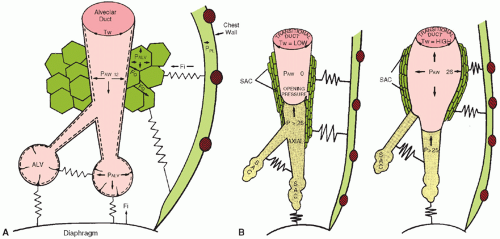
FIGURE 28.5 A: A mature alveolar duct and alveoli. Dotted line, surfactant; PALV, alveolar pressure; PAW, airway pressure; PPL, pleural pressure; Fi, tissue force (stretched springs) acting inward; Fo, tissue force directed outward; Tw, wall tension or recoil pressure. B: The end-expiratory airway pressure (PAW) equals zero in an immature distal airway (left). The saccules (SAC) and airways contain fluid (shaded area). The axial airway is concave at the air-liquid interface as a result of the surface tension forces. The peripheral SACs are collapsed or fluid filled. The lax tissues are represented by relaxed springs. The inspiratory airway pressure (PAW) is equal to 26 cm H2O (right). The distended distal airway has a high wall tension (TW). The liquid front has been pushed peripherally, but the SACs are still not inflated. From Thibeault DW, Lang MJ. Mechanisms and pathobiologic effects of barotrauma. In: Merritt TA, Northway WH Jr, Boynton BR, eds. Bronchopulmonary dysplasia. Contemporary issues in fetal neonatal medicine. Boston, MA: Blackwell Scientific Publishers, 1988:82, with permission.


FIGURE 28.6 Laryngoscopy for endotracheal intubation.


Endotracheal Intubation


Route

Orotracheal and nasotracheal intubation may be used for prolonged mechanical ventilation of term and premature infants. The principal advantage of the nasal route is the stabilization of the tube afforded by the close fit within the naris, but the nasal passages may limit the size of tube that can be used. Necrosis of the nasal septum or the alae nasi can occur if circulation is impaired because the tube is too large. Orotracheal intubation is more easily and quickly accomplished and is indicated for delivery room and emergency situations. It is the preferred route for prolonged mechanical ventilation (Fig. 28.6).

The endotracheal tube should allow a small air leak between the tube and the glottis. A tube that fits too snugly within the trachea is likely to cause pressure necrosis of the mucosa. If too large a leak is allowed, it may be difficult to achieve sufficient pressure for ventilation of noncompliant lungs. A tube with a 2.5-mm inner diameter usually fits infants weighing less than 1,000 g; a 3-mm tube for 1,000 to 2,000 g; a 3.5-mm tube for 2,000 to 3,000 g; and 3.5- to 4.0-mm tube for larger infants.


Positioning

The length of the trachea from the vocal cords to the carina varies from about 3.6 cm in the smallest premature infants to 6 cm in large, term infants. Optimal positioning for the tip of an endotracheal tube is in the middle of the trachea, in which it is least subject to dislodgment into the pharynx or displacement into a bronchus. The proper depth of insertion of an endotracheal tube, as determined by postmortem and radiographic measurements, is related to body weight (40,41). Suggested depths of insertion for orotracheal intubation are given in Table 28.2.

Immediately after intubation, the position of the tube should be confirmed by inspection and auscultation. Identification of humidity in the tube or of CO2—by a sensitive color detector— helps confirm endotracheal tube placement. Two common errors
of tube placement are intubation of the esophagus and intubation of the right mainstem bronchus. Auscultation, although helpful, is not reliable because breath sounds are well transmitted in a small chest. A chest radiograph should be obtained to confirm tube placement.


TABLE 28.2 Depth of Insertion of an Orotracheal Tube from the Lips of a Premature Infant


Infant Weight (kg)


Depth of Insertion (cm)


1.0


7


2.0


8


3.0


9


4.0


10


Indications for Assisted Ventilation

The decision for initiation of assisted ventilation should be individualized. Factors to consider include underlying disease and its expected natural history, birth weight, gestational age, postnatal age, chest radiographic appearance, progression of clinical signs, serial arterial blood gas tension measurements, and pH measurements. The criteria indicating a need for mechanical ventilation are difficult to define, and there is lack of unanimity about a particular threshold for PaO2, partial pressure of CO2 (PaCO2), or FiO2. In general, the PaO2 should be maintained at or above 50 mm Hg because of reasonable oxyhemoglobin saturation at this level, but the maximal level of inspired O2 dictating intubation and application of assisted ventilation remains controversial. A trend of rising PaCO2 with concomitant decrease in pH or onset of apnea indicates a need for mechanical assistance. After assisted ventilation is initiated, the generally accepted goals are to maintain the PaO2 between 45 and 70 mm Hg, PaCO2 between 45 and 60 mm Hg, and pH at 7.25 or more, minimizing PIP and FiO2, and optimizing MAP (PAW[gas]) and PEEP. These general guidelines must be interpreted and often must be modified to provide optimal support for the individual patient (42).

Because acute lung disease is usually more severe and protracted in more immature infants, criteria for intervention for infants weighing less than 1,000 g differ from those for larger or older infants. For example, a 750-g infant with RDS has a high probability of developing apnea, fatigue, or both, and most of these infants require assisted ventilation even if the FiO2 need is less than 40%. A 2,500 g, 36-week-old infant with RDS has greater muscular and caloric reserve and is able to sustain rapid ventilatory rates and higher respiratory work for several days without assistance. Infants of gestational age 35 to 39 weeks and older than 24 hours who develop respiratory failure with RDS may benefit from surfactant treatment (43).


Physiologic Considerations

An understanding of the effects of mechanical ventilation on the lungs requires knowledge of the interplay among thoracic mechanics, including pulmonary compliance and airway resistance, lung volumes, respiratory control mechanisms, and alveolar gas exchange.

Lung compliance, that is change in lung volume per unit pressure change, in units of mL/cm H2O depends on the elastic properties of the tissue, which are influenced by the lung volume and abnormalities such as tissue inflammation and edema. Compliance is low if there is alveolar collapse or overdistention. Expansion from alveolar collapse requires inflation pressures of 12 to 20 cm H2O in preterm infants with RDS to achieve tidal volumes of 3 to 5 mL/kg. The lungs of infants with RDS have areas of collapse and overexpansion, and there is nonuniformity of compliance. Other conditions, such as pneumothorax, lobar atelectasis or consolidation, and pulmonary edema, decrease compliance. The most relevant measure of compliance, specific compliance, is calculated by normalizing compliance by EEV, the functional equivalent of FRC measured during positive-pressure ventilation. Very low or high values for EEV will reduce compliance. Changes in compliance, EEV, and gas exchange are not concordant, at least not during RDS. This has limited the value of bedside measurements of compliance, particularly without concomitant measurements in EEV. Chest wall compliance usually is high and does not present a problem to mechanical ventilation.

Airway resistance (cm H2O/L/s) is inversely related to the fourth power of the radius during laminar air flow. Airway resistance is high in infants, increasing with low lung volumes and with obstruction of the airway. High rates of air flow increase resistance by producing turbulence in the airways.

Resistance and compliance determine the rate at which lung areas inflate and deflate. An increase in airway resistance increases the time required for air to reach the alveoli; a decrease in compliance results in less time required to reach equilibrium. The product of resistance and compliance is the pulmonary time constant. Changes in resistance or compliance can alter the pattern or distribution of ventilation, and recognition of the variations in the time constant (e.g., short with poor compliance, prolonged with increased airway resistance) helps determine respirator settings. Unfortunately, a single time constant does not exist for all lung areas during complex pulmonary disorders. Thus, all conventional positive-pressure ventilators produce areas of overinflation and underinflation of gas exchanging areas, each contributing to suboptimal gas exchange.

Because RDS should result in a short time constant, short inspiratory times are permissible, and MAP should be increased to improve oxygenation. With meconium aspiration or airway edema, the time constant is slower, and sufficient time for expiration is important to avoid gas trapping, overdistention of the lungs, and possible air leak. If the expiratory time (TE) is shorter than the time constant of the lung for expiration, overdistention results. If the overall time constant for the lung is longer than the imposed ventilator inspiratory time (TI), inadequate ventilation could result. Unequal time constants coexisting in different parts of the lung are most likely to occur if pulmonary abnormalities are unevenly distributed, as in pneumonia, meconium aspiration, pulmonary interstitial emphysema, pneumothorax, or CLD, in which case the optimal TI or TE becomes difficult to determine.

The circulatory effects of mechanically applied pressure to the alveoli are important. Normal breathing results in negative intrapleural pressure that enhances venous return and cardiac output. Positive-pressure breathing can impede venous return and may diminish cardiac output. Pressure during inspiration decreases the pulmonary capillary circulation as long as alveolar pressure exceeds capillary pressure and can affect total pulmonary blood flow and hence gas exchange.


Lung Volume Measurements During Mechanical Ventilation

Application of hot wire anemometry or pneumotachography to timecycled pressure-limited ventilation systems allows measurement of inspiratory and expiratory tidal volumes, minute ventilation (VE) and air leak around the ETT. Tidal volume can be correlated with PIP, inspiratory gas flow rate, TI, and PEEP. Knowledge of tidal volume from bedside measurements helps allow determination of the optimal PIP to achieve optimal tidal volume. For many clinical situations, this value is between 3 and 6 mL/kg. This knowledge allows minimizing PIP, which, if excessive, otherwise may induce or exacerbate the small airway injury of CLD. However, knowledge of tidal volume and VE does not provide knowledge of distribution of inspired ventilation and of (image) matching. Distribution of tidal volume may vary with the associated PIP; low PIP can
result in tidal volume distribution only to already overinflated lung regions, resulting in worsened (image) matching, development or exacerbation of high (image) areas, and worsening of CO2 retention, despite normal or elevated (image)

Many ventilators now have pulmonary graphic systems to monitor breath-by-breath pressure-volume and flow-volume loops. These graphics can be helpful in evaluating the extent of airway instability during inspiration and especially expiration. Pressure-volume curves may be helpful in detecting overdistention and/or air trapping or small airway obstruction. One limitation in the use of pulmonary graphics is the considerable breath-to-breath variability in the pattern exhibited, probably more than exists with assisted ventilation of adults, and the previously mentioned air leak around the ETT. Flow waveforms can be helpful in determining the appropriate TI. There is as yet no strong evidence suggesting that a clinically important outcome can be modified in neonates with reliance on interpretation of these patterns of airflow and pressure; however, they can be a useful adjunct to care.

Automated bedside EEV measurements are now available. The devices utilize helium dilution methodology, and early trials demonstrated safe use and reproducible measurements. Although not in routine clinical use, they may allow a more individualized approach to application of PEEP in infants.


Investigation of Newer Forms of Assisted Ventilation

The gold standard approach to the introduction of new therapies in medicine is the performance of prospective, definitive, randomized clinical trials (RCTs) with predetermined clinically relevant primary outcomes. This approach has been followed belatedly but now enthusiastically in neonatology. Part of the acceptance of the results of RCTs devolves from effective blinding of the treatment group assignment, and the widespread agreement that the control group received the best available therapy known at the time. When these principles are applied to trials testing new approaches in assisted ventilation, there are several obvious limitations.

It is difficult to conduct a blinded clinical study of two different types of assisted ventilation, especially if different ventilators are used. It is difficult to agree on what constitutes optimal standard of care to be used for the control group. This applies not only to the minutiae of management of the ventilation device itself but also to the ancillary supportive care. Even if agreement about standards of care is achieved, it is difficult to sustain adherence to these standards of care.

Contemporary ventilators designed for application to neonates are very complex devices. This complexity mandates a balance in the performance of clinical trials between the rigid prescriptions of ventilator management versus the “field level” studies with greater tolerance for physician-specific practices. The latter study design may have perhaps more applicability to broad populations but runs the risk that center-to-center variance in approach and in outcome may be greater, potentially diluting the signal of interest. Although these issues are inherent in all prospective definitive trials, they are particularly acute when applied to testing modes of assisted ventilation in neonates.

Identifying a clinically relevant outcome can be difficult. Death is the obvious outcome of interest, but for most populations of neonates treated with assisted ventilation, it may be an insensitive marker and not informative for other important outcomes. Utilizing a surrogate marker for death (e.g., need for extracorporeal membrane oxygenation [ECMO]) has worked well for clinical trials of near-term infants but will not work well for babies ineligible for ECMO (those <34 to 35 weeks of gestation). For preterm infants, evidence of pulmonary injury (i.e., CLD) in combination with death is commonly used. However, even these outcomes pose problems because of the poorly understood pathogenesis of CLD and the role of factors other than assisted ventilation that contributes to its development and severity. Combined outcomes melded into one also have the problem that reduction in one part of the primary outcome (death) may increase the incidence of the adverse-associated outcome. Some studies testing different forms of assisted ventilation were plagued by the problem that to qualify for the study, infants first were treated with one form of assisted ventilation for a variable period before the application of different patterns of ventilation could be undertaken. More recent trials, especially those testing highfrequency ventilation (HFV) (44,45,46), have largely avoided this problem. Some studies, even those purporting to show a benefit from an experimental form of ventilation, may demonstrate the benefit because the control patients demonstrated a higher than expected rate of adverse outcomes. Successful recruitment of sufficiently large populations in sufficiently short time and the ability to stratify them in a meaningful way can also prove to be difficult. This is increasingly true, given the role of economics in the decision regarding whether eligible patients for definitive clinical trials are moved from one center to another to participate in those trials (47).

Despite these limitations and others, advances have occurred in the application of assisted ventilation using RCTs. RCTs will remain valuable to better understand the limitations and strengths of the many different patterns of assisted ventilation.


Conventional Ventilation


Available Modes and Details of Use

Several manipulations of applied pressure allow for an increase in PaO2. The physician decides whether to increase FiO2 or PAW[gas] by considering the prior settings and balancing the possible harmful effects of increasing PAW[gas] against those of increasing FiO2, recognizing that threshold limits are arbitrary. If the FiO2 is approaching 1, and SpO2 or PaO2 are unacceptably low, then other options must be invoked. During time-cycled, pressure-limited ventilation, if PEEP is already 6 to 7 cm H2O, the PIP or the TI is increased. The use of PEEP helps to maintain patent small airways and prevent collapse to airlessness of those alveoli already open. Inspiratory pressures of greater than 15 cm H2O usually are required to open collapsed or fluid-filled acinar areas. A combination of an increase in the TI with 6 cm H2O PEEP may be helpful during the initial phase of assisted ventilation for RDS. With subsequent opening of air spaces, the optimal TI may need to be decreased. The use of an end-inspiratory pause or plateau should improve the distribution of inspired gas if there are regional differences in airway resistance. However, if the alveolar pressure exceeds capillary pressure, there will be tamponade of the pulmonary circulation and development of high (image) areas (48).

Hypoxemia may persist during all combinations of ventilator settings in some conditions, and other underlying abnormalities should be suspected. The clinician should always consider the degree of air leak around the endotracheal tube when adjusting pressure and flow rates. Echocardiographic evaluation of the infant for coexisting pulmonary vascular hypertension or structural or functional heart disease is then indicated. Other management considerations are listed in Table 28.3.

If there is airway obstruction, as may occur with CLD or meconium aspiration, optimal ventilator settings may differ from those used for RDS. Because there is a relatively long time constant, the gas flow rate should not be too rapid, and there should be adequate time for expiration. One of the multiple available newer modes of assisted ventilation may then be useful.


Patient-Triggered Ventilation

The goal of patient-triggered ventilation is to maximize the efficiency of spontaneous breathing efforts although minimizing the risk of insufficient ventilation or trauma to airways (49). All patterns of patient-triggered ventilation require a rapidly responding sensor and transducer that can detect the onset of spontaneous
inspiratory effort and provide the mechanical initiation of machineassisted ventilation during the early phase of the infant’s inspiration. Currently used methods to signal the initiation of inspiratory effort are listed in Table 28.4. The current methodology allows transduction to be accomplished in as short a time as 30 to 50 milliseconds, approximately one-tenth the duration of the inspiratory phase of a spontaneous respiratory cycle (Fig. 28.7) (50). The means by which this signal is provided and the addition of other subtle but potentially important changes in the capabilities of particular ventilators differentiate one type of conventional neonatal ventilator from another (51). A list of available modes and their theoretical advantages is found (Table 28.5).


TABLE 28.3 Management Considerations


  • Utilize alternative mode of ventilation (e.g., HFV, SIMV, A/C, PRVC)


  • Raise the hematocrit to 45%—50% with packed erythrocyte transfusions


  • Reposition the baby into prone position if supine or into left or right lateral positions


  • Use of sedation with or without paralysis; if the infant is paralyzed, discontinue its use


  • Change to a larger size endotracheal tube to diminish air leak


  • Consider repeated doses of exogenous surfactant beyond 24 h of age


  • Administration of diuretics


  • Reevaluate hemodynamic status and treat appropriately


  • Undertake trial of NO


  • Consider use of corticosteroids


A/C, assist/control; HFV, high-frequency ventilation; IMV, intermittent mandatory ventilation; SIMV, synchronous intermittent mandatory ventilation; PRVC, pressure-regulated volume control.


Although it is not clear that modern means of patient-triggered ventilation have achieved their optimum, these methods have already gained widespread acceptance for three reasons. These include the clinical impression that infants are more comfortable and less distressed although being ventilated with patient-triggered ventilation; there may be at least modest improvements in pulmonary gas exchange during patient-triggered ventilation, and there appears to be decreased need for sedation and muscle relaxation. Even with patient-triggered ventilation, it is important to recognize the pitfalls that may occur when the synchronized intermittent mandatory ventilation (SIMV) rates are too high or when the patient is allowed to breathe in the assist/control mode (Fig. 28.8) (52). With assist/control, hyperventilation may occur, especially if the sensor for initiation of respiration is inappropriately sensitive and triggers ventilator breaths that are not associated with patient inspiratory effort. If the goal is to avoid breaths triggered late in inspiration or during expiration, then flow-triggering systems are less prone to auto triggering and have a shorter and more consistent response time than do impedance-triggered systems (53). The “Pressure-Regulated Volume Control” mode available in some ventilators has proven useful in correcting gas exchange problems in larger preterm infants or term infants, although air leak with neonatal endotracheal tube makes this mode less effective in some patients.


TABLE 28.4 Available Mechanisms for Detecting Onset of Respiration


Advantage


Disadvantage


Body movement measurement


No dead space


Position dependent


Transthoracic impedance


No dead space


Position dependent


Airway pressure sensors


Little dead space


Speed


Airflow sensors


Pneumotachography


Sensitive


Added dead space


Hotwire anemometer


Sensitive


Too sensitive


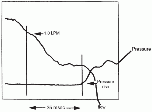
FIGURE 28.7 System response time, also known as trigger delay. The flow change trigger is set at 1.0 LPM. It took 25 milliseconds from the time this threshold was reached (vertical line on the left) until there was a measurable rise in airway pressure (vertical line on the right). From Donn SM, Sinha SK. Controversies in patient-triggered ventilation. Clin Perinatol 1998;25:49, with permission.

However, the set tidal volume is arbitrary because of loss of volume to the compliant breathing circuit leading to inaccuracy of its measurement (51). If end-tidal CO2 is measured with the ventilator, then a second measure of tidal volume (VT) is made at the breathing tube, which may be more reflective of actual VT. No definitive clinical trial of this variable flow mode of ventilation compared to conventional patterns of ventilation in the treatment of low-birthweight (LBW) infants has been reported.

A limitation of many devices is that the individual breaths generated by the ventilator are monomorphic. Proportional assist ventilation may be one way to overcome the problem and provide improved individualization of support. With proportional assist ventilation, the relationship between patient-induced inspiratory effort and ventilator response is interactive. During proportional assist ventilation, the ventilator amplifies the patient’s effort throughout the inspiratory phase of the cycle. With each spontaneously generated breath, the patient can individualize the
machine-initiated tidal volume and flow patterns. Sensors monitor instantaneous flow rate and volume of gas from ventilator to patient; the applied pressure then changes according to the equation of motion. This system may allow for both greater patient comfort and reduction of peak airway pressure required to sustain ventilation, with less likelihood of overventilation compared to assist/control modes (52).


TABLE 28.5 Modes of Assisted Ventilation Via Endotracheal Tube


Modes


Theoretical Advantages


IMV


Pressure- and time-limited breaths


Fresh gas flow allowing efforts at spontaneous breathing


SIMV


Synchronized, controllable number of assisted breaths


A/C


Every spontaneous breath assisted with a monomorphic wave form (with a set TI and PIP)


AC + VG as above but Vt is set, and PIP adjusts in an attempt to maintain that Vt


PAV


Respiratory pressure is servocontrolled and is proportional to endogenous volume and flow


PSV


Inspiratory time and flow are variable but PIP is still fixed


PSV + VG


Time variable inspiration


Constant flow


Automatic attempt to deliver same tidal volume


NAVA


Patient determines RR, tidal volume, inspiratory time


Reduces asynchrony


IMV, intermittent mandatory ventilation; SIMV, synchronized intermittent mandatory ventilation; A/C, assist/control; PAV, proportional assist ventilation; PSV, pressure support ventilation; VG, volume guarantee; NAVA, neurally adjusted ventilatory assist.


FIGURE 28.8 Tidal volume tracings (Inspiration = Upward) demonstrating three patterns of ventilator interaction with spontaneous breathing. In this illustration, tidal volume of spontaneous breaths is less than that of ventilator breaths. A: Asynchronous intermittent mandatory ventilation (IMV) with ventilator breaths delivered during spontaneous expiration. During IMV, ventilator breaths occur at a constant rate, with random timing with respect to spontaneous breaths. B: Synchronous intermittent mandatory ventilation (SIMV) with ventilator breaths delivered early in selected spontaneous inspirations. During SIMV, ventilator breaths occur more irregularly, but the ventilator delivers the set rate synchronously with spontaneous breaths. C: Assist/control mode, with ventilatory breaths delivered early in all spontaneous inspirations. The assist/control mode delivers ventilatory breaths synchronously with all spontaneous breaths and may lead to increased ventilation. From Cleary JP, Bernstein G, Mannino FL, et al. Improved oxygenation during synchronized intermittent mandatory ventilation in neonates with respiratory distress syndrome: a randomized, crossover study. J Pediatr 1995;126:407, with permission.

Neurally adjusted ventilatory assist (NAVA) is a relatively new ventilator modality that is currently available for the neonatal and infant population. NAVA assists ventilation by capturing and analyzing the electrical activity of the diaphragm (Edi) via electrodes embedded in a feeding tube (Edi catheter). NAVA utilizes the Edi to trigger the ventilator, allowing the patient to determine their respiratory rate, tidal volume, and inspiratory time. Although studies remain limited, this method of ventilation appears to be safe and effective and reduce asynchrony (54,55,56).

No studies have been reported to date that measured a clinically important outcome after a significant duration of exposure to a patient interactive device compared to another similar device. One study utilizing a crossover design, in infants with RDS, examined the effect of pressure support ventilation plus volume guarantee, a volume targeted form of ventilation. Results of measurements of EEV and of minute ventilation are shown in Figures 28.9 and 28.10 (53). No obvious improvement could be detected in VE, a/A, or EEV when PCO2 was controlled.

The ability to reduce assisted ventilatory support in a controlled manner with patient interaction devices is improved compared to that with ventilator models available previously. Decremental changes in back-up rate, PIP, and FiO2 can be accomplished smoothly and cumulatively reduce the reliance on the “trial-anderror” approach to reduction in ventilatory support necessary with older generation devices. One established adjunctive therapy for VLBW infants is the use of methylxanthine therapy. Routine use of this medical therapy reduces the need for reintubation and reintroduction of assisted ventilation (57).


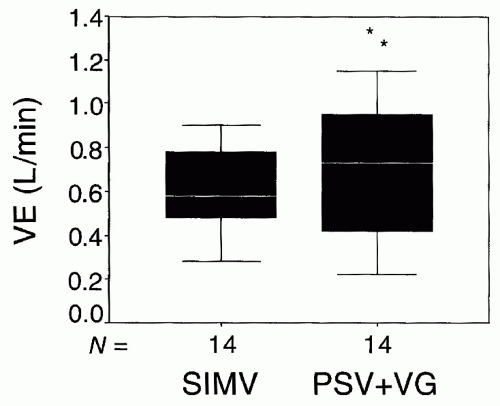
FIGURE 28.9 The relationship between the VE and the mode of ventilation (SIMV or PSV + VG). The difference between the two modes is significant (*p = 0.012). From Olsen SL, Thibeault DW, Truog WE. Crossover trial comparing pressure support with synchronized intermittent mandatory ventilation. J Perinatol 2002;22:461, with permission.

A new modality that will be very useful in the future is the automated control of inspired oxygen, currently not available for every ventilator and/or every country. Few studies indicate that it is possible to safely and efficaciously automatically adjust the FiO2, to maintain a predetermined SpO2 within an intended range (58,59).


Pressure Regulation versus Volume Guidance

It is helpful to think of current modes of conventional rate-assisted ventilation as either pressure controlled or volume targeted (60

Only gold members can continue reading. Log In or Register to continue

Stay updated, free articles. Join our Telegram channel

May 30, 2016 | Posted by in PEDIATRICS | Comments Off on Principles of Management of Respiratory Problems

Full access? Get Clinical Tree

Get Clinical Tree app for offline access