Dermatologic Conditions


Dermatologic Conditions


James G. Dinulos


▪ INTRODUCTION

As an organ, the skin provides essential functions for survival and can be an indicator of overall newborn health (1). At no other time in life are these roles more evident than in a newborn infant who is adjusting to life outside the uterus. Abnormalities in the skin may be extremely distressing to parents, and it is important for medical providers to be able to distinguish a worrisome from a banal newborn skin disorder. To assist in this process, this chapter reviews basics of skin structure and development, techniques of skin examination and recognition, and management of important newborn skin disorders.


▪ SKIN DEVELOPMENT, STRUCTURE, AND FUNCTION

The skin is composed of three anatomic layers (epidermis, dermis, and fat) and three adnexal structures (hair, nails, and glands). The formation of these skin layers and structures from primitive embryonic tissues occurs in a predictable and sequential manner. Most structural development is complete by 24 weeks of gestation; however, complete functional activity of the skin, such as epidermal barrier development, is not achieved until after birth (1,2) (Fig. 51.1).

The epidermis plays critical functions in fluid homeostasis and in protection from infections, toxins, and adverse effects of ultraviolet radiation (3). The outermost layer, the stratum corneum, provides most of this protection. Lamellar bilayers composed of hydrophobic lipids, principally fatty acids, cholesterol, and ceramides, are “cemented” between multiple layers of tightly knit protein and keratin-rich cornified cells (3,4). These lipids and proteins protect by creating an impermeable barrier and by providing an acidic and xeric environment that impedes microbial invasion. The epidermis is not merely a static barrier, however, as keratinocytes actively produce antibacterial products, including cytokines, lipid breakdown products, and cationic antimicrobial peptides that provide an important link between the innate and adaptive immune systems (5,6). Recent evidence highlights the importance of the dynamic relationship between cutaneous microbial communities and host immunologic responses (6).

Prematurity and a number of skin disorders disrupt epidermal permeability barrier function and may allow for massive transepidermal water loss (TEWL) (7). Premature infants can lose up to 30% of their total body weight in 24 hours, as their rate of TEWL may be 10 to 15 times greater than in a full-term infant (8). Such significant fluid losses can cause hypotension, electrolyte imbalance, and increased caloric demands and may contribute to intraventricular hemorrhage (IVH) and necrotizing enterocolitis. Epidermal barrier development is accelerated by exposure to the dry, extrauterine environment (8). This barrier maturation typically takes 2 to 4 weeks and may be delayed for 8 weeks in extremely premature infants. Thus, premature infants are particularly vulnerable to infections and toxins, particularly during the first week of life when approximately two-thirds of all neonatal deaths worldwide occur.

During development, skin and its structures are thought to migrate along the “lines of Blaschko” (9). Most authorities believe that Blaschko lines are an expression of epidermal and not dermal migration. Conditions that follow these lines are thought to result from a single postzygotic mutation of a single clone of cells or from a mutation on the X chromosome that is brought about by X inactivation (10). Incontinentia pigmenti (IP) and linear epidermal nevi are two examples of conditions that occur along lines of Blaschko.

Newborn skin may look and feel different depending on the newborn’s gestational age. Premature infants less than 32 weeks of estimated gestational age (EGA) have skin that appears thin and transparent. Infants born after 40 weeks of EGA have thicker skin with wrinkles and peeling. Most term infants have a pasty substance on their skin, referred to as vernix caseosa. Vernix caseosa is a substance composed of sloughed corneocytes, shed lanugo hair, and lipids (11). The role of this substance is not fully understood, although it is thought to assist in passage through the birth canal and have an important role in skin hydration and innate immune defense (12). Most premature and postmature infants have little vernix.

Skin adnexal structures (hair, nails, and glands) can provide valuable clues to the overall well-being of the newborn. Most infants are born with hair on the scalp, and some have hair on the body. Typically, hair in the newborn undergoes “physiologic” shedding at approximately 3 months of age.


▪ EXAMINATION OF NEWBORN SKIN

Newborns should be examined with an adequate light source, preferably natural sunlight. Sidelighting and magnification are helpful to accentuate surface topography. Handheld devices applied directly to the skin such as a dermatoscope can be helpful, especially to examine pigmented lesions. The environment should be kept warm to minimize reactive vascular changes as a result of temperature. Table 51.1 summarizes the steps of a newborn skin examination and an approach to dermatologic diagnosis. Table 51.2 lists primary lesions, secondary lesions, special configurations, and distributions of skin lesions.


▪ SCALY RASHES

Rashes are common in the first month of life, affecting 80% of newborns (13). During the first week of life, the vast majority of term and postterm infants will have superficial peeling or desquamation of skin; it rarely occurs in those under 35 weeks of gestation. Deeper skin peeling that results in denuded skin and continuous peeling for longer than 1 week are potential warning signs of an underlying systemic process. Scales should be examined for quality (firm, adherent), color (white, yellow), and associated inflammation (redness), features that help in the identification of a variety of skin diseases.


Inflammatory Rashes


Diaper Dermatitis

Diaper dermatitis is common in newborns. It is characterized by a pink to red scaly rash localized to the diaper area and is caused by prolonged exposure to urine and stool combined with the frictional forces of the diaper. This irritant contact dermatitis should be distinguished from numerous other dermatologic conditions affecting the diaper area. Irritant contact dermatitis affects skin contacting the diaper, such as the pubis, penis, labia majora, scrotum, upper thighs, and buttocks. Generally, the creases are spared. Continued irritant exposure may produce painful fissures and ulcerations (Jacquet erosive dermatitis), especially in premature infants (e-Fig. 51.1). Involved skin is susceptible to secondary infection, primarily with Candida albicans, Staphylococcus aureus, and Streptococcus pyogenes. C. albicans causes pustules on a red base (“beefy-red”) that, after rupture, form erosions surrounded by fine superficial white scale. Frequently, the inguinal and gluteal folds are involved. S. aureus and S. pyogenes cause a superficial skin
infection called impetigo, which is characterized by pustules on a red base that form yellow crusts when ruptured. Certain types of S. aureus produce an epidermolytic toxin, causing superficial bullae. However, secondary bacterial infection with S. aureus and S. pyogenes may be characterized primarily by superficial erosions and crusting. Streptococcal infection is malodorous, which may be used to distinguish it from Candida infection.


FIGURE 51.1 Anatomy of the skin. From Fletcher MA. Physical diagnosis in neonatology. Philadelphia, PA: Lippincott-Raven Publishers, 1998:108.

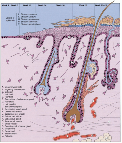
Factors involved in the genesis of diaper dermatitis include increased skin wetness (e.g., sweat, urine, and stool) of the stratum corneum (14), which makes the skin more susceptible to friction from the diaper material, and irritation from elevated pH caused by urinary ammonia and from activation of fecal proteases and lipases in the alkaline milieu (15,16). When alkaline urine combines with feces, the potential for irritation is compounded. Breastfed infants have been noted to have stools with lower pH, which may account for their decreased incidence of diaper dermatitis.

The differential diagnosis of irritant diaper dermatitis includes psoriasis, seborrheic dermatitis, secondary syphilis, histiocytosis, acrodermatitis enteropathica (AE), and cystic fibrosis. In those conditions, however, unlike diaper dermatitis, skin outside the diaper area is usually involved.


TABLE 51.1 Steps in a Newborn Skin Examination


  1. Assess skin integrity.


    1. Skin breakdown


    2. Texture


    3. Hydration


    4. Swelling


  2. Determine primary and secondary skin lesions (see Table 51.2).


  3. Determine the location, configuration, and distribution of the skin lesions.


  4. Evaluate mucous membranes, palms, soles, hair, and nails.


  5. If there is a rash, determine whether it is fixed or evolving.


  6. Develop a differential diagnosis.


  7. Perform appropriate diagnostic tests.


  8. Consider referral to a specialist.


Treatment of diaper dermatitis should be targeted toward reducing skin wetness, minimizing contact of the skin with urine and feces, and eradicating infectious organisms. Skin wetness and irritation can be minimized in several ways. Ultra-absorbent diapers have been shown to be superior to cloth diapers in decreasing skin wetness and maintaining an acidic skin pH (17,18). Frequent diaper changes also help to minimize wetness. Barrier ointments (e.g., containing petrolatum) help keep urine and feces from contacting the skin. Other commercial products containing zinc and vitamins A and D also may be effective. The skin should be gently cleansed with water or, if necessary, a mild nonalkaline soap before barrier ointment reapplication. Complete removal of barrier ointment with diaper changes is not necessary and, if attempted, may further exacerbate skin injury. When diaper dermatitis is unusually severe or recalcitrant or when risk factors are likely to be ongoing (e.g., malabsorption syndromes), a thick layer of pectin-based paste without alcohol, followed by a barrier ointment or zinc oxide ointment, may prove efficacious. Cholestyramine compounded in Aquaphor helps neutralize bile acids and is effective in erosive dermatitis (19). Commercially available diaper wipes can exacerbate irritation and should be reserved for healthy-appearing skin or for circumstances in which soap and water are not available.


TABLE 51.2 Primary and Secondary Skin Lesions, Configurations, and Locations


  1. Primary lesions


    1. Macule (<0.5 cm) and patch (>0.5 cm)—well-circumscribed flat lesion


    2. Papule (<0.5 cm) and plaque (>0.5 cm)—well-circumscribed raised lesion


    3. Vesicle (<0.5 cm) and bullae (>0.5 cm)—fluid-filled raised lesion


    4. Pustule—well-circumscribed raised fluid-filled lesion containing leukocytes


    5. Nodule (>0.5 cm)—deep-seated raised lesion


    6. Tumor—a large nodule


    7. Wheal (hive)—raised transient (<24 h) lesion


    8. Cyst—fluid-filled nodule


    9. Telangiectasia—dilated dermal blood vessel


    10. Petechiae—areas of extravasated red blood cells resulting from broken capillaries


  2. Secondary lesions


    1. Atrophy—depressed lesion


    2. Erosion—superficial break in the skin


    3. Ulceration—full thickness (i.e., break in the skin through the dermis)


    4. Scale—flakes of skin


    5. Crust—scale with serum


    6. Fissure—linear erosion


    7. Scar—fibrosis of the dermis


  3. Configuration


    1. Blaschko—embryologic lines of skin cell migration (i.e., whorled)


    2. Linear—straight


    3. Annular—round with normal skin in central “ring”


    4. Iris—round with a dark center


    5. Nummular—round, “coin shaped”


  4. Location


    1. Symmetric—both sides of the body


    2. Localized—one area


    3. Widespread—generalized


Mild, low-potency topical corticosteroids such as 1% hydrocortisone ointment can be applied if inflammation is significant; however, higher-potency topical steroids should be avoided because of the risk of cutaneous atrophy, striae, adrenal suppression, and Cushing syndrome. Combination antifungal-steroid compounds such as Lotrisone and Mycolog II have no use in treating neonatal skin conditions involving the diaper area. Each contains a potent topical corticosteroid (betamethasone dipropionate, and triamcinolone 0.1%, respectively) that when placed under diaper occlusion can cause cutaneous and systemic side effects. Use of powders also should be avoided. Fungal or bacterial superinfection can be managed with appropriate topical antimicrobial agents (20). For more severe bacterial and fungal infections, oral agents may be considered. Topical antifungal agents such as nystatin, clotrimazole, miconazole, or ketoconazole can be safely applied to newborn skin. Bacterial infection generally responds to mupirocin or bacitracin. Neomycin carries a higher risk of allergic contact sensitization and should be avoided.


Intertrigo

Intertrigo is a symmetric, red, moist, macerated eruption that occurs in the skin folds. It is thought to be as a result of excessive sweating and close approximation of the skin surfaces. Yeast and bacterial superinfection are common (see “Diaper Dermatitis”). The most effective treatment is exposure of skin to air. Barrier creams and occlusive ointments can exacerbate intertrigo. A light application of 0.5% to 1% hydrocortisone cream two to three times a day, pimecrolimus, or tacrolimus can help. Concurrent bacterial and/or yeast infections must be treated in a manner similar to irritant contact diaper dermatitis.


Seborrheic Dermatitis

Seborrheic dermatitis is characterized by red skin with white-yellow waxy scale, occurring on the scalp, on the eyebrows, and in the intertriginous areas, especially in the posterior auricular, neck, axillary, and inguinal folds. Typical scale is absent in the intertriginous areas. The eruption begins at 2 to 3 weeks of age and in some infants becomes widespread over the ensuing months. The vast majority of patients with seborrheic dermatitis improve significantly during the first year of life. Persistent and severe seborrheic dermatitis may be associated with human immunodeficiency virus (HIV) infection and other underlying systemic diseases.

Alterations in fatty acid metabolism, nutrition, and/or immunity, and infection with Pityrosporum ovale have been thought to contribute to seborrheic dermatitis, but no firm cause has been established.

Atopic dermatitis, psoriasis, Langerhans cell histiocytosis (LCH), scabies, and dermatophyte infections are the principal conditions that must be excluded. In general, atopic dermatitis produces itching and psoriasis is more difficult to control than is seborrheic dermatitis. Sometimes, several visits with careful observation and assessment of the therapeutic response are required to establish the proper diagnosis; skin biopsy sometimes becomes necessary for diagnosis.

When infants have limited involvement, no specific therapy is required other than mild shampoo and/or mineral oil to gently remove the scale. For more adherent, thick scalp scale, warm olive oil covered with a warm moist towel assists in removing scale. Care
must be taken not to remove hair, when removing scale, since permanent alopecia can occur with repeated hair removal. Selenium sulfide shampoo or 2% ketoconazole shampoo may be helpful but should be kept out of the eyes. Ketoconazole 2% cream, hydrocortisone 0.5% to 1% (lotion, cream, ointment), pimecrolimus cream, and tacrolimus ointment applied once to twice daily are alternative therapies.


Psoriasis

Psoriasis can produce various cutaneous patterns such as guttate (teardrop), papules, pustules, and plaques. Most infants with psoriasis develop thick, pink plaques with firmly adherent thick white (micaceous) scale in a distribution similar to seborrheic dermatitis. Psoriasis in the newborn period is uncommon, and congenital psoriasis is rare. When newborns are affected, they tend to have localized plaques, on the scalp, diaper area, or hands. One can also see isolated nail and nail-fold involvement. Rarely, newborns develop generalized pustular erythroderma with fever.

The cause of psoriasis is unknown, but genetic influences play an important role because children with two psoriatic parents have an approximately 50% lifetime risk (21). Infection (S. pyogenes), cold weather, emotional stress, medications (beta-blockers, antimalarials), and withdrawal from systemic corticosteroids have all been shown to provoke psoriatic flares. Psoriasis improves with therapies targeting T lymphocytes and cytokines produced with T-cell activation (22).

A family history of psoriasis and “soft signs” of psoriasis (e.g., pink skin in the gluteal cleft, nail pitting, geographic tongue, and rash in the umbilicus) are useful aids to diagnoses.

Medium-strength topical corticosteroids, tacrolimus, and pimecrolimus are the mainstay of therapy for infantile psoriasis. Short-contact therapy (application for <30 minutes, followed by washing) with anthralin is a safe therapy, but may be limiting because of skin irritation. Calcipotriol may be applied, but to limited areas because of risk of vitamin D toxicity. Tar ointments are effective but have an unpleasant odor, and stain the skin, and may not be well accepted by caregivers. Biologic treatments targeting T lymphocytes and associated cytokines improve psoriasis in adults, but have not been studied in infants.


Atopic Dermatitis

Newborns with atopic dermatitis develop red, itchy papules and plaques involving the forehead, cheeks, and flexural surfaces, with relative sparing of the diaper area; there may be widespread generalized redness and scaling. Well-defined criteria exist to diagnose atopic dermatitis (23). In the newborn, atopic dermatitis is recognized when an infant has the typical rash and a family history of atopy, asthma, or atopic dermatitis. In the young infant, pruritus may not be evident because of lack of scratching. Many infants are colonized with S. aureus, and all are susceptible to viral infections (e.g., herpes simplex virus [HSV], smallpox virus, molluscum contagiosum virus, and human papillomavirus).

Although up to 30% of infants with atopic dermatitis have concurrent food hypersensitivity, food allergy does not cause atopic dermatitis. Rather, atopic dermatitis is a complex disorder subject to genetic, immunologic, and environmental influences (23). Atopic dermatitis must be differentiated from seborrhea, psoriasis, fungal infection, and scabies, and the treatment is similar to other noninfectious inflammatory conditions. Moisturizers, low-potency topical steroids, tacrolimus, pimecrolimus, avoidance of cutaneous irritation, and treatment of secondary infections are the fundamental elements of therapy.


Erythroderma

Erythroderma is a generalized red eruption as a result of a number of inherited and acquired conditions. Inflammatory conditions (atopic dermatitis, seborrheic dermatitis, mastocytosis), infectious conditions (syphilis, herpes, S. aureus), metabolic conditions (methylmalonic aciduria, maple syrup urine disease, cobalamin deficiency), genetic skin diseases (ichthyosis, Netherton syndrome, nonbullous ichthyosiform erythroderma), and immunodeficiency (severe combined immunodeficiency, common variable hypogammaglobulinemia) are examples of conditions that can result in erythroderma. These infants have a compromised epidermal permeability barrier and are at risk for hypothermia, dehydration, and sepsis. Diagnostic evaluation should be directed by history and physical examination. Typically, a skin biopsy is not helpful during acute erythroderma. Management focuses on fluid and temperature homeostasis, utilizing intravenous fluids and emollients, and placing infants in isolettes with warm humidified air. The caloric requirements of the infant are dramatically increased, and parenteral nutrition is sometimes required. Emollients applied two to three times daily assist in healing the skin barrier. Fissures should be treated with topical antibiotics such as mupirocin or bacitracin. Infants with signs of skin infection (e.g., crusting, oozing, malodorous areas) and sepsis should be cultured and treated with appropriate systemic antibiotics.


Neoplastic


Langerhans Cell Histiocytosis

LCH is a malignant condition as a result of abnormal clones of Langerhans cells. Langerhans cells are skin antigen-presenting cells, derived from the bone marrow. Infants with LCH can develop a range of cutaneous findings, including crusted papules, pustules, vesicles, bullae, petechiae, purpura, and nodules. Lesions can occur in the “seborrhea areas” (scalp, face, and flexural areas) and extremities, including the palms and soles. Many infants with LCH develop lesions limited to the skin. Cutaneous features of LCH often precede systemic signs (e.g., fever, hepatosplenomegaly, lymphadenopathy, anemia), however, making early diagnosis in more aggressive forms of LCH more difficult.

Acral vesicles and bullae must be differentiated from bullous impetigo, scabies, and syphilis. LCH in the seborrheic areas can mimic benign inflammatory conditions such as intertrigo, seborrhea, psoriasis, and atopic dermatitis.

All infants with LCH should have an evaluation for systemic involvement (hematologic, pulmonary, hepatic, renal, and skeletal), and they should be followed closely, because extracutaneous relapses may occur months to years after diagnosis. Topical corticosteroids and moisturizers can be used for cutaneous lesions; however, the rash typically responds poorly. Infants with systemic involvement should be followed by pediatric hematology and oncology.


Genetic Skin Diseases


Collodion Baby

Infants born with a thick “saran-wrap” covering of skin (collodion membrane) are referred to as collodion babies (Fig. 51.2). In the early newborn period, this covering can produce respiratory distress, painful cracks and fissures, and temperature and fluid instability. Newborns frequently require oxygen, a humidified air isolette, and liberal applications of emollients. The eyes should be meticulously cared for because desiccation may cause corneal scarring. The collodion membrane sloughs over a period of 2 to 4 weeks. The vast majority of infants who present with a collodion membrane have lamellar ichthyosis. Other causes of collodion membrane include trichothiodystrophy, Sjögren-Larsson syndrome, Conradi-Hunermann syndrome, Gaucher disease (type IIB), and Refsum disease. Ten to fifteen percent of infants have no underlying disorder and have normal skin when the collodion membrane resolves.


Harlequin Ichthyosis

Infants with harlequin ichthyosis are born with an extremely thick and cracked, armor-like covering with pronounced eclabium and ectropion (Fig. 51.3). Harlequin infants are exceedingly rare and usually die during infancy; however, there are some long-term
survivors (24). This condition is inherited in an autosomal recessive manner and is known to be caused by the ABCA12 gene, which is important for normal epidermal development. Treatment with a systemic retinoid may increase survival of these infants (24).


FIGURE 51.2 Collodion baby. Lamellar desquamation of the newborn. A and C: The infant appears encased in a covering resembling dried collodion. The face is relatively immobile, and growth of the cartilaginous nose has been restricted. Mild ectropion is present. B: The ears are distorted with fixation to the head. D: Within days after birth, the skin cracks and peels in thick sheets or scales, leaving hemorrhagic fissures and an erythematous base. From Fletcher MA. Physical diagnosis in neonatology. Philadelphia, PA: Lippincott-Raven Publishers, 1998:130, Fig. 33a-d.


Ichthyosis

Ichthyosis is a term utilized to describe inherited and acquired conditions that produce “fish-like” scales. There are different clinical forms, including ichthyosis vulgaris, X-linked ichthyosis, and lamellar ichthyosis. Ichthyosis vulgaris, the most common form, is inherited in an autosomal dominant manner and usually manifests after 3 months of age. Ichthyosis vulgaris can be associated with atopic dermatitis. Infants with ichthyosis vulgaris often have fine light-colored scale that becomes thicker on the lower extremities. X-linked ichthyosis occurs with an incidence of 1:6,000 males. Most infants show signs of this form of ichthyosis by 3 months of age. The skin develops thick brown firmly adherent scale with relative sparing of the flexural regions. X-linked ichthyosis is caused by a mutation in the steroid
sulfatase gene, resulting in decreased shedding of keratinocytes. Infants with X-linked ichthyosis are frequently born after prolonged labor, because absence of steroid sulfatase can lead to increased fetal productions of DHEAS and decreased placental estrogen. Undescended testes, testicular cancer, and cataracts are associated with X-linked ichthyosis. Autosomal recessive congenital ichthyosis is a rare form of ichthyosis that can present with fine white scale to thick plate-like scale (see section Harlequin Ichthyosis). Approximately half of the cases are due to mutations in transglutaminase-1. Six other genes have been described. These genes are important in keratinocyte proliferation and differentiation (25). Sometimes, infants with autosomal recessive ichthyosis are born with a saran-wrap covering of skin referred to as a collodion membrane (see section on “Collodion Membrane”). Newborns with ichthyosis are primarily managed with petroleum-based emollients, such as Vaseline and Aquaphor. A thinner lotion may be preferable, if a thicker emollient is difficult to apply. Keratolytic emollients containing lactic and salicylic acid should be avoided, because they can be systemically absorbed. Infants with severe forms of ichthyosis can benefit from topical and/or systemic retinoids, but these medications should be used in consultation with a dermatologist. Genetic counseling is essential for families of infants with ichthyosis.


FIGURE 51.3 Harlequin fetus. A: There is total involvement with tight restriction in movement including respiratory excursions. B: The internal mucous membranes are normal. Note the ectropion of the eyes and eclabium of the mouth, the restricted nasal and eye growth, and hypoplasia of the digits and nails. From Fletcher MA. Physical diagnosis in neonatology. Philadelphia, PA: Lippincott-Raven Publishers, 1998:130, Fig. 34A and B.


Ectodermal Dysplasia

Ectodermal dysplasia is a term that refers to conditions resulting in abnormalities of the skin and adnexal structures (teeth, hair, and nails). There are many different forms of ectodermal dysplasia. Hypohidrotic ectodermal dysplasia, hidrotic ectodermal dysplasia, keratosis ichthyosis, and deafness (KID) are examples. Infants with ectodermal dysplasia are difficult to diagnose in the newborn period, because teeth are rarely present at birth and most infants have sparse hair and thin nails. Unexplained fevers may be the first clue to hypohidrotic ectodermal dysplasia. There is no specific therapy for ectodermal dysplasia. Skin health should be maintained with mild soaps and meticulous applications of bland emollients to prevent skin infection. Families should be referred to a dermatologist and geneticist to assist in diagnosis and genetic counseling.


Netherton Syndrome

Netherton syndrome is a condition that causes progressive and diffuse cutaneous findings. Newborns are frequently born prematurely and fail to thrive. Typically, there is erythroderma in the newborn period. Newborns with extensive skin barrier compromise are at risk for dehydration and sepsis. The rash can appear very similar to atopic dermatitis with significant involvement of the face and scalp. Infants develop sparse scalp hair as a result of hair fragility. Netherton syndrome is diagnosed after finding a characteristic hair shaft abnormality referred to as trichorrhexis invaginata (“bamboo hair” or “ball-and-socket hair”). Only 30% of hairs may be involved, and typically, trichorrhexis invaginata occurs after 3 months of age, making early diagnosis of Netherton syndrome difficult. There are no specific laboratory abnormalities, except most infants have elevated levels of immunoglobulin E (IgE). A mutation in SPINK5, encoding a serine protease inhibitor (LEKTI), has been shown to cause Netherton syndrome in some patients (26). Patients are managed with topical corticosteroids, emollients, and antihistamines. Tacrolimus and pimecrolimus should be used with caution in infants with Netherton syndrome, because high serum levels of tacrolimus have been detected in some infants. Serum levels of tacrolimus should be checked in infants treated with this medication. Special attention should be given to nutrition, because these patients tend to have problems with growth.


Metabolic and Nutritional Disorders

There are a number of metabolic diseases and nutritional deficiencies that can cause diffuse red dry flaky skin in the newborn period (27). Examples include maple syrup urine disease, carbamoyl phosphate synthetase deficiency, argininosuccinic aciduria, propionic acidemia, methylmalonic aciduria, cystic fibrosis, biotinidase deficiency (multiple carboxylase deficiency), and essential fatty acid deficiency. AE (zinc deficiency) produces a unique rash and will be discussed in more detail.


Acrodermatitis Enteropathica

AE is caused by zinc deficiency (27). This condition results from decreased zinc intake, poor gastrointestinal absorption, and/or increased metabolic needs. Infants develop a tetrad of diarrhea, periorificial and acral vesiculobullous dermatitis, alopecia, and apathy. Autosomal recessive AE is as a result of an altered gastrointestinal zinc transport mechanism and develops within the first several months of life (28). Usually, the dermatitis is the first sign (e-Fig. 51.2). Acquired forms of AE are more common and result from low or absent levels of zinc in the breast milk, decreased gastrointestinal absorption (as a result of diarrhea), or increased metabolic requirements. Absent or low zinc in the breast milk occurs because of altered transfer of zinc from maternal serum. Mothers usually have normal serum zinc levels. Breastfed premature infants can develop symptoms when their nutritional requirements rise. Premature infants and those with HIV infection develop more severe disease. A low plasma zinc level (<65 mg/dL) is diagnostic, although AE can occur in the setting of normal zinc levels. Administration of zinc gluconate or sulfate at a dose of 5 mg/kg/d divided into two doses results in rapid improvement of symptoms in 2 to 4 days and may prevent death. Metabolic disorders such as cystic fibrosis may also produce a similar rash (29).


Connective Tissue Disorders


Neonatal Lupus Erythematosus

Neonatal lupus erythematosus (NLE) is a connective tissue disorder resulting in an annular, red, scaly rash and/or congenital heart block in newborns. Except for the rash, most infants thrive and appear well. Affected newborns develop sharply demarcated plaques with central atrophy and peripheral white scale. The rash can be purpuric and appear lacy. Typically, the head, neck, and periorbital areas (raccoon eyes) are involved. Skin biopsy shows vacuolar interface dermatitis with perivascular and perifollicular lymphocytes. Increased mucin is found within the superficial and deep dermis. Less than 5% of newborns develop involvement of other organ systems. Mothers with systemic lupus erythematosus (SLE) have a 30% chance of delivering a newborn with NLE. Infants born to mothers with SLE should be followed closely for congenital heart block, because not all infants develop cutaneous findings. Anti-Ro antibody is found in 98% of infants with NLE. Newborns with NLE should receive special protection from ultraviolet radiation. Topical corticosteroids are effective for skin lesions. Infants with NLE have an excellent prognosis with most symptoms resolving by 1 year of age.


Infections


Staphylococcal Scalded Skin Syndrome

Newborns can acutely develop widespread red skin referred to as erythroderma (see “Erythroderma” section). Bacterial exotoxins are an important cause of diffuse erythroderma in the newborn period (30). These extracellular toxins can be produced at a focus of infection or colonization, and oftentimes, the site of bacterial replication is inconspicuous. Toxins can act locally, as in bullous impetigo, or can cause widespread clinical signs as a result of hematogenous spread, as in scalded skin syndrome.

Staphylococcal scalded skin syndrome (SSSS) is a staphylococcal epidermolytic toxin-mediated disease characterized by painful, diffuse redness of the skin with accentuation in flexural and periorificial areas (e-Fig. 51.3). In 2 to 5 days, the skin develops fine flaky desquamation, especially in the flexural
surfaces. Severely affected newborns can develop widespread sterile, flaccid blisters that easily rupture with gentle shear forces (Nikolsky sign), leaving large areas of open, weeping skin. Many newborns develop a distinctive radial crusting and fissuring around the eyes, mouth, and nose. Widespread skin breakdown compromises the epidermal permeability barrier, putting newborns at risk for sepsis, fluid and electrolyte imbalance, and temperature instability.

SSSS is caused predominantly by phage group II staphylococci, particularly strains 71 and 55; occasionally, a group I or III isolate is involved. Foci of infection include the nasopharynx, umbilicus, urinary tract, a cutaneous wound, and the conjunctivae. Bacteria produce epidermolytic (i.e., exfoliative or exfoliatin) toxins A or B, which enter the bloodstream. Rarely, disease has also been transmitted through breastfeeding. The disease severity is related to the toxin load in the blood, rather than the site of infection or colonization. Reduced renal clearance of the toxin is thought to put newborns at risk for SSSS. The epidermolytic toxins produce superficial blisters by binding to an epidermal cadherin called desmoglein 1. SSSS may be mistaken for a number of other disorders, including scarlet fever, bullous impetigo, epidermolysis bullosa (EB), diffuse cutaneous mastocytosis, familial peeling skin syndrome with eosinophilia, epidermolytic hyperkeratosis, drug eruption, erythema multiforme, and drug-induced toxic epidermal necrolysis (TEN; Lyell disease). TEN often can be distinguished by a history of drug ingestion, presence of the Nikolsky sign only at sites of erythema, and absence of perioral crusting. The differentiation of TEN from SSSS may occasionally require a skin biopsy: TEN results in full-thickness epidermal necrosis, with a blister cleavage plane in the lowermost epidermis. Distinguishing between these conditions is particularly important because mortality rates are relatively higher with TEN and avoidance of the offending drug is crucial to preventing a recurrence. Recovery usually is rapid once appropriate antibiotic therapy is begun. Parenteral therapy with nafcillin, oxacillin, vancomycin, or clindamycin should be administered promptly. General principles include minimizing handling of the infant and use of emollients (e.g., petrolatum and Vaseline gauze) and semiocclusive dressings to provide lubrication and minimize pain. Healing occurs without scarring in 10 to 14 days.

Only gold members can continue reading. Log In or Register to continue

Stay updated, free articles. Join our Telegram channel

May 30, 2016 | Posted by in PEDIATRICS | Comments Off on Dermatologic Conditions

Full access? Get Clinical Tree

Get Clinical Tree app for offline access