Physical Assessment and Classification


Physical Assessment and Classification


Michael R. Narvey

Mhairi G. MacDonald


The newborn is unable to cooperate with the examiner but requires a complete systematic examination that by necessity is flexible in approach. During a quiet moment, one may appreciate the heart sounds or the clarity of breath sounds. Similarly, during an awake, active period, simple observation may yield a plethora of information regarding the neurologic status of the infant. In the acutely ill infant, delaying the complete examination until such time that the infant may be handled safely is prudent. Inspection, palpation, percussion, and auscultation are all important tools for examination at any age and are incorporated into the neonatal assessment.

Antenatal ultrasound or MRI screening for fetal abnormalities has provided physicians with the capability of preparing for the delivery of newborns with anticipated problems (see Chapter 11). For example, antenatal discovery of congenital diaphragmatic hernia allows planned delivery in a tertiary/quaternary care hospital with neonatal staff present at delivery. Despite improvements in triaging of deliveries, the physical examination at birth remains a critical tool in planning the management of all newborns. In the above example, the diagnosis alone is insufficient to plan detailed clinical management; vital signs, work of breathing, color, and the presence of other anomalies at birth must be determined. In other cases, significant anomalies may be present but their impact on the newborn not appreciated until reexamination after completion of the fetal-neonatal physiologic transition period.

Physical assessment in neonates serves to determine anatomic normality but may present a challenge in determining which findings will be transient, which are variations of normal, and which are markers of major malformations or syndromes. For example, in extremely premature infants with Down syndrome, the typical findings may be absent at birth but with further growth and maturation become clearly detectable.

There is considerable overlap between normal processes associated with physiologic changes occurring during the postnatal transition period and those of pathologic states. Once the examiner has determined that findings represent a disease process, it is necessary to decide just how ill the infant is or is likely to become. To that end, a number of acuity of illness scores have been developed that range from the very simple to complex systems that include physiologic monitoring and laboratory values (1,2,3,4). The primary advantage of such scoring systems is in forcing a systematic and quantitative assessment that can be compared among observers and over time. Such scores have been used as indicators of mortality risk (4).

The first neonatal examination occurs immediately after birth in the assigning of Apgar scores at 1 and 5 minutes of age and every 5 minutes thereafter until the total is above 7. Scoring is based on assigning values of 0, 1, or 2 for observations of color, heart rate, respiratory effort, tone, and muscle activity. At this time, priority is given to determination of gender and identification of any major abnormalities. If neither provides cause for concern, a more thorough examination may be completed within the first 24 hours, after initial transition, and during the first bath. Infants discharged prior to 48 hours of postnatal age should be reexamined within 48 hours of discharge by a health professional competent in newborn assessment (5).

After being transitioned to the community, the following systems should be evaluated in follow-up visits for abnormalities that may not have presented while in hospital:


  • Eyes to rule out cataracts


  • Hips for instability


  • Hearing (if not screened in hospital)


  • Skin for hyperbilirubinemia

The following is a brief discussion of the steps for assessing the newborn infant and interpreting some of the findings. For comprehensive, detailed, coverage of this topic, the reader is referred to an excellent textbook edited by M.A. Fletcher (5).


▪ NEWBORN HISTORY

Unless an infant is critically ill, the newborn exam should be preceded by a review of the maternal chart to identify details including those outlined below, that may raise one’s index of suspicion for certain conditions.

Important elements of the maternal history include the following:


  • Age, gravidity, parity, premature births/outcomes, prior fetal losses, and fertility issues


  • Illnesses before/during pregnancy, family history, consanguinity, extent of prenatal care


  • Drug, alcohol, and tobacco use; medications used whether prescription or not


  • Prenatal testing, especially for hepatitis, human immunodeficiency virus (HIV), and group B streptococcus


  • Labor: Duration, assessments of fetal health, medications administered, mode of delivery


  • Social information, educational level, vocation, and ethnic or racial background


Examination of the Placenta and Cord

For most of the 20th century, if the placenta and cord were examined at all, it was to determine the cause of perinatal demise. In fact, however, information derived from the examination of the placenta and cord may be equally or more important, in individual cases, than the gestational history in planning the clinical management of an infant born alive. The umbilical cord is assessed for appearance, length and diameter, number of vessels, and insertion site. The cord is a uniform ivory, ranging in length from 30 to 100 cm; a shorter cord suggests decreased fetal movement and a reason for fetal distress, failed descent, or avulsion. Deep green staining of the cord is a sign of prior fetal distress reflecting the passage of meconium at least several hours prior to delivery. Superficial staining reflects very recent passage of meconium. Longer cords are more likely to result in fetal entanglement or prolapse. At term, the cord diameter is an average of 1.5 cm and is relatively uniform throughout its length, without strictures. If the base of the umbilical cord itself is especially broad or remains fluctuant after vascular pulsations have stopped, there may be a herniation of abdominal contents into the cord.

At birth, the presence of two arteries and a single vein should be identified. Single umbilical arteries occur in approximately 1% of pregnancies, with nearly 10% of identified cases having another congenital malformation. A thin cord with a paucity of Wharton jelly is present in neonates with intrauterine growth restriction (IUGR) and may be compressed more easily by fetal parts. The cord should be examined over its entire length for the presence of true or false knots (Fig. 18.1A and B). Several features of the placenta can be readily assessed on gross examination at the time of delivery. There should be a uniform thickness and density throughout. Depressions and adherent clots or changes in firmness on the maternal surface suggest abruption or infarction. The normal placenta has only a slight odor of fresh blood.

The color of the fetal surface changes with gestational age (GA), but pallor or plethora suggests aberrations in fetal blood volume or hemoglobin level. Elevated bilirubin in the amniotic fluid stains the placenta bright yellow. Both old blood and meconium will discolor
the fetal surface greenish-brown. If either meconium passage or bleeding occurred more than 1 day prior to delivery, it can be difficult to differentiate the two by gross examination.


FIGURE 18.1 True and false knots in the umbilical cord. From Fletcher MA. Physical diagnosis in the neonatology. Philadelphia, PA: Lippincott-Raven Publishers, 1998:74.

The fetal surface should be examined for cloudiness of fetal membranes, which suggests an inflammatory reaction but not necessarily due to infection. Nodules on the fetal surface of the amnion (amnion nodosum) indicate prolonged, extreme oligohydramnios. The nodules are usually only a few millimeters in diameter and are slightly raised round or oval plaques that have a shiny surface and leave a depression when picked off (Fig. 18.2). Fetal pulmonary hypoplasia is highly probable in this setting and is a key finding in renal agenesis. The presence of amniotic nodules suggests futility if resuscitation is under way.

In multiple gestations with a single placenta, the dividing membranes should be assessed. With a dichorionic placenta or completely separate placentas and same gender twins, one cannot say if they are identical or fraternal from the placental examination. Monochorionicity has traditionally been viewed as pathognomonic for identical twins; however, a recent report refutes this assertion (6). Just as monozygotic twins can have separate placentas, dizygotic twins can have tightly fused placentas that appear to be one mass. If there are any remnants of vessels seen in translucent dividing membranes held to a light, there are four membrane layers. If there is only a transparent membrane with no chorionic remnants, it is likely to contain only amnion (Fig. 18.3A and B).


FIGURE 18.2 A close-up view of amnion nodosum shows how superficial and variably sized the nodules are. Any infant born with this finding would have marked compression facies and probably severe pulmonary hypoplasia. Renal agenesis is the most common reason for severe oligohydramnios. From Fletcher MA. Physical diagnosis in the neonatology. Philadelphia, PA: Lippincott-Raven Publishers, 1998:83.

For clinical situations in which gross and microscopic examination by a pathologist may offer additional important diagnostic information. See Tables 18.1 and 18.2.


▪ GESTATIONAL AGE ASSESSMENT

Determination of GA and birth weight is required both for determining normalcy and also for accurate reporting of health statistics. First-trimester ultrasonography has improved the accuracy of pregnancy dating; however, after this period or if there has been no prenatal care, physical assessment remains the primary clinical determinant of GA.

GA is noted in completed weeks after the onset of the last menstrual period (LMP). Accurate determination is critical for discussing outcomes for a given infant based on the appropriate comparative group. The American College of Obstetricians and Gynecologists has recently endorsed a new classification for infants born between 37 and 42 weeks of gestation, developed at a workshop in 2012 (7):


  • Early term: Between 37 weeks, 0 days and 38 weeks 6 days


  • Full term: Between 39 weeks, 0 days and 40 weeks 6 days


  • Late term: Between 41 weeks, 0 days and 41 weeks 6 days


  • Postterm: 42 weeks and beyond

The primary reason for this change in terminology is the unacceptably high rate of unnecessary early deliveries, by induction or cesarean section, when it is well recognized that the risks for the fetus born before 39 weeks and after 40 weeks are significantly higher than the risks for fetuses born between 39 and 40 weeks. However, it is clearly emphasized that the early-term group is placed at increased risk primarily as the result of induced or surgical delivery (i.e., labor not initiated by the fetus), so that the risks and benefits must be carefully weighed.


Gestational age should never be rounded up, so that an infant born at 24 weeks and 6 days is classified as 24, not 25 weeks GA. Postdates is an obstetric term meaning a pregnancy that has continued to any time after the expected date of confinement but is not necessarily postterm.


Assessment Techniques

Estimation of GA by physical examination is possible because there is a predictable pattern of physical changes that occur throughout gestation. The most popular score for GA assessment was originally developed in part by Saint-Anne-Dargassies (8), Amiel-Tison (9), and Dubowitz et al. (10). The original cohort upon which this system was based was of an older GA, and, therefore, assessments at GA below 28 weeks were frequently inaccurate. Modification by Ballard et al. (11), which included infants with GAs ≥26 weeks, purportedly improved reliability but yielded a 1- to 2-week error,


in infants under 1,500 g (12,13,14,15). A final modification produced the New Ballard Score (NBS), which claims to improve accuracy of age assessment to within 1 week by the inclusion of larger numbers of infants between 20 and 26 weeks in the study cohort (Table 18.3) (16). A recent study of infants born between 24 and 27 weeks refutes this claim, showing a persistent miscalculation by up to 2 weeks (17). Some have attributed the tendency to overestimate true GA to accelerated neurologic maturity secondary to in utero stressors (12,16). In a multicenter study, infants who were small for their GA had consistent overestimation of their GA secondary to higher neurologic scoring (12). It is for this reason that maternal LMP and early ultrasound remain the gold standard for determining GA, especially at the edge of viability when plans for care depend on the best estimates of GA (see also Chapter 11). In their absence, the NBS remains the best method available to estimate GA but may be influenced by such additional factors as:


FIGURE 18.3 Dividing membranes. A: Membrane with chorionic remnants visible. A single layer of amnion (arrow) that has torn away is completely transparent. When there are visible remnants in a dividing membrane, the placenta is dichorionic. B: Transparent dividing membrane in a monochorionic diamnionic placenta. There are no remnants of chorionic tissue. From Fletcher MA. Physical diagnosis in the neonatology. Philadelphia, PA: Lippincott-Raven Publishers, 1998:88.


TABLE 18.1 Salient Features of the Placenta in Maternal Disorders


Disorders


Salient Gross Features


Salient Microscopic Features


Comment


Toxemia of pregnancy


Low weight, infarcts (>5%), retroplacental hematoma


Villi: accelerated maturation, prominent cytotrophoblastic hyperplasia, thickening of trophoblastic basement membrane; maternal arteries-acute atherosis (fibrinoid necrosis, lipid macrophages); thrombosis of vessels may be present


Placenta may be normal in mild cases; severe lesions tend to occur in severe cases; additional sections from the membranes and maternal surface of the placenta may be needed to get adequate sample of maternal arteries; HELLP syndrome shows similar but more severe lesions


Maternal hypertension


Low weight, infarcts (5%), retroplacental hematoma


Villi: same as in toxemia, except for less prominence of syncytial knots; maternal arteries—intimal hyperplasia and medial thickening


Toxemia of pregnancy may be superimposed on hypertension


Maternal diabetes


Increased weight, generalized pallor, high incidence of single umbilical artery, localized pallor due to fetal artery thrombosis


Villi: edema, variable maturity (normal, delayed, or accelerated) and vascularity obliterative endarteritis, thrombosis of fetal stem arteries


Maternal vascular lesions are not seen in diabetes without hypertension or toxemia of pregnancy; the placenta may be normal; lesions are less severe and less frequent in gestational diabetes


Abortion


Torsion, stricture and true knot of the cord, massive subchorial thrombosis, partial hydatidiform mole, maternal floor infarction


Villi: (a) normal or (b) stromal fibrosis with prominent trophoblast or (c) hydropic change with hypo- or avascularity of villi or (d) hydatidiform change when partial mole or (e) hypoplastic villi


In hydropic change, there is no circumferential trophoblastic hyperplasia: DNA analysis of the placental tissue by flow cytometry and/or image cytometry should be done if hydatidiform change is present


Premature labor and delivery


Placental findings related to associated obstetrical or maternal condition such as abruptio placentae, preeclampsia, hypertension, diabetes, chorioamnionitis


Placental findings in otherwise normal pregnancies; variable villous maturation (delayed, accelerated, or normal for the gestational age), higher incidence of fibrinoid necrosis of villi


In many cases, there is no detectable associated condition; the placenta may be normal for the gestational age


Postmaturity


Heavy placenta, infarct, and calcification (not more frequent than in term placentas)


Villi: stromal fibrosis, prominent trophoblast, thickened trophoblastic basement membrane, obliterative endarteritis of fetal stem arteries, variable vascularity (normal, hypovascular)


Some cases may be misdiagnosed due to incorrect calculation of the gestational age


Polyhydramnios


Seen in association with fetal malformations (e.g., esophageal atresia), twin transfusion syndrome, maternal diabetes, and chorangioma


No histologic changes related to polyhydramnios


Placental findings are related to the maternal or fetal condition present in the particular case (e.g., vascular anastomoses in twin transfusion syndrome)


Premature, preterm, and prolonged rupture of membranes


Retroplacental hematoma tends to occur more frequently in patients with rupture of membranes


Acute chorioamnionitis


Cultures should be taken in the delivery room in all of these cases


Maternal fever


Chorioamnionitis, villitis, and funisitis related to specific viruses, bacteria, fungi, or parasites are the main lesions seen in the placenta


Malarial parasites may be present in the intervillous space in cases of maternal malaria; placenta may not show any lesion in some cases of maternal fever


Maternal substance abuse


Low weight, retroplacental hematoma, premature rupture of membranes, and meconium staining are seen with higher frequency in different types of substance abuse


Chorioamnionitis, villitis, hypovascularity of villi, and stromal fibrosis


Placenta may be normal; the lesions will vary depending on the type of substance abused


Abruptio placentae


Retroplacental hematoma with/without infarction of the overlying placental territory


Lesions associated with preeclampsia and hypertension seen when these conditions are present


Retroplacental hematoma is present in only 30% of cases of abruption placentae; the blood clot may become detached and not recognized; depression on the maternal surface, with or without an accompanying blood clot can be considered evidence of retroplacental hematoma


Systemic lupus erythematosus (SLE)


Infarct (>25%), retroplacental hematoma


Premature aging of villi, acute atherosis of maternal arteries, and obliterative changes in fetal stem arteries may occur


Acute atherosis with thrombosis also can occur in women with lupus anticoagulant in the absence of SLE; preeclampsia may be superimposed on SLE; therefore the vascular lesions probably reflect the former


From Joshi VV. Handbook of placental pathology. New York: Igaku-Shoin, 1994.


TABLE 18.2 Salient Features of the Placenta in Fetal Disorders


Disorder


Salient Gross Features


Salient Microscopic Features


Comment


Twin birth with MoDi placenta


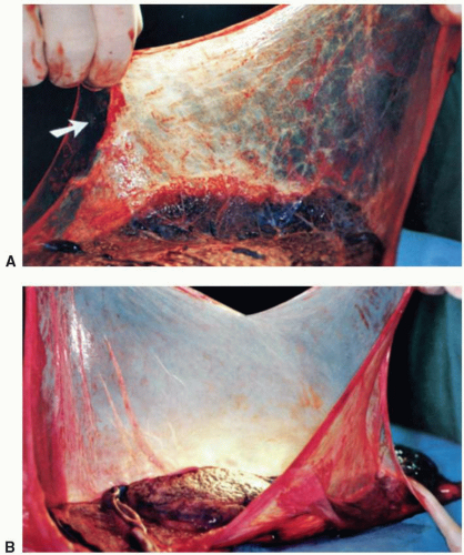
Single placental disk, two amniotic sacs, of same or dissimilar size, and dividing septus attached to the fetal surface are seen; velamentous or marginal insertion and a single umbilical artery are more frequent; vascular anastomoses are present in 85% to 100% of cases; pallor of the donor and congestion of the recipient territory are seen when there is twin transfusion syndrome; amnion nodosum may be present in the donor territory.


Histologic findings reflecting the gross findings are seen; the dividing septum in composed of two amnions only, without intervening chorion.


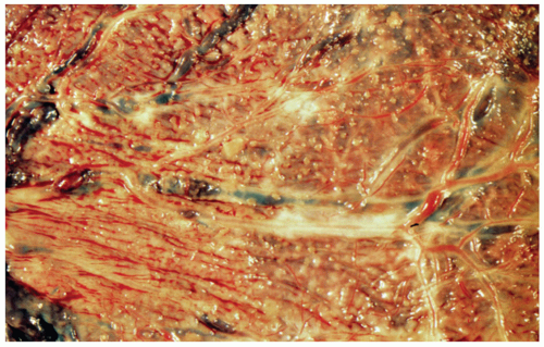
Superficial vascular anastomoses can be visualized by naked eye examination; injection studies may be done to confirm these findings; injection studies are the only way to demonstrate deeper arteriovenous anastomoses via a shared lobule. The twin transfusion syndrome is seen in 15% to 30% of twins with MoDi placenta.


Twin birth with DiDi placenta


Two placental disks may be completely separate or fused, resembling the MoDi placenta; the fusion may be partial, with fusion of only a portion of membranes; a dividing septum is present in fused placentas. Vascular anastomoses are extremely rare.


Dividing septum shows chorion between the two amnions.


Twin transfusion is extremely rare; the pathology of separate DiDi placentas is same as that of singleton placenta.a


Twin birth with MoMo placenta


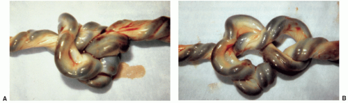
Only one amniotic sac and a single placement disk amniotic fold representing disrupted septum of a previously DiDi placenta may be present; vascular anastomoses are common; findings of twin transfusion syndrome described above may be seen; the cords often become entangled.


No specific features other than vascular anastomoses.


Demonstration of vascular anastomoses should be done when twin transfusion syndrome is done when twin transfusion syndrome is present. This is the rarest type of twin placement with a high incidence of fetal morbidity


Vanishing twins


Plaques of previllous fibrin, embryonic remnant on the membranes, and a second amniotic sac, with or without an embryo may be found; the embryonic remnant appears as a flattened yellow plaque, with or without ocular pigment.


The embryonic remnant shows autolyzed embryonic tissues.


There are few reports describing the pathologic features of the placenta; the placenta should be carefully examined for detection of the embryonic remnant; the twin may be lost via vaginal bleeding.


Fetus papyraceous/fetus compressus (FP/FC)


FP/FC representing a dead twin is identifiable as a plaque of dehydrated remnant, with or without identifiable fetal parts; umbilical cord torsion or massive infarction may cause fetal death.


Histologic findings reflect the gross abnormalities; autolyzed fetal tissues are seen in the sections from FP/FC.


Careful gross examination is essential to detect FP/FC, roentgenograms may be taken to show the skeleton of FP/FC; the causes of fetal death may not be evident.


Acardiac twin


MoMo placenta with artery-to-artery and vein-to-vein anastomoses between the viable twin and acardiac twin placental territories.


No specific microscopic features other than vascular anastomoses


Vascular anastomoses should be sought; the acardiac twin has no heart or a severely malformed heart; other malformations may also be present.


Intrauterine growth restriction (IUGR)


IUGR is associated with maternal factors (preeclampsia, chronic renal disease, substance abuse, etc.), fetal factors (severe congential anomalies chromosomal disorders, intrauterine infarctions, etc.), and placental factors (extrachorial placenta, velamentous insertion of cord, maternal floor infarctions, VUE, extensive infarction or perivillous fibrin deposition, etc.); placenta findings related to the maternal, fetal, and placental factors are seen.


Histologic findings related to fetal, maternal and placental factors are seen.


The placenta is small, which may be a reflection rather than a cause of IUGR; the cause of IUGR may not be evident may be normal except for its size.


Erythroblastosis fetalis


Enlarged weight ans size, pallor, intervillous thrombi


Villi; immature with persistent cytotrophoblast, numerous normoblasts in capillaries, villous edema, hemosiderin in chorionic macrophages


Severity of placental changes is related to severity of fetal anemia; the fetus is hydropic


Nonimmunologic hydrops fetalis (NIHF)


The causes of NIHF include genetic and metabolic disorders, chromosomal abnormalities, cardiac and pulmonary anomalies, thalassemia, fetomaternal hemorrhage, fetal infection, fetal tumors, arrhythmias congenital nephritic syndrome; placental findings related to the disorders are present.


Histologic findings related to the associated condition are seen.


In approximately 22% of NIHF cases, no associated condition can be found.


Chromosomal disorders (trisomy 13, 18, 21)


Small placenta, high incidence of single umbilical artery


Villi: delayed maturation, hypovascularity, large, atypical Hofbauer or trophoblastic cells.


Karyotyping may be done on the chorion or amnion.


Metabolic disorders


Large hydropic placenta


Vacuoles in the syncytiotrophoblasts, intermediate trophoblast, Hofbauer cells, endothelium and fetal WBCs in the villous capillaries.


The vacuoles represent the accumulated metabolite, which is dissolved during processing; electron microscopy may give clues regarding the precise diagnosis (e.g., Niemann-Pick disease glycogenosis type IV); biochemical study of snap-frozen fresh placental tissue is essential for definitive demonstration of enzyme deficiency.


Antepartum stillbirth, intrauterine fetal death (IUFD)


Massive infarction retroplacental hematoma, large chorangioma, true knot, torsion or stricture of the cord, nuchal cord, intrauterine infection, maternal floor infarction, and extensive perivillous fibrin deposition are placental lesions than may lead to IUFD.


Histologic findings reflecting these placental lesions are seen; assessment of the interval between IUFD and delivery can be made on the basis of histologic findings.


Maternal conditions (e.g., preeclampsia) and fetal conditions (e.g., erythroblastosia fetalis) may also cause IUFD; certain placental abnormalities such as stromal fibrosis, hypovascularity thrombosis of villous edema are secondary to IUFD; confined placental mosaicism may occur in rare cases.


a There may still be significant fetal-to-placental transfusion after delivery of the first-born twin, but true twin-to-twin transfusion is so unlikely as to necessitate looking for a different etiology should this be a consideration.


MoDi, monochorionic diamnionic; DiDi, dichorionic diamnionic; MoMo, monochorionic, monoamnionic.


From Joshi VV. Handbook of placental pathology. New York: Igaku-Shoin, 1994, with permission.


TABLE 18.3 Neonatal Oral Findings


Finding


White (%)


Nonwhite (%)


Comments


Palatal cysts (e.g., Bohn nodules, Epstein pearls)


73-85


65-79


Yellow-white elevated cysts 1 mm in diameter; nests of epithelial cells in the midpalatal raphe at the fusion points of the soft and hard palates


Alveolar or gingival cysts


54


40


Appear similar to palatal cysts


Alveolar lymphangioma


0


4


Blue-domed, fluid-filled cysts in posterior regions; no more than one per quadrant; may cause discomfort during feeding if cysts are large


Alveolar eruption of cysts with or without teeth


<0.1


<0.1


Clear, fluid-filled cysts; mandibular central incisor; rates range from 1:2,500 in Hong Kong to 1:3,392 in Canada


Leukoedema


11


43


Filmy, white hue of mucosa, nonblanching; of no significance compared with thrush


Median alveolar notch


16


26


Reduces when teeth erupt or persists as notch between central incisors


Ankyloglossia


˜2


˜2


Male-to-female ratio of 3:1; lingual frenum prevents protrusion of the tongue, extends to papillated surface of the tongue, or causes fissure in the tip


Commissural lip pits


1


3


Blind-ended pits at corners of the mouth; autosomally dominant; associated with preauricular pits; medial pits more syndromic


Thrush


Adherent white plaques on tongue and buccal and palatal surfaces; will scrape off; caused by Candida sp.


Bifid uvula


<1


<1


Associated with submucous cleft palate


Ranula


<<1


<<1


Cyst of sublingual salivary gland


Epulis


<<1


<<1


Large, pedunculated cyst of incisor region


From Fletcher MA. Physical diagnosis in neonatology. Philadelphia, PA: Lippincott-Raven Publishers, 1998.


  • Maternal medications or drugs


  • Position in utero


  • Sleep state


  • Significant hypertonia or hypotonia

Finally, examination as soon as possible after initial stabilization or by 12 hours of life increases the accuracy of assessment in gestations shorter than 28 weeks (16).


Neuromuscular Maturity

Tone increases in a caudocephalad direction to a pattern of full flexion at term (Fig. 18.4). To properly assess tone, the infant must be in an unrestrained resting state.

The square window is assessed by flexing the wrist and measuring the minimal angle between the palm and flexor surface of the forearm. With advancing GA, this angle decreases, and it is worthwhile noting that this progression proceeds at a slower rate ex utero than if the fetus had continued to develop undisturbed.

The scarf sign, indicative of shoulder and superior axial tone, is assessed by pulling the arm across the chest to encircle the neck as a scarf and observing the position of the elbow in relation to the midline. The “Owls test” is demonstrated in Figure 18.5. Factors that may lead to increased GA score by limiting mobility include marked obesity, chest wall edema, and shoulder girdle hypertonicity, while conditions causing generalized hypotonia have the opposite effect.

Arm recoil is assessed by placing the infant supine with the arm fully flexed at the elbow. After holding this position for a few seconds, the forearm is fully extended and then released. The degree to which the arm returns to a flexed state is noted. Similar to the scarf sign, conditions affecting muscle tone can have significant impact on this score.

To determine the popliteal angle, the hips are flexed and the thighs brought up alongside the abdomen. While keeping the pelvis flat, the knee is then extended as far as possible to estimate the popliteal angle. Frank breech positioning in utero may yield a greater angle than expected for a particular GA.

In contrast to the above test, in the heel-to-ear maneuver, the legs are held together over the abdomen and pressed as far as possible toward the ears without lifting the pelvis from the table. The angle made by an arc from the back of the heel to the table decreases with maturity.


Physical Maturity

Skin varies from nearly transparent in the premature infant to opaque with cracking in the postmature newborn.

Lanugo, which is the fine light-colored hair (as distinct from dark body hair seen in infants of medium to dark complexion) evenly distributed over the body, first appears at 19 to 20 weeks of GA and becomes maximally apparent at 27 to 28 weeks. After this point, lanugo begins shedding from areas of greatest contact.

Assessment of the plantar surface of the foot includes measuring the foot because its length reliably corresponds to early GA (Fig. 18.6). With normal muscle activity and uterine compression, creases develop in the sole, progressing from the toes toward the heel. Neuromotor impairments affecting the lower extremities may lead to decreased and/or deep vertical sole creasing, while the opposite can be seen in the presence of oligohydramnios.

The breast develops with an increase in color, stippling of the areola, and increase in the volume of the breast tissue. Volume may be affected by the nutritional state of the fetus and is less consistent with advancing GA than is areolar development, which is, therefore, a better measure of maturity.

With advancing GA, the number of ear folds and firmness of the ear cartilage increase. Extrinsic pressure may impair this process and yield a lower score than expected for a particular GA. Unfusing of the eyelids may begin as early as 22 weeks, with complete unfusing by 28 weeks at the latest (16).


FIGURE 18.4 Assessment of maturity by the expanded Ballard score. From Donovan EF, Tyson JE, Ehrenkranz RA, et al. Inaccuracy of Ballard scores before 28 weeks’ gestation. J Pediatr 1998;135:147-152.

The progression of the testes into the scrotum is a reliable marker of GA. The testes are high in the scrotum at 36 weeks and fully descended by 40 weeks. The presence of a mature scrotal sac (pendulous, rugose) indicates that testicular descent has occurred even if the sac is empty at the time of birth, as may occur due to an in utero vascular compromise late in gestation.

Much like the breast volume, the labia majora may appear underdeveloped in the setting of poor in utero nutrition. The clitoris, however, approaches term size well before 38 weeks and thus appears disproportionately large in premature females (18,19). The appearance of a pigmented vertical line, the linea nigra, originating above the pubis, directed toward the umbilicus suggests a GA of at least 36 weeks.


▪ GROWTH


Measurement Techniques

Weight classification coupled with GA helps to determine levels of risk for neonatal and long-term morbidity and mortality. Weights are classified as low birth weight (LBW) if less than 2,500 g; very LBW if less than 1,500 g, and extremely low birth weight (ELBW) if less than 1,000 g.


FIGURE 18.5 The “Owl” test in a premature newborn. The chin of the premature infant will pass posterior to the shoulder with gentle passive positioning.


FIGURE 18.6 Foot length at birth. Reprinted from Hall JG, Froster-Iskenius UG, Allanson JE. Handbook of normal physical measurements. Oxford, UK: Oxford University Press, 1989, with permission. Data from Merlob P, Sivan Y, Reisner SH. Ratio of crown-rump distance to total length in preterm infants. J Med Genet 1986;23:338-340.

Crown-heel length is most subject to variability and may require repeat measurements if not congruent with weight or head circumference. Proper positioning involves full extension of the supine infant with the top of the head and bottom of the feet both at 90-degree angles to the horizontal. Anomalies of the lower extremities make accurate measurement of crown-heel length impossible; however, the crown-rump measurement may still be feasible. Crown-rump length is measured with the infant supine and the hips flexed 90 degrees. Infants with congenital dwarfism may be classified as those with a short trunk, short legs, or both. These subtypes can be readily differentiated by the crown-rump to total length ratio. From 27 to 41 weeks of gestation, the value is fairly consistent at 0.665 ± 0.027 (20). Proportional reductions in length of the upper and lower body yield a normal ratio. The ratio is increased if the legs are shortened to a greater degree and decreased if the trunk is foreshortened. Standards for separate lengths of the upper and lower limbs are available (21,22).

The occipital-frontal head circumference (OFC) is the largest dimension around the head obtained with a tape placed snugly above the ears. Head circumference undergoes a marked increase during the last trimester, averaging 25 cm at 28 weeks and 35 cm at term (23). The average head circumference is 0.5 cm greater in male, compared with female, neonates (24). Due to greater reliability of repeated measurements, paper rather than reusable cloth tape measures should be used (25). Molding seen after prolonged breech positioning can lead to an OFC measurement that is as much as 2 cm higher than it will be after molding resolves.

If the OFC differs from length by more than one quartile, the cause should be sought because head size in part reflects brain growth. The most frequent reason for a head percentile to exceed that of length is familial and typically demonstrates a pattern of following a persistently higher but consistent growth curve through childhood. In contrast, pathologic macrocephaly tends to cross to higher percentile curves as it progresses. A decreased rate of head growth, manifested by a flat curve or by dropping to a lower percentile, may indicate poor brain growth, atrophy, or premature suture fusion (craniosynostosis [CS]).


Interpretation of Growth Parameters—See also Chapter 23

Interpretation of growth parameters requires plotting the measurements on percentile charts based on data from a similar population. If birth weight falls between the 10th and 90th percentiles for a given GA, the infant is appropriate for gestational age (AGA); if less than the 10th percentile, the infant is small for gestational age (SGA); and if above the 90th percentile, the infant is large for gestational age (LGA). Some literature cites the 3rd and 97th percentiles as outer limits, but for most clinical purposes, this broader range under selects for some at-risk infants, particularly in the lower weight range. AGA infants born at term are at lowest risk for problems associated with neonatal mortality and morbidity.

Infants are considered symmetric if the three parameters of weight, length, and head circumference fall within 25 percentile points of each other. The infant is asymmetric if the parameters are on different curves more than 25 percentile points apart, usually with the weight on a curve lower than those of the head circumference or length. If an infant has either a slowing of intrauterine growth rate documented by serial fetal sonography or a presumed slowing by very low weight for length measurements, he or she is classified as having IUGR. All infants who fall below the 10th percentile for weight are both SGA and IUGR. Infants above the 10th percentile are AGA but may be IUGR, such as the infant who demonstrates a deceleration in growth from the 50th to the 20th percentile during the last trimester due to maternal hypertension. A full-term newborn who has suffered IUGR and is SGA secondary to neonatal thyrotoxicosis is shown in Figure 18.7. Newborns with this condition are also at increased risk of premature fusion of the cranial sutures.


FIGURE 18.7 A full-term SGA newborn with thyrotoxicosis. Premature fusion of the cranial sutures was not detected at birth but presented in early infancy.

Infants who are SGA, IUGR, or LGA are at risk for perinatal and long-term problems. Problems encountered by LGA infants include the following:


  • Iatrogenic prematurity due to overestimation of GA based upon size in utero


  • Increased requirement for delivery by cesarean section


  • Pulmonary hypertension


  • Shoulder dystocia


  • Birth injuries (brachial plexus, fractures, cephalohematoma)


  • Ecchymoses with increased risk of hyperbilirubinemia from RBC degradation


  • Increased risk of cyanotic heart disease particularly transposition of the great vessels


  • The complications associated with poorly controlled maternal diabetes or maternal obesity, including hypoglycemia


  • Local fat necrosis associated with instrumented delivery


  • Polycythemia with hyperviscosity syndrome


  • Seizures


  • Renal vein thrombosis


  • Increased total blood volume


  • Poor feeding


▪ EXAMINATION


Examination Conditions

A routine neonatal examination, normally 5 to 10 minutes in duration, should take place in a quiet, warm environment. Lighting should be such that skin markings and color are visible and may require dimming in order to encourage eye opening. If an infant is ill, the examination should focus on those findings that are important for determining course of management, with a comprehensive exam being deferred until the clinical condition is more stable. Healthy infants tolerate lengthy examination poorly and an ill infant even less so.

Performing the examination in the presence of one or both parents offers an opportunity to explain any relevant findings and answer any questions they may have.


General Assessment

The specifics of neonatal examination are discussed in the following sections. Some systems that are discussed in more detail in other chapters are given less emphasis in this chapter than they would merit in an actual examination.


Inspection

Inspection begins from a sufficient distance to visualize the infant as a whole. An immediate assessment of wellness can come from simply noting the state, color, respiratory effort, posture, and spontaneous activity. Even simple observations of spontaneous movement patterns can suggest future neurologic deficits or well-being (26).


State

Important indicators of infant well-being are the states or levels of arousal that the infant achieves throughout the examination and the day as described by the parents or nursing staff. One categorization of states listed here was originally defined by Prechtl and Beintema (26); modifications have been made but are not clinically important for general assessments (28,29,30,31):

May 30, 2016 | Posted by in PEDIATRICS | Comments Off on Physical Assessment and Classification

Full access? Get Clinical Tree

Get Clinical Tree app for offline access