Fluid and Electrolyte Management


Fluid and Electrolyte Management


Edward F. Bell

Jeffrey L. Segar

William Oh


The constancy of the internal environment is the condition for free and independent life: the mechanism that makes it possible is that which assured the maintenance, with the internal environment, of all the conditions necessary for the life of the elements.

—Claude Bernard


From: Lectures on the Phenomena of Life Common to Animals and Plants (1878), translated by Hebbel E. Hoff, Roger Guillemin and Lucienne Guillemin (1974)

Infants who are born prematurely or who are critically ill cannot regulate their own intake of fluids and nutrients. Often, enteral feeding is limited by feeding intolerance or medical conditions that preclude or limit use of the gastrointestinal tract for feeding. In other cases, the infant develops disordered fluid and electrolyte balance as a primary result of an underlying illness. In all these situations, water and electrolytes must be provided by prescription of the health provider. Providing the correct amounts of water and electrolytes helps to assure the infant’s healthy recovery.

The objectives of fluid and electrolyte management include (a) the provision of maintenance needs so as to maintain normal balance of these essential substances during growth and recovery from disease; (b) repair of acute deficits of water and electrolytes; (c) correction of abnormal concentration, volume, and pH relationships; and (d) replacement of ongoing abnormal losses. A subsidiary aim in the first days of life is to allow successful transition from the aquatic environment of the fetus into the arid extrauterine milieu. Except during this unique period of life, the principles of fluid and electrolyte management in the neonatal period are relatively similar to those established for older children, save for some variations and specific features of body composition, insensible water loss (IWL), renal function, and the neuroendocrine control of fluid and electrolyte balance.

Appropriate fluid therapy of newborns requires the clinician to understand the normal physiologic mechanisms that govern water and electrolyte balance and the variations in these mechanisms that occur with development as well as in sick or premature infants. The clinician should develop a systematic approach to the estimation of fluid and electrolyte requirements for correction of deficits and replacement of ongoing losses, both normal and abnormal. Finally, the results of fluid and electrolyte management must be carefully monitored so that the intakes of water and electrolytes can be adjusted as needed.


▪ BODY COMPOSITION OF THE FETUS AND NEWBORN INFANT


Changes in Body Water during Growth

Water is, by weight, the major constituent of the body. The total body water (TBW) is divided into two major compartments, intracellular water (ICW) and extracellular water (ECW). The ECW is further divided into the interstitial water and the plasma volume, the latter component being the intravascular component of the ECW (Fig. 19.1).

Body fluid compartments change with development (1). It has been estimated that TBW is 94% of the body weight during the 3rd month of fetal life. As gestation progresses, the TBW per kilogram declines. By 24 weeks, the TBW is approximately 86%, and by term, it is about 78% of the body weight (Fig. 19.2). There also are characteristic changes in the partition of body water between ECW and ICW during development. ECW decreases from 59% of the body weight at 24 weeks of gestation to about 44% at term, and ICW increases from 27% to 34% of the body weight during the same period (Table 19.1) (1,2,3,4,5,6). Infants born prematurely thus have higher TBW and ECW per kilogram than do their term counterparts (7,8,9), and small-for-gestational-age infants have higher TBW per kilogram than do appropriate-for-gestational-age infants (8).


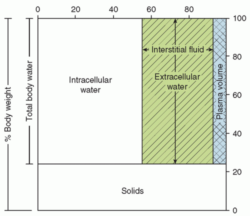
FIGURE 19.1 Distribution of body water in a term newborn infant.

After birth, TBW per kilogram of body weight continues to fall, due primarily to a contraction of the ECW (4,7,9,10,11). This mobilization of extracellular fluid occurs in conjunction with the alterations in renal function that take place following birth (12,13), which occur as a result of increasing renal blood flow, glomerular filtration rate, and expression and activity of the epithelial transporters involved in renal tubular function (14,15,16,17). It has also been suggested that atrial natriuretic peptide (ANP) plays a role in the postnatal contraction of the ECW (11). Various studies have shown an increase, decrease, or no change in the ICW after birth. ICW increases roughly in proportion to body weight in the first weeks of postnatal life (4,7,9). Thereafter, ICW increases faster than does body weight and by 3 months exceeds ECW (Fig. 19.2) (1,4). These
postnatal changes in body water and its partition between ECW and ICW are influenced by developmental changes in neuroendocrine systems as well as the intake of water and electrolytes (18,19). Failure to allow the normal postnatal contraction of ECW in premature infants, often a result of inappropriately high water or electrolyte intake, may increase the risk of significant patent ductus arteriosus (PDA) (20), necrotizing enterocolitis (NEC) (21,22,23), and bronchopulmonary dysplasia (24,25). In the past, lack of postnatal weight loss has been associated with increased risk of bronchopulmonary dysplasia in very-low-birth-weight infants, but with improved early nutrition, postnatal weight loss is not inevitable, as the reduction in ECW is presumably offset by gain in lean body mass (26,27).


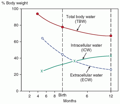
FIGURE 19.2 Changes in body water during gestation and infancy. Adapted from Friis-Hansen B. Changes in body water compartments during growth. Acta Paediatr Suppl 1957;46:1-68, with permission.


TABLE 19.1 Changes in Body Water and Electrolyte Composition during Intrauterine and Early Postnatal Life


Gestational Age (Wk)


Component


24


28


32


36


40


1-4 wk after term birth


Total body water (%)


86


84


82


80


78


74


Extracellular water (%)


59


56


52


48


44


41


Intracellular water (%)


27


28


30


32


34


33


Sodium (mEq/kg)


99


91


85


80


77


73


Potassium (mEq/kg)


40


41


40


41


41


42


Chloride (mEq/kg)


70


67


62


56


51


48


Data from References (2,3,4,5,6).


Solute Distribution in Body Fluids

Each body water compartment has a different electrolyte composition (Fig. 19.3) (28). The major cation in the blood plasma is sodium. Potassium, calcium, and magnesium constitute the balance of the cation fraction. The primary anion is chloride, with protein, bicarbonate, and some undetermined anions constituting the balance of the anions. The interstitial fluid (i.e., nonplasma ECW) has a solute composition that is similar to plasma except that it has a lower protein content. The ICW contains potassium and magnesium as its primary cations, and phosphate, both organic and inorganic, is the major anion, with bicarbonate contributing a smaller fraction.

The electrolyte composition of the body fluids of the newborn infant is largely determined by gestational age. Premature infants contain more sodium and chloride per kilogram of body weight than do term infants (2,3,5,6) because of their larger ECW (Table 19.1). Total body potassium content largely reflects ICW and is similar or slightly lower per kilogram of body weight in premature infants than at term (5,6). These concepts are important in the management of fluid and electrolyte therapy for newborn infants.

In the fetus, fluid and electrolyte balance depends on maternal homeostasis and placental exchange. Thus, fluid and electrolyte status at birth is influenced by the management of maternal fluid and electrolytes in labor (29,30).


▪ INSENSIBLE WATER LOSS

The loss of water by evaporation from the skin and respiratory tract is known as IWL. About 30% of IWL normally occurs through the respiratory tract as moisture in expired gas (31,32,33), with the remaining 70% lost through the skin. IWL depends more on surface area (m2) than weight, but it is commonly expressed per kilogram (kg) because weight is more easily determined than is area.

IWL is a function of energy expenditure, though a number of additional factors are known to influence IWL in a predictable manner (Table 19.2) (31,32,33,34,35,36,37,38,39,40,41,42,43,44,45,46,47,48,49,50,51,52,53,54,55). When expressed per kilogram of body weight, IWL is inversely proportional to birth weight and gestation age (Figs. 19.4 and 19.5) (51,54). In other words, smaller, more immature infants have larger IWL per kilogram (Table 19.3). The same is true if IWL is expressed per square meter of body surface (43). Therefore, although the greater IWL of smaller premature infants is partly due to the increased ratio of surface area (skin and respiratory tract) to body weight, it also is thought to be related to their thinner skin, greater skin blood flow, larger body water per kilogram of body weight, and higher respiratory rate. Because skin permeability to water varies inversely with gestational age, the degree of immaturity is an important determinant of cutaneous IWL independent of birth weight.


Factors That Increase Insensible Water Loss

An increase in minute ventilation, as may occur with cardiac disease, pulmonary dysfunction, or metabolic acidosis, increases the respiratory IWL (44), as long as the water vapor pressure is less in the inspired than in the expired gas. Preterm infants receiving mechanical ventilation breathing gas mixtures that have been warmed (31.5°C) and humidified (100%) have about a 30% decrease in respiratory IWL compared to nonintubated infants (32). Environmental temperature above the neutral thermal zone increases IWL in proportion to the increment in temperature (31,36,52). This effect can occur even without a rise in body temperature. In contrast, a subneutral environmental temperature is not associated with reduced IWL, although metabolic heat production is increased (36). Increased body temperature, whether caused by fever or environmental overheating, elevates IWL (31,52).

Skin breakdown or injury disrupts the barrier against cutaneous evaporation and raises IWL. Skin trauma from thermal, chemical, or mechanical injury is common among critically ill, small premature infants. Such injury may result from removal of tape and adherent monitoring devices or from prolonged skin exposure to disinfectant solutions. IWL also is increased in conjunction with the skin manifestations of essential fatty acid deficiency, a potential problem in infants receiving fat-free parenteral nutrition. Congenital skin defects, such as those seen in gastroschisis, omphalocele, and neural tube defects, are associated with increased IWL until surgically corrected.

Use of nonionizing radiant energy, in the form of either a radiant warmer or phototherapy, has been shown to increase IWL by about 50% (37,38,40,42,45,50,54). For infants in incubators with controlled air temperature, the increase in IWL with overhead phototherapy is most likely a result of increased body temperature because of the warmer incubator walls (50). For infants in incubators operated to control skin temperature, the rise in IWL with phototherapy can be explained by the lower absolute humidity resulting from the reduced air temperature that accompanies the warming of the incubator walls by the phototherapy. The impact on IWL of phototherapy delivered by fiberoptic blankets or pads is not known but is probably negligible unless the blanket produces a warmer or moister microenvironment around the infant. Investigators using direct measurements of transepidermal and respiratory water loss have obtained conflicting results regarding the effect of overhead phototherapy on IWL. One group (42) found an increase in transepidermal water loss with phototherapy, but another did not (56,57). In contrast to conventional phototherapy, light-emitting diode (LED) phototherapy does not result in changes in transepidermal IWL (58).

If an infant’s IWL is measured at the same skin temperature under a radiant warmer and in an incubator, the IWL is higher (by about 50%) under the radiant warmer. IWL is higher because absolute humidity (water vapor pressure) is lower under the radiant warmer than in the incubator (38). This may be true even though relative humidity is higher under the radiant warmer (37,38), because the lower air temperature with the radiant warmer means that the saturation pressure of water vapor is considerably lower than in the incubator (Table 19.4). This finding has been confirmed
using direct measurements of transepidermal water loss (45). It is now understood that the higher IWL with radiant warmers arises from the lower ambient water vapor pressure and not from higher air velocity or a direct effect of nonionizing radiation on the skin. The same phenomenon explains the effect of phototherapy on IWL of infants in incubators operated by skin temperature servocontrol. The effects on IWL of radiant warmers and phototherapy are additive; the IWL with the combination is approximately twice as large as in an incubator without phototherapy (37,40).


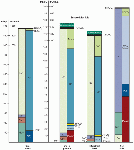
FIGURE 19.3 Ion distribution in seawater, blood plasma, and interstitial fluid, and in the intracellular fluid compartment. The similarity between seawater and extracellular fluid is striking. As noted by Macallum (28): “The vertebrate kidney, therefore, by its control over the concentration of the inorganic elements in the blood plasma and by its maintenance therein of the paleo-ratios, has thus perpetuated in the blood of vertebrates the ocean water of the early Cambrian if not of the late Proterozoic.”

Increased motor activity and crying increase IWL by up to 70% (31,39,55). This effect may be partly due to elevated minute ventilation.


Factors That Reduce Insensible Water Loss

Increasing the humidity or water vapor pressure of inspired gas reduces respiratory IWL. The inspired humidity is raised by humidifying the air-oxygen mixture delivered to a head hood or directly to the infant’s upper airway (e.g., via nasal cannula, face mask, or
endotracheal tube) if respiratory support is required. If the temperature and water content of the inspired and expired gas are the same, the respiratory IWL will be entirely eliminated. Increasing ambient humidity, for example in an incubator, reduces total IWL, but respiratory IWL is decreased more than is cutaneous IWL (31); a threefold increase in ambient water vapor pressure, from an average of 7 to 25 mm Hg, resulted in a 30% reduction in total IWL. Increasing ambient humidity is facilitated by the design of certain recent models of incubators. The use of incubator humidification systems should not be overlooked as a way of reducing IWL and total fluid requirements (59). Modern incubators allow adjustment of the ambient humidity, which, in turn, affects IWL and water requirement.


TABLE 19.2 Factors Affecting Insensible Water Loss in Newborn Infants


Factor


Effect on IWL


Level of maturity (43,51,54)


Inversely proportional to birth weight and gestational age (Fig. 19.4)


Respiratory distress (hyperpnea) (44)


Respiratory IWL increases with rising minute ventilation when dry air is being breathed


Environmental temperature above neutral thermal zone (36)


Increased in proportion to increment in temperature


Elevated body temperature (52)


Increased by up to 300%


Skin breakdown or injury


Increased by uncertain magnitude


Congenital skin defect (e.g., gastroschisis, omphalocele, neural tube defect)


Increased by uncertain magnitude until surgically corrected


Radiant warmer (37,38,45,54)


Increased by about 50%


Phototherapy (40,42,50,54)


Increased by about 50%


Motor activity and crying (31,39,55)


Increased by up to 70%


High ambient or inspired humidity (31,32)


Reduced by 30% when ambient vapor pressure is increased by 200%


Plastic heat shield (35,38)


Reduced by 30%-70%


Plastic blanket (34,35,48) or chamber (34,41)


Reduced by 30%-70%


Semipermeable membrane (46,47)


Reduced by 50%


Topical agents (49)


Reduced by 50%


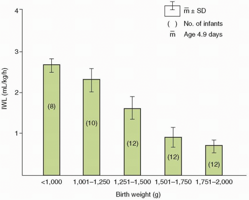
FIGURE 19.4 Relation between insensible water loss (IWL) and birth weight of 5-day-old (mean) infants in incubators. Data from Wu PY, Hodgman JE. Insensible water loss in preterm infants: changes with postnatal development and non-ionizing radiant energy. Pediatrics 1974;54:704-712, as redrawn in Shaffer SG, Weismann DN. Fluid requirements in the preterm infant. Clin Perinatol 1992;19:233-250, with permission.


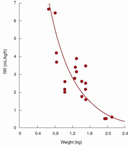
FIGURE 19.5 Insensible water loss (IWL) as a function of birth weight in premature infants nursed under radiant warmers. Adapted from Costarino AT, Jr., Gruskay JA, Corcoran L, et al. Sodium restriction versus daily maintenance replacement in very low birth weight premature neonates: a randomized, blind therapeutic trial. J Pediatr 1992;120:99-106.

Plexiglas heat shields are effective in reducing the IWL of small premature infants in incubators (38), especially if the ends are at least partially enclosed to decrease air movement near the skin. Plexiglas heat shields are not effective for infants under radiant warmers (35,38), because Plexiglas is opaque to the infrared energy produced by the radiant heaters. Thin barriers of saran and other materials reduce IWL of infants under radiant warmers while allowing the infrared heat to reach the skin (35). These heat shields presumably reduce IWL by limiting air movement and raising water vapor pressure near the infant’s body surface.

Thin plastic blankets have been found to reduce IWL by 30% to 70% for infants under radiant warmers and in incubators (34,35,48). Chambers made of thin plastic material also reduce IWL by a similar amount (34,41). Semipermeable membranes (46,47) and water-proof topical agents (49) reduce IWL from the covered areas by an average of approximately 50%.

Knowledge of these factors that affect IWL is essential for estimating the water intake required by newborn infants and for
making appropriate adjustments in water intake with changes in care. Of all infants, premature and critically ill infants are the ones whose IWL is most profoundly influenced by these factors. This is especially true for the extremely premature infant. However, these are exactly the infants for whom precise maintenance of fluid and electrolyte balance is most important and for whom the margin for error is smallest.


TABLE 19.3 Average Insensible Water Loss (mL/kg/d) of Premature Infants in Incubators


Birth Weight Range (kg)


Age (d)


0.50-0.75


0.75-1.00


1.00-1.25


1.25-1.50


1.50-1.75


1.75-2.00


0-7


100a


65


55


40


20


15


7-14


80


60


50


40


30


20


a Insensible water loss (mL/kg/d).


Data from References (43

Only gold members can continue reading. Log In or Register to continue

Stay updated, free articles. Join our Telegram channel

May 30, 2016 | Posted by in PEDIATRICS | Comments Off on Fluid and Electrolyte Management

Full access? Get Clinical Tree

Get Clinical Tree app for offline access