Jaundice


Jaundice


Jeffrey M. Maisels

Jon F. Watchko


Jaundice is the most common and one of the most vexing problems that can occur in the newborn. As Hansen points out in an elegant historical review (1), “neonatal jaundice must have been noticed by caregivers throughout the centuries …,” but the first documented scientific description of neonatal jaundice occurred in the latter part of the 18th century when Baumes was awarded a prize from the University of Paris for his description of the clinical course of jaundice in 10 infants (1,2). Although most jaundiced infants are otherwise perfectly healthy, they make us anxious because bilirubin is potentially toxic to the central nervous system (CNS).

Jaundice occurs when the liver cannot clear a sufficient amount of bilirubin from the plasma. When the problem is excessive bilirubin formation or limited uptake and conjugation, unconjugated (i.e., indirect-reacting) bilirubin appears in the blood. When bilirubin glucuronide excretion is impaired (i.e., cholestasis), conjugated monoglucuronide and diglucuronide (i.e., direct-reacting bilirubin) accumulate in plasma and, because of their solubility, also appear in the urine. There is also a fourth bilirubin fraction (unconjugated, monoglucuronide, and diglucuronide are the first three) known as &dgr;-bilirubin, which is formed nonenzymatically from conjugated bilirubin and reacts directly with the diazo reagent (3).

In most jaundiced neonates, only unconjugated bilirubin is found in the blood, and the accumulated bilirubin is distributed by the circulation throughout the body and produces clinical jaundice. It generally is assumed that to cross intact cell membrane barriers, the bilirubin must be free, or dissociated, from its albumin binding.


▪ FORMATION, STRUCTURE, AND PROPERTIES OF BILIRUBIN

Bilirubin is the end product of the catabolism of iron protoporphyrin or heme, of which the major source is circulating hemoglobin (Fig. 32.1). The formation of bilirubin from hemoglobin involves removal of the iron and protein moieties, followed by an oxidative process catalyzed by the enzyme microsomal heme oxygenase, an enzyme found in the reticuloendothelial system as well as many other tissues. The &agr;-methane bridge of the heme porphyrin ring is opened, and carbon monoxide (CO) and biliverdin are formed. One molecule of CO and biliverdin (and, subsequently, bilirubin) is formed for each molecule of heme degraded (Fig. 32.1).

It is likely that the prevalent structure of bilirubin in plasma has the ridge-tile conformation shown in e-Figure 32.1, because it is consistent with the biologic properties of bilirubin. In this conformation, the bilirubin polar groups of the molecule are involved in intramolecular hydrogen bonding that restricts solvation and renders the pigment nearly insoluble in water at pH 7.4 but soluble in nonpolar solvents such as chloroform (4). Under these circumstances, bilirubin behaves like other lipophilic substances—it is difficult to excrete but crosses biologic membranes, such as the placenta, blood-brain barrier (BBB), and hepatocyte plasma membrane, easily (4,5,6). The addition of methanol or ethanol interferes with hydrogen bonding and results in an immediate diazo reaction, the basis for measurement of indirect-reacting bilirubin by the van den Bergh test (3).


▪ FETAL BILIRUBIN METABOLISM

Bilirubin can be detected in normal amniotic fluid after about 12 weeks of gestation, but it disappears by 36 to 37 weeks’ gestation. The ability of human fetal liver to remove bilirubin from the circulation and to conjugate it is severely limited. Between 17 and 30 weeks of gestation, uridine diphosphoglucuronosyl transferase 1A1 (UGT1A1) activity in fetal liver is only 0.1% of adult values, but it increases 10-fold to 1% of adult values between 30 and 40 weeks’ gestation. After birth, activity increases exponentially, reaching adult levels by 6 to 14 weeks of postnatal life independent of gestation (6).

The major route of fetal bilirubin excretion is across the placenta. Because virtually all the fetal plasma bilirubin is unconjugated, it is readily transferred across the placenta to the maternal circulation, where it is excreted by the maternal liver. Thus, the newborn rarely is born jaundiced, except in the presence of severe hemolytic disease, when there may be accumulation of unconjugated bilirubin in the fetus. Conjugated bilirubin is not transferred across the placenta, and it also may accumulate in the fetal plasma and other tissues.


Maternal Hyperbilirubinemia and Its Effect on the Fetus

Obstetricians and pediatricians occasionally are confronted with a pregnant mother who has hyperbilirubinemia as a result of hemolytic anemia or liver disease. Reported cases in the literature provide evidence for transfer of unconjugated bilirubin from the mother to her fetus, but no clear guidelines for management (7,8,9).

It is possible that prolonged exposure of the fetus to a modest degree of unconjugated hyperbilirubinemia in utero could lead to neurologic damage (9).


▪ NEONATAL BILIRUBIN METABOLISM


Bilirubin Production

The normal destruction of circulating erythrocytes accounts for about 75% of the daily bilirubin production in the newborn. Senescent erythrocytes are removed and destroyed in the reticuloendothelial system, where the heme is catabolized and converted to bilirubin (Fig. 32.1). The catabolism of 1 g of hemoglobin yields 35 mg of bilirubin.

A significant contribution (25% or more) to the daily production of bilirubin in the neonate comes from sources other than effete erythrocytes (see Fig. 32.1). This bilirubin consists of two major components:


  • A nonerythropoietic component resulting from the turnover of nonhemoglobin heme protein and free heme, primarily in the liver


  • An erythropoietic component arising primarily from ineffective erythropoiesis and the destruction of immature erythrocyte precursors, either in the bone marrow or soon after release into the circulation


Transport and Hepatic Uptake of Bilirubin

Once bilirubin leaves the reticuloendothelial system, it is transported in the plasma bound tightly but reversibly to albumin at primary (high-affinity) as well as secondary (low-affinity) binding sites. Although the magnitude of the affinity constant at the primary binding site remains a source of debate (5,6), the concentrations of free or unbound bilirubin in plasma are very low (in the nmol range), even in the presence of significant hyperbilirubinemia.

The parenchymal cells of the liver have a selective and highly efficient capacity for removing unconjugated bilirubin from the plasma. When the bilirubin-albumin complex reaches the plasma membrane of the hepatocyte, a proportion of the bilirubin, but not the albumin, is transferred across the cell membrane into the hepatocyte, a process that potentially involves four different transport proteins (10). In the hepatocyte, bilirubin is bound principally to
ligandin and possibly other cytosolic-binding proteins (Fig. 32.1). A network of intracellular microsomal membranes may also play an important role in the transfer of bilirubin within the cell and to the endoplasmic reticulum.


FIGURE 32.1 Neonatal bile pigment metabolism. RBC, erythrocytes; R.E., reticuloendothelial.


Conjugation and Excretion of Bilirubin

Because of its hydrogen-bonded conformation (see Formation, Structure, and Properties of Bilirubin above), unconjugated (i.e., indirectreacting) bilirubin is nonpolar and insoluble in aqueous solutions at pH 7.4 and must be converted to its water-soluble conjugate (i.e., direct reacting bilirubin) before it can be excreted (see Fig. 32.1). This is achieved when bilirubin is combined enzymatically with a sugar, glucuronic acid, producing bilirubin monoglucuronide and diglucuronide pigments that are more water soluble and sufficiently polar to be excreted into the bile or filtered through the kidney.

The presence of elevated bilirubin concentrations in utero prematurely induces bilirubin uridine diphosphoglucuronate glucuronosyltransferase 1A1 (UGT1A1) activity, which suggests that bilirubin plays an important role in the initiation of its own conjugation after birth (11).


Structure and Function of the Uridine Diphosphoglucuronate Glucuronosyltransferase 1A1 Gene

The process of conjugation is catalyzed by a specific hepatic enzyme isoform (1A1) belonging to the UGT family of enzymes. These enzymes metabolize endogenous compounds and various food chemicals in most tissues. Although the UGT1 family contains several isoforms, only the A1 isoform (UGT1A1) participates in the conjugation of bilirubin (12). The glucuronosyltransferase enzyme is synthesized in the hepatocyte, and its structure is determined by the UGT1A1 gene (Fig. 32.2).

The gene encoding the UGT1 enzyme is located on chromosome 2 at 2q37 (13) and consists of 4 common exons and 13 variable exons (Fig. 32.2) (13). The gene also has a noncoding promoter area, which is an upstream regulatory region controlling gene expression. The UGT1A1 promoter contains a TATAA box, which is a deoxyribonucleic acid (DNA) sequence of thymine (T) and adenine (A). Mutations in the UGT1A1 exon or its promoter will affect bilirubin conjugation. Examples of this effect are seen in Gilbert syndrome and the Crigler-Najjar syndromes (see Pathologic Causes of Jaundice: Decreased Bilirubin Clearance below).


Transfer of Bilirubin into Bile and Intestinal Transport

After conjugation, bilirubin is excreted rapidly into the bile canaliculi by the liver cell, a process that requires metabolic work for the active transport of bilirubin across a large concentration gradient (5,6). Interference with this process is probably responsible for the hyperbilirubinemia associated with hepatocellular disorders such as hepatitis.

Once in the small intestine, conjugated bilirubin is not reabsorbed. In the healthy adult, it is largely reduced by the action of colonic bacteria to a series of colorless tetrapyrroles, collectively known as urobilinogen, and an insignificant amount is hydrolyzed to unconjugated bilirubin and reabsorbed by way of the enterohepatic circulation. In the newborn, however, this enterohepatic circulation of bilirubin is significant and important (see Jaundice in the Healthy Newborn: Physiologic Jaundice below). In addition, in conditions involving high plasma bilirubin levels and poor hepatic excretion, there is a gradient for unconjugated bilirubin from the plasma to the intestinal lumen, and significant amounts of unconjugated bilirubin may be cleared by diffusion across the intestinal wall (6). Figure 32.1 summarizes bile pigment metabolism in the newborn.


▪ PHYSIOLOGIC MECHANISMS OF NEONATAL JAUNDICE

At any time in the infant’s first few days after birth, the serum bilirubin level reflects a combination of the effects of bilirubin production, conjugation, and enterohepatic circulation. Using measurements of blood carboxyhemoglobin (COHb) corrected for ambient CO (COHbc) as an index of bilirubin production and high-performance liquid chromatography (HPLC) measurements of conjugated bilirubin, Kaplan et al. (14) demonstrated that an imbalance between bilirubin production and conjugation is fundamental in the pathogenesis of neonatal bilirubinemia. Small alterations in these two processes together with the enterohepatic circulation account for the fact that more than 80% of term, near-term, and late preterm infants are jaundiced in the first week (15,16). Table 32.1 lists the mechanisms responsible for the bilirubinemia that occurs in these newborns.


▪ BILIRUBIN TOXICITY


Kernicterus


Pathology

The first description of kernicterus (or brain jaundice) in newborns was provided by Hervieux in 1847 (1) and in 1875, Orth observed bilirubin pigment at autopsy in the brains of infants who were severely jaundiced. Schmorl (1) subsequently described two forms of “brain icterus,” the first “characterized by a diffuse yellow coloration of the entire brain substance” and a second form in which “the jaundiced
coloration appears to be completely circumscribed and … limited to the so-called ‘kern’ or nuclear region of the brain (1).”


FIGURE 32.2 Schematic of the UG1A1 gene. The uppermost panel represents the entire UGT1A gene complex encompassing: (a) the A1 exon, (b) nine additional exons that encode functional proteins (exons 3-10, 13), (c) three pseudogenes (exons 2P,11Pm12P), and (d) the common domain exon 2-5 sequence shared across all UGT1A transcripts. The UTG1A1 locus and common exons 2-5 are shown in middle panel including the upstream (i) phenobarbital-responsive enhancer module (PBREM) encompassing six nuclear receptor motifs (and hypomorphic variant UGT1A1*60) and (ii) TATA box promoter sequences. Lower panels show wild-type UTG1A1*1 and UGT1A1*28, UGT1A1*37, and UGT1A1*6 variant alleles and relevant change in expression function. Adapted from Clarke DJ, Moghrabi N, Monaghan G, et al. Genetic defects of the UDP-glucuronosyltransferase-1 (UGT1) gene that cause familial nonhemolytic unconjugated hyperbilirubinemias. Clin Chim Acta 1997;166:63-74, with permission from Elsevier Science; Perera MA, Innocenti F, Ratain MJ. Pharmacogenetic testing for uridine diphosphate glucuronosyltransferase 1A1 polymorphisms. Are we there yet? Pharmacotherapy 2008;28:755-768, with permission from Pharmacotherapy; Li Y, Buckely D, Wang S, et al. Genetic polymorphisms in the TATA box and upstream phenobarbital-responsive enhancer module of the UGT1A1 promoter have combined effects on UDP-glucuronosyltransferase 1A1 transcription mediated by constitutive androstane receptor, pregnane X receptor, or glucocorticoid receptor in human liver. Drug Metab Dispos 2009;37:1978-1986, with permission.


TABLE 32.1 Physiologic Mechanisms of Neonatal Jaundice (6) (see Fig. 32.1)


Increased bilirubin load on liver cell (newborn vs. adult values)


8-10 mg/kg/d vs. 3.8 mg/kg/d (17)


Increased erythrocyte volume


Hematocrit 56% ± 5.5% (SD), vs. 41% ± 2.5% (female), 47% ± 3.0% (male)


Decreased erythrocyte survival


Approximately 80 vs. 110-120 d


Increased early labeled bilirubin—bilirubin produced by turnover of erythrocyte precursors in the bone marrow and from heme protein and free heme (Fig. 32.1)


25% vs. 15% of bilirubin production (17)


Increased enterohepatic circulation of bilirubin (6)


Few bacteria [which convert bilirubin to urobilinogen] in small and large bowel and more &bgr;-glucuronidase. Conjugated bilirubin not converted to urobilinogen but hydrolyzed to unconjugated bilirubin that is reabsorbed.


Decreased hepatic uptake of bilirubin from plasma


Decreased ligandin


Decreased bilirubin conjugation


Decreased uridine diphosphoglucuronosyl transferase activity (approximately 1% of adult values at term)


Defective bilirubin excretion


Excretion impaired but not normally rate limiting. With hemolysis can get increase in conjugated bilirubin.


Topography

Full-term infants who die of kernicterus demonstrate bilirubin staining in a characteristic distribution (Table 32.2), although a variety of patterns have been described, grossly and microscopically (2,18). Kernicteric premature infants and Gunn rats with inherited UGT1A1 deficiency display a similar topography of neuronal damage (see Table 32.2) (2,18). Those regions most commonly affected are the basal ganglia, particularly the subthalamic nucleus and the globus pallidus; the hippocampus; the geniculate bodies; various brainstem nuclei, including the inferior colliculus, oculomotor, vestibular, cochlear, and inferior olivary nuclei; and the cerebellum, especially the dentate nucleus and the vermis (2,18). Ahdab-Barmada has provided a detailed review of the neuropathology of kernicterus, and its anatomic, cytologic, and histologic characteristics (18).


Gross Anatomy

Yellow staining of the brain occurs when it is exposed to elevated levels of bilirubin. There can be some confusion regarding the diagnosis of kernicterus in the presence of yellow discoloration of CNS tissue. Table 32.3 lists the three patterns of bilirubin staining of the brain seen in the newborn (18), only one of which characterizes kernicterus. Ahdab-Barmada (18) emphasizes that the diagnosis of kernicterus should only be applied when bilirubin-stained neurons show microscopic damage.


TABLE 32.2 Comparative Neuropathology of Kernicterus


Topography of Lesions


Full-Term Infants, Hyperbilirubinemia


Homozygous Gunn Rats


Premature Infants, Low Bilirubin Levels


Globus pallidus


+


+


+


Subthalamus


+


+


+


Hypothalamus


+


Horn of Ammon


+


+


+


Reticular zone of the substantia nigra


+


+


+


Cranial nerve nuclei


+


+


+


Reticular formation


+


+


Central pontine nuclei


Interstitial nucleus


Locus caeruleus


+


+


Lateral cuneate nucleus of the medulla


+


+


+


Cerebellum


Dentate nuclei


+


+


Nuclei of roof of fourth ventricle


+


+


+


Purkinje cells


+


+


Spinal cord


+


+


+


+, Yellow pigment present; -, yellow pigment absent.


From Ahdab-Barmada M, Moossy J. The neuropathology of kernicterus in the premature neonate: diagnostic problems. J Neuropathol Exp Neurol 1984;43:45-56, with permission.


Histology and Cytology

e-Table 32.1 summarizes the cytopathology of kernicterus. The unique topographic pattern of nuclear involvement as described above (see Topography) in combination with the bright yelloworange staining of these brain nuclei, together with evidence of neuronal damage and degeneration within the nuclei, is required before a postmortem diagnosis of kernicterus can be made (18).

Autopsies on jaundiced infants reveal bilirubin staining of the aorta, pleural fluid, and ascitic fluid, or a generalized yellow cast throughout the viscera. The staining usually is not considered a sign of tissue damage unless other cytologic changes are found (19). Bilirubin staining also can be found in necrotic tissue anywhere in the body and has been described in the gastrointestinal tract, lungs (hyaline membranes), kidney, adrenals, and gonads. In infants with hemolytic disease, bile plugs commonly are found in the canaliculi between the hepatocytes, especially in the periportal areas. The kidneys may show bilirubin-stained tubular casts, bilirubin crystals in the small vessels or in edematous interstitium, and renal tubular necrosis. The bilirubin infarcts (i.e., patches of yellow staining in the renal medulla) are probably the result of focal areas of acute tubular necrosis that have been stained by bilirubin (19).

Neuronal necrosis is the dominant histopathologic feature after 7 to 10 days of postnatal life (see e-Table 32.1). For the most part, its distribution corresponds with the distribution of bilirubin staining, although there are some exceptions to this rule. For example, intense staining develops in the olivary and dentate nuclei, but there is little neuronal necrosis in these regions. The important areas of neuronal injury (as opposed to staining) include the basal ganglia, brainstem oculomotor nuclei, and brainstem auditory (cochlear) nuclei (20). The involvement of these regions explains some of the clinical sequelae of bilirubin encephalopathy (see Clinical Features of Bilirubin Encephalopathy below).


TABLE 32.3 Patterns of Bilirubin Staining of the Brain in Hyperbilirubinemia


Diffuse yellow staining of areas that normally lack a BBB, for example, leptomeninges, ependyma, choroid plexus, CSF


Diffuse yellow staining of brain tissues in areas where BBB integrity has been compromised (as can occur following hypoxic ischemic encephalopathy, PVL, ischemic cerebral infarct)


Yellow staining of specific neuronal groups (kernicterus)


From Ahdab-Barmada M. The neuropathology of kernicterus: definitions and debate. In: Maisels MJ, Watchko JF, eds. Neonatal jaundice. London, UK: Harwood Academic, 2000:75-88.


As e-Table 32.2 shows, the neuropathology of kernicterus is different from that of hypoxic ischemic encephalopathy. Even though hypoxic ischemic insults may predispose the brain to bilirubin deposition in some low-birth-weight (LBW) infants, in others the typical histologic features of kernicterus will be found.


Mechanisms by Which Bilirubin Enters the Brain

Bratlid (38) has provided a simplified scheme, not involving membrane transporters for bilirubin entry into the brain, its binding to neuronal cell membranes, and the potential clinical signs that may follow (see Fig. 32.4). Under normal circumstances, bilirubin can enter the brain unaccompanied by albumin. Clinical confirmation of this fact is provided by the observation that even modest elevations of serum bilirubin can sometimes produce clinical and electrophysiologic alterations in healthy full-term infants as demonstrated by changes in behavior (84), characteristics of the cry (85), and changes in the brainstem auditory-evoked response (BAER) (86). These symptoms disappear as the bilirubin level decreases (86,87).

The probability of toxic levels of bilirubin entering the brain increases when the serum level of unbound pigment increases, as depicted in the center panel of Figure 32.4. Finally, if the BBB is disrupted, both albumin and bilirubin can enter the brain, but even in the presence of a damaged BBB, more bilirubin than albumin, on a molar basis, seems to be deposited (78,80). In all of these situations, acidosis will increase deposition of bilirubin in brain cells.


TABLE 32.5 Population-Based Estimates of Kernicterus Incidence


Author(s)


Country


Years


Ascertainment Case Definition


No. of Cases


Denominator


Rate


Bjerre and Ebbesen (90)


Denmark


1994-2002


Registry; voluntary reports; ≥35 wk of gestation; TSB ≥ 31.1 mg/dL; symptoms of chronic bilirubin encephalopathy


8


576,000


1.4/100, 000


Bjerre et al. (91)


Denmark


2002-2005


National laboratory information system linked to medical reports; ≥35 wk of gestation and ≤28 d of age; TSB ≥ 26.5 mg/dL and advanced phase symptoms of bilirubin encephalopathy


1


249,308


0.4/100, 000a


Manning et al. (92)


United Kingdom


2003-2005


Voluntary reports; ≥35 wk of gestation and <1 mo of age; TSB ≥ 30 mg/dL; death OR typical sequelae at 12-month follow-up


7


1,500,052


0.46/100,000


Sgro et al. (93)


Canada


2002-2004


Surveillance program; voluntary reports; ≥35 wk of gestation and ≤60 d of age TSB ≥ 25 mg/dL and/or exchange transfusion AND clinically important neurologic abnormalities at final discharge


13


640,000


2/100,000


Sgro et al. (94)


Canada


2007-2008


Surveillance program; voluntary reports; ≥35 wk of gestation at birth; TSB > 425 &mgr;mol/L (≥24.8 mg/dL) or exchange transfusion AND two or more signs/symptoms of kernicterus; OR abnormal MRI with history of hyperbilirubinemia


17a,b


740,000


2.3/100,000b


Burke et al. (95)


United States


1998-2005


Hospital discharge abstracts; ≤30 d of age; ICD-9 for kernicterus AND CPT for phototherapy or exchange transfusion


436


Not stated


2.7/100,000


Brooks et al. (96)


California


1988-1997


State registry for developmental services; ICD-9 for kernicterus


25c


5,697,147


0.49/100,000


a Ten cases identified, 3 normal at 12-19 mo. Six cases lost to follow-up. Depending on how many of these were normal, rate could range from 1.5 to 2.3/100,000.

b Based on these estimates, Canada (annual births 370,000) could expect about 8 cases per year and the United States (annual births 4 million) about 20 cases per year. In the United States, there are 8,000-10,000 new cases of cerebral palsy every year.

c Based on these estimates, Canada (annual births 370,000) could expect about 8 cases per year and the United States (annual births 4 million) about 20 cases per year. In the United States, there are 8,000-10,000 new cases of cerebral palsy every year.


TSB, total serum bilirubin; MRI, magnetic resonance imaging; ICD-9, International Statistical Classification of Diseases-Ninth Revision; CPT, Current Procedural Terminology. Modified from Burgos AE, Flaherman VJ, Newman TB. Screening and follow-up for neonatal hyperbilirubinemia: a review. Clin Pediatr 2012;51(1):7.


Oxidation of Bilirubin in the Brain

There is some evidence that mitochondria in the brain and other tissues contain a bilirubin oxidase that converts bilirubin to biliverdin and other nontoxic products (88,89). Although the specificity and clinical relevance of this putative enzyme remain to be established, Hansen showed that it is not bilirubin oxidase (ECI, 3.5) but, tentatively, a member of the cytochrome P-450 oxidases (89).


▪ ACUTE BILIRUBIN ENCEPHALOPATHY AND KERNICTERUS


Incidence

Acute bilirubin encephalopathy and kernicterus continue to be seen throughout the world with population-based estimates of incidence in Europe and North America ranging from 0.5 to 2.4 cases per 100,000 live births (Table 32.5) while the incidence in low- and middleincome countries could be as high as 73/100,000 per 100,000 live births (97). The early descriptions of bilirubin encephalopathy and kernicterus involved infants with Rh hemolytic disease (2), a condition now rarely seen in the Western world (97) but still prevalent elsewhere. In countries where the neonatal mortality rate is greater than 5/1,000 live births, Bhutani et al. (97) estimate that 373,300 live births were affected by Rh disease in 2010 (a rate of 277/100,000 live births). In high-resource countries, the majority of infants who now develop kernicterus are not those with Rh disease and many of them have no documented evidence of hemolytic disease (98). Many are term and late preterm infants who have been discharged from the nursery as “healthy newborns” yet have returned to a pediatrician’s office, clinic, or an emergency department with TSB levels often exceeding 30 mg/dL (50,99) and have gone on to develop the classic neurodevelopmental findings associated with kernicterus (50,99). Others, more difficult to identify, have an unanticipated precipitous
increase in TSB while still in the hospital or soon after discharge and present with acute bilirubin encephalopathy (98,100).

Glucose-6-phosphate dehydrogenase (G6PD) deficiency is an important cause of the hyperbilirubinemia in some of these infants (98,101). Some authors have suggested that in the 1970s and 1980s, kernicterus had essentially disappeared in the United States (102) but experienced a resurgence in the 1990s. Ebbesen (103) found no cases of kernicterus in Denmark for two decades prior to 1994 while six cases were diagnosed between 1994 and 1998. Brooks et al. (96) used the data from the California Department of Developmental Services to identify kernicterus cases in children born between 1988 and 1997 and the death certificate data from the U.S. National Center for Health Statistics to identify kernicterus mortality from 1979 to 2006. There was no significant trend in kernicterus incidence from 1988 to 1997 (p = 0.77) and no increase in kernicterus mortality from 1979 to 2006. Burke et al. (95) found a 70% decline in neonatal hospitalizations with a diagnosis of kernicterus from 1988 to 2005. These findings do not support the suggestion of a resurgence of kernicterus in the United States.


Terminology

Although originally a pathologic diagnosis characterized by bilirubin staining of the brainstem nuclei and cerebellum, the term kernicterus has come to be used interchangeably with both the acute and chronic findings of bilirubin encephalopathy. Bilirubin encephalopathy describes the clinical CNS findings caused by bilirubin toxicity to the basal ganglia and various brainstem nuclei. To avoid confusion and encourage greater consistency in the literature, the AAP recommends (50) that the term acute bilirubin encephalopathy be used to describe the acute manifestations of bilirubin toxicity seen in the first weeks after birth and that the term kernicterus be reserved for the chronic and permanent clinical sequelae of bilirubin toxicity. This is the terminology that is used in this chapter. Recently, the term bilirubin-induced neurologic dysfunction or BIND has been used to describe the intensity of the clinical features associated with acute bilirubin encephalopathy and to provide a scoring system that quantifies the severity of the clinical manifestations and their association with developmental outcome (104). Others have used the term BIND to describe children who have subtle neurodevelopmental disabilities, thought to be due to bilirubin neurotoxicity, but who do not show the classical findings of kernicterus (105).


Acute Bilirubin Encephalopathy

In classic acute bilirubin encephalopathy, markedly jaundiced infants progress through the three fairly distinct clinical phases (20) listed in Table 32.6. The hypertonia involves the extensor muscle groups, and most infants exhibit backward arching of the neck (retrocollis) and trunk (opisthotonos) (Fig. 32.5). Additional findings include fever that might be caused by diencephalic involvement and apnea, seen most often in preterm newborns (106). The advanced phase is characterized by pronounced retrocollis-opisthotonos, shrill cry, refusal to feed, apnea, fever, deep stupor to coma, and sometimes seizures and death (105). Subsequently, usually after 1 week, hypertonia subsides and is replaced by hypotonia.


TABLE 32.6 Major Clinical Features of Acute Bilirubin Encephalopathy


Initial phase


Slight stupor (“lethargic,” “sleepy”)


Slight hypotonia, paucity of movement


Poor sucking; slightly high-pitched cry


Intermediate phase


Moderate stupor—irritable


Tone variable—usually increased; some with retrocollis-opisthotonos


Minimal feeding; high-pitched cry


Advanced phase


Deep stupor to coma


Tone usually increased; some with retrocollis-opisthotonos


No feeding; shrill cry


From Volpe JJ. Neurology of the newborn, 5th ed. Philadelphia, PA: WB Saunders, 2008, with permission.


FIGURE 32.5 This infant presented at age 30 days with a serum bilirubin level of 30 mg/dL (513 mmol/L) secondary to the Crigler-Najjar syndrome type I. He demonstrates retrocollis and opisthotonos, signs of the intermediate to advanced stage of acute bilirubin encephalopathy.

Infants who manifest hypertonia during the second phase almost always develop the clinical features of chronic bilirubin encephalopathy although an emergent exchange transfusion might, in some cases, reverse the CNS changes (107,108). Some infants manifest none of the signs of acute bilirubin encephalopathy but, nevertheless, go on to develop the classical features of kernicterus (109,110). Table 32.7 shows the diversity observed in the clinical presentation of acute bilirubin encephalopathy.


Chronic Bilirubin Encephalopathy: Kernicterus


Clinical Features

The classic sequelae of postkernicteric encephalopathy are listed in Table 32.8 (20). Shapiro suggests that kernicterus be categorized into four main categories based on location and the predominant features of the clinical presentation (Table 32.9).


Extrapyramidal Disturbances

Athetosis (i.e., involuntary, sinuous, writhing movements) may develop as early as 18 months of age but may be delayed until as late as 8 or 9 years of age (111). If sufficiently severe, athetosis may prevent useful limb function. These movements are described as “uncontrollable, purposeless, involuntary, and incoordinate. They may be rapid and jerky (choreiform), slow and worm-like (orthodox athetosis), or so slowed by hypertonicity that the patient may assume momentarily fixed attitudes with stiffness of the extremities (dystonia) (112).” Severely affected children also may have dysarthria, facial grimacing, drooling, and difficulty chewing and swallowing.


TABLE 32.7 Occurrence of Clinical Features in Acute Bilirubin Encephalopathy


Clinical Features


Percent (%) of Cases


No definite neurologic signs


15


Equivocal neurologic signs


20-30


Definite neurologic signs


55-65


Adapted from Volpe JJ. Neurology of the newborn, 5th ed. Philadelphia, PA: WB Saunders, 2008;15:248.


TABLE 32.8 Major Clinical Features of Chronic Postkernicteric Bilirubin Encephalopathy


Extrapyramidal abnormalities, especially athetosis


Gaze abnormalities, especially of upward gaze


Auditory disturbance, especially sensorineural hearing loss


Intellectual deficits, but minority in mentally retarded range


From Volpe JJ. Neurology of the newborn, 4th ed. Philadelphia, PA: WB Saunders, 2001, with permission.


Auditory Abnormalities

Some degree of hearing loss is often found in children with kernicterus. Pathologic studies and studies of BAERs indicate that injury to the brainstem, specifically to the cochlear nuclei, is the principal cause of hearing loss, although occasional studies suggest possible involvement of the peripheral auditory system as well (48,113).

Hearing loss is generally most severe in the high frequencies, and an association between moderate hyperbilirubinemia and subsequent sensorineural hearing loss has been described in LBW infants (see Clinical Sequelae of Hyperbilirubinemia below).

The auditory neuropathy spectrum disorder (ANSD), or auditory neuropathy/dyssynchrony, is increasingly associated with chronic bilirubin encephalopathy and can be the predominant finding in some infants although most also have abnormalities of tone or movement (105). This condition is functionally defined as abnormal or absent BAER with normal inner ear function. Thus, otoacoustic emissions (OAEs), which test the mechanical integrity of the inner ear, and cochlear microphonic responses, which test the integrity of the outer hair cells of the inner ear, are normal, while the ascending auditory pathway in the nerve or brainstem (BAER) is abnormal (105,113). These infants have auditory processing problems due to dyssynchrony in the auditory nerve and/or brainstem auditory pathways causing auditory problems and neural, rather than sensory, hearing loss (105,113). They may have minimal or no hearing loss but have difficulty in processing sounds. As described by Shapiro (105), the severity of auditory impairment varies from difficulty in “understanding speech in noisy environments … to profound deafness (105).” He notes that “children with ANSD have a severe disruption in the temporal coding of speech and an inability to cope with the dynamics of speech.” More than 50% of cases of ANSD are the result of hyperbilirubinemia (105).


TABLE 32.9 Proposed Classification of Kernicterus by Location


Kernicterus Subtype


Description


Classic kernicterus


Classic triad or tetrad of (a) auditory neuropathy/auditory dyssynchrony ± hearing loss or deafness, (b) neuromotor symptoms, for example, dystonia, hypertonia ± athetosis, (c) oculomotor paresis of upward gaze, and (d) enamel dysplasia of the deciduous teeth. Note that the oculomotor and dental criteria may be variably present or absent


Auditory kernicterus


Predominantly auditory symptoms, that is, auditory neuropathy/auditory dyssynchrony with minimal motor symptoms


Motor kernicterus


Predominately motor symptoms, for example, dystonia ± athetosis with minimal auditory symptoms


Subtle kernicterus or bilirubininduced neurologic dysfunction (BIND)


Subtle neurodevelopmental disabilities without classical findings or kernicterus that, after careful evaluation and consideration, appear to be due to bilirubin neurotoxicity. These may include disturbances of sensory and sensorimotor integration, central auditory processing, coordination, and muscle tone


From Shapiro SM. Chronic bilirubin encephalopathy: diagnosis and outcome. Semin Fetal Neonatal Med 2010;15(3):157-163, with permission.


Gaze Abnormalities

There may be limitation of upward gaze and other gaze abnormalities although full vertical eye movements during the doll’s eye maneuver are attained in most affected children. This suggests that the lesion is above the level of the oculomotor nuclei (20). Some patients have paralytic gaze palsies. Supranuclear palsies can be explained by bilirubin deposition and neuronal injury in the rostral midbrain, and nuclear palsies can be explained by damage to the oculomotor nuclei (114).


Dental Dysplasia

Approximately 75% of children with posticteric encephalopathy have some degree of dental enamel hypoplasia. A smaller percentage have green discoloration of the teeth.


Magnetic Resonance Imaging (MRI)

Magnetic resonance imaging (MRI) in infants with acute bilirubin encephalopathy and/or kernicterus often shows a characteristic and almost pathognomonic image. The typical finding is a bilateral, symmetric, high-intensity signal in the globus pallidus seen initially on T1- and later on T2-weighted images (105) (Fig. 32.6), although it should be noted that similar findings in the globus pallidus have been described in mitochondrial disorders, in pyruvate dehydrogenase deficiency (115), and in early T1-weighted images of healthy neonates (116). High signal intensity can also be seen (less easily) in the subthalamic nucleus. Similar changes in the auditory brainstem nuclei and cerebellum have also been reported
but are usually too small to be seen on routine MRI scans (105). It should be noted, however, that a normal MRI does not exclude the diagnosis of kernicterus (105) nor does an abnormal MRI exclude normal developmental outcome (108).


FIGURE 32.6 MRI scan of a 21-month-old male infant who had erythroblastosis fetalis and presented with extreme hyperbilirubinemia and clinical signs of kernicterus at age 54 hours. Note the symmetric, abnormally high-intensity signal from the area of the globus pallidus on both sides (arrows). From Grobler JM, Mercer MJ. Kernicterus associated with elevated predominantly direct-reacting bilirubin. S Afr Med J 1997;87:146, with permission.


▪ CLINICAL SEQUELAE OF HYPERBILIRUBINEMIA


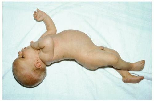
Bilirubin Levels and Developmental Outcome

Although there is no doubt about the relationship between elevated TSB levels and brain damage, the ability of a single peak bilirubin level to predict long-term neurodevelopmental outcomes is relatively poor (117,118,119). In addition, our conclusions regarding hyperbilirubinemia and neurodevelopmental outcomes are limited by the quality of the published data. In many case reports of kernicterus, analyzed in detail by Ip et al. (117,118), it is impossible to know if the reported peak TSB levels (measured in some cases more than 7 days after birth) were true peak levels. In addition, many cohort studies have significant problems with dropouts and with blinding of the examiners (117,118,120). For a detailed analysis of the published literature from 1966 to 2001 dealing with these issues, see the evidence reports of Ip et al. (117,118).

Hsia et al. (121) and Mollison and Cutbush (122) first established the link between bilirubin levels and brain damage in the early 1950s, when they demonstrated that the risk of kernicterus in infants with Rh hemolytic disease increased dramatically with rising bilirubin levels and that exchange transfusion could markedly reduce the risk. Subsequent studies suggested that, in untreated infants with hemolytic disease, the incidence of kernicterus was much higher than the incidence in markedly jaundiced infants without hemolytic disease (123). Starting in 1967 and continuing into the late 1970s, reports from the Collaborative Perinatal Project (CPP), a study of 53,000 pregnant women and their offspring, linked moderate elevations of neonatal serum bilirubin to lower developmental scores, lower IQ scores, and an increased risk of neurologic abnormalities. These findings occurred at TSB levels previously presumed to be safe and suggested that acute bilirubin encephalopathy or classic kernicterus was only the most obvious and extreme manifestation of a spectrum of bilirubin toxicity. At the other end of the spectrum might lie more subtle forms of neurotoxicity that occur at much lower bilirubin levels and in the absence of any obvious abnormal clinical findings in the neonatal period (105,118,124,125).


Effect of Hemolysis

Newman et al. (126) evaluated 140 term and late preterm infants with TSB levels ≥25 mg/dL. One hundred and thirty-two (94%) infants were followed for at least 2 years and 82 (59%) for 5 years with blinded neurodevelopmental evaluations. They found no difference in neurodevelopmental outcomes between the hyperbilirubinemic infants and controls. But nine children in the hyperbilirubinemia group who had positive direct antiglobulin tests (DATs) had full-scale IQ scores that were, on average, 17.8 points below those with a negative DAT. In a reanalysis of the data from the Collaborative Perinatal Project, Kuzniewicz and Newman (127) found that the maximum TSB level was not a significant predictor of IQ scores, but there was a significant interaction term of -6.7 full-scale IQ points for those with a positive DAT and a TSB ≥25 mg/dL. In a study of full-term Turkish infants (128), those who had DAT-positive ABO incompatibility or Rh immunization (vs. infants without hemolytic disease) had a greater risk of neurologic abnormalities and lower IQ scores when their indirect reacting bilirubin levels exceeded 20 mg/dL (342 &mgr;mol/L). Two hundred and fortynine newborns were admitted to the Cairo University Children’s Hospital with TSB levels of ≥25 mg/dL (129), and 44 (17.7%) had evidence of moderate or severe acute bilirubin encephalopathy (ABE). The presence of Rh incompatibility (OR 48.6) and sepsis (OR 20.6) increased the risk of neurotoxicity. These data reinforce the belief that isoimmune hemolysis is an important risk factor in bilirubin-dependent brain damage although the mechanism for this association remains to be elucidated.

The hemolysis associated with G6PD deficiency and hyperbilirubinemia is also considered to be an additional risk factor for poor developmental outcome (130), although an analysis of the cases in the U.S. Kernicterus Registry did not find this to be the case (119).


Infants without Hemolysis

Two issues in neonatal medicine that consistently generate controversy are the relationship between hyperbilirubinemia and adverse developmental outcome in nonhemolyzing newborns and the indications for treating these infants. These issues are addressed in multiple studies, and the reader is referred to extensive reviews for details of the individual studies (117,118,123,131,132). Although in many studies of otherwise-healthy neonates without hemolytic disease, there is no convincing demonstration of any adverse effect of TSB levels below 25 mg/dL on IQs, definite neurologic abnormalities, or sensorineural hearing loss (117,118,123,126,131,132,133,134), some older (135), and more recent studies (136,137,138,139) have raised the possibility of subtle developmental abnormalities in some infants with modest degrees of hyperbilirubinemia.

No studies have looked specifically at infants who are 35 to 37 weeks of gestation, although the data analyzed by Ip et al. (117,118) include infants of 34 weeks and older. In the very large CPP, the study population included all infants with birth weights ≥2,500 g (123,135). Presumably, some of these infants were in the 34- to 37-weeks’ gestational age category. When they combined both abnormal and suspicious neurologic examination results, Newman and Klebanoff (135), in their analysis of the CPP, did demonstrate a significant increase in abnormalities associated with increasing bilirubin levels. The “suspicious” abnormalities included nonspecific gait abnormalities, awkwardness, an equivocal Babinski reflex, abnormal cremasteric reflex, abnormal abdominal reflex, failure of stereognosis, questionable hypotonia, and gaze abnormalities. When the abnormal and suspiciously abnormal children were combined, the risk of abnormalities increased from 14.9% for those whose TSB levels were less than 10.0 mg/dL (171 &mgr;mol/L) to 22.4% for those whose TSB levels exceeded 20 mg/dL (340 &mgr;mol/L). Because 41,324 infants were enrolled in this study, these differences were statistically highly significant, but this finding should be kept in perspective. Even if the relationship between these findings is causal, we have no evidence that the use of a bilirubin-lowering intervention, such as phototherapy, at these low bilirubin levels would affect the outcome. Finally, as the authors point out (135), even if bilirubin levels had been prevented from exceeding 10 mg/dL (171 &mgr;mol/L) in every infant, the expected rate of abnormal or suspicious neurologic examination results would only decline from 15.13% to 14.85%.

Less-reassuring information is provided by a study of a group of Israeli army draftees (n = 1,948) in which their preinduction psychological and physical examinations at age 17 years were matched with their newborn bilirubin levels. Seidman et al. (140) found an association between the risk of an IQ below 85 and a TSB level higher than 20 mg/dL (342 &mgr;mol/L) in full-term boys (but not with girls) with a negative Coombs test (p = 0.01). No association was found between bilirubin levels and mean IQ score, the risk of physical or neurologic abnormality, or hearing loss. Three more recent studies identified “soft signs” of neurologic dysfunction in infants exposed to moderate levels of bilirubin (137,138,139). Dutch investigators (139) noted that five of eight (63%) infants at age 1 year who, as newborns, had TSB levels between 19.6 and 26 mg/dL (335 and 444 &mgr;mol/L) demonstrated minor abnormalities in muscle tone and posture compared with 0 of 20 control infants (p < 0.001). This group subsequently evaluated 42 healthy term infants whose neonatal TSB levels were ≥12.9 mg/dL (137), and 68 controls. A neurologic examination at age 18 months with the
examiner blinded to the history and TSB level found no difference in the incidence of minor neurologic dysfunction (MND) between the two groups, but a TSB level of ≥17.5 mg/dL was associated with an increased risk of complex MND (adjusted OR 4.21, 95% CI: 1.02 to 17.37). In a German study, children at 7 years of age whose neonatal TSB levels were greater than 20 mg/dL (342 &mgr;mol/L) scored significantly worse on a scale designed to measure choreiform and athetoid movements. Eight of sixteen (50%) children in the hyperbilirubinemia group versus 3 of 18 (17%) in the control group had abnormal scores (data not found in the original paper but kindly provided by the authors) (120,138).

In Milwaukee, 39/93 infants whose TSB levels as neonates were greater than 20 mg/dL were followed to ages 2.5 to 3.5 years and compared with nonjaundiced controls (141). No differences were observed between the two groups with regard to the Bayley mental or psychomotor development indices, expressive or receptive speech, abnormal hearing, or minor neurologic abnormalities. The sample size was small, however, and in every one of these areas, the jaundiced infants performed worse. Although the differences did not achieve statistical significance, the possibility of a type II error is high.

In Northern California, Newman et al. (126) identified 140 infants with neonatal TSB levels of ≥25 mg/dL, 10 of whom had TSB levels of ≥30 mg/dL, and 492 randomly selected controls. One hundred and thirty-six infants received phototherapy, and five had exchange transfusions. One hundred and thirty-two (94%) infants were followed for at least 2 years and 82 (59%) for 5 years with blinded neurodevelopmental evaluations by child psychologists and neurologists and detailed developmental and behavioral evaluations using parent questionnaires. There was no difference in neurodevelopmental outcomes, between the hyperbilirubinemic infants and controls although, as noted above (see Effect of Hemolysis), hyperbilirubinemic infants with a positive DAT had lower cognitive scores but did not have more neurologic or behavioral problems.

Wong et al. (134) studied 99 full-term Chinese neonates with nonhemolytic TSB levels of 16.8 to 29.2 mg/dL. All received phototherapy and three an exchange transfusion. These infants had regular physical, neurologic, visual, and auditory assessments every 3 to 6 months until the age of 3 years. All except two infants with mild motor delay had normal neurodevelopmental status at age 3 years. Most recently, Vandborg et al. (133) had parents complete the Ages and Stages Questionnaire in 162 infants, ≥35 weeks of gestation and aged 1 to 5 years, whose neonatal TSB levels were ≥25 mg/dL. When compared with 146 controls, they found no evidence of developmental delay in the hyperbilirubinemic infants.


Duration of Exposure to Hyperbilirubinemia

It is intuitive to assume that the longer one is exposed to a potential toxin, the more likely it is for the toxin to have an effect. In the study from Turkey mentioned above (128), the risk of neurologic damage increased from 2.3% to 18.7% and 26% in those infants exposed to indirect bilirubin level of ≥20 mg/dL for less than 6 hours, 6 to 11 hours, and ≥12 hours, respectively. In a study of 83 term and late preterm infants exposed to indirect bilirubin levels of ≥15 mg/dL (142), there was a linear increase in abnormal neurologic findings from those exposed for less than 1 day (5%) to 65% in those exposed for ≥6 days. These and other data (143) suggest that the duration of hyperbilirubinemia is related to the risk of long-term neurodevelopmental outcome although this was not seen in the NICHHD collaborative phototherapy study (144) where no association was found between IQ and duration of exposure to bilirubin.


Comorbid Factors and Outcome

Hyperbilirubinemia is more likely to cause damage in the presence of the neurotoxicity risk factors (130,145) or if the infant is sick or unstable (see Table 32.10).


TABLE 32.10 Clinical and Laboratory Factors That Increase the Risk of Neurotoxicity in the Presence of Rising Bilirubin Levels


Isoimmune hemolytic disease


G6PD deficiency


Sepsis


Acidosis


Lower gestational age


Clinical instability such as apnea and bradycardia, hypotension, need for mechanical ventilation


Serum albumin < 2.5 g/dL


Bilirubin-Binding Capacity, Kernicterus, and Developmental Outcome

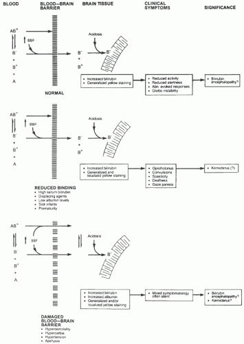
Because bilirubin that is “free” or loosely bound to albumin is more likely to cross the BBB (see Bilirubin Chemistry and Neurotoxicity and Blood-Brain and Blood-Cerebrospinal Fluid Barriers above), our ability to predict the risk of bilirubin encephalopathy should be improved by measurement of unbound bilirubin or the reserve albumin-binding capacity (119). Wennberg et al. (119) have summarized the limited clinical data in term and preterm infants that suggest the measurement of free bilirubin is better than TSB as a predictor of bilirubin toxicity and, if nothing else, would reduce the risk of unnecessary intervention. Unfortunately, there are, currently, no bilirubin-binding tests in routine clinical use in the United States, although a semiautomated peroxidase method has been used in Japan (119). In addition, there are problems with the accuracy and standardization of free bilirubin measurements and the potential for interference from photoisomers (146).


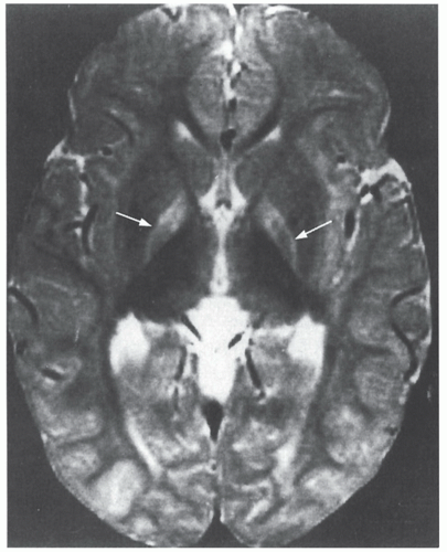
Hearing Loss and Audiometric-Evoked Responses

The BAER test is an accurate and noninvasive means of assessing the functional status of the auditory nerve and the brainstem auditory pathway. e-Figure 32-2 shows the BAER tracing of a normal full-term infant. The three positive waveforms labeled in the figure are those most easily identified in the neonate (87). The latency for wave I represents the peripheral conduction time. Latency of waves III and V and the interpeak latency of waves I to III, III to V, and I to V all represent measurements of central conduction time. The interpeak latency I to V is referred to as the brainstem conduction time. Reports also include amplitudes of the waveforms. These may decrease or be lost in response to various insults (20,113).

Several studies have documented a relationship between TSB levels and the BAER (87), and the acute changes seen in the BAER can be reversed by lowering the TSB level with phototherapy or exchange transfusion (147). Abnormalities of the BAER are more closely related to unbound bilirubin levels than to TSB level (45,148).

As noted above (Kernicterus, Auditory Abnormalities), deficits in central hearing, speech, and language can occur in the absence of pure-tone hearing loss, and these may be manifestations of auditory neuropathy or dyssynchrony (149). This entity is defined as an abnormal or absent BAER with normal inner ear function as tested by cochlear microphonic responses or OAEs (149). Remarkably, in a follow-up of 36 children with the Crigler-Najjar syndrome, none had evidence of sensorineural hearing loss (150).


Cry Analysis

An abnormal cry is a sign of neurologic distress and is associated with acute bilirubin encephalopathy (20). Modest degrees of hyperbilirubinemia also affect the infant’s cry (87).


Infant Behavior

Investigators have used the Brazelton Neonatal Behavioral Assessment Scale to evaluate the effect of hyperbilirubinemia on infant
behavior. Most studies show some effect, although several are confounded by the use of phototherapy (87). Jaundiced infants score lower than do controls in habituation, orientation, motor performance, regulation of state, and autonomic stability (87,151).


Hyperbilirubinemia, Autism Spectrum Disorder, and Attention Deficit Disorder

Some authors have identified an association between hyperbilirubinemia, autism, and attention deficit disorder (ADD) (152,153), but others have not (154). In a population-based cohort of infants ≥35 weeks in Nova Scotia, Jangaard et al. (136) identified 3,779 infants (6.7%) with TSB levels of ≥13.5 mg/dL (moderate hyperbilirubinemia) and 348 (0.6%) with TSB ≥19 mg/dL (severe hyperbilirubinemia). When compared with nonjaundiced controls, there was no increase in cerebral palsy, developmental delay, or deafness in infants with TSB ≥19 mg/dL, but those with moderate hyperbilirubinemia had an increased risk of developmental delay (adjusted RR: 1.6; 95% CI: 1.3 to 2.0). There was also a significant increase in the risk of attention deficit disorder in those exposed to a TSB of ≥19 mg/dL (adjusted RR: 1.9; 95% CI: 1.1 to 3.3) and a nonsignificant increase in the risk of autism (adjusted RR: 1.6; 95% CI: 1.0 to 2.5). In response to this study, Kuzniewicz et al. (155) evaluated the data from the Northern California Kaiser Permanente Medical Care Program of infants ≥34 weeks. They found no association between TSB levels and a diagnosis of ADD. Using the same database, Croen et al. (154) compared 338 children with the diagnoses of autism spectrum disorder (ASD) with 1,817 controls and also found no association between neonatal hyperbilirubinemia and ASD. The association between ASD and hyperbilirubinemia described by Maimburg et al. (152) was subsequently retracted (156). In their comprehensive review and metaanalysis, Gardener et al. (153) evaluated the association with ASD of over 60 perinatal and neonatal factors. Some 17 risk factors including hyperbilirubinemia were associated with an increased risk of ASD. They conclude that “There is insufficient evidence to implicate any one perinatal or neonatal factor in autism etiology, although there is some evidence to suggest that exposure to a broad class of conditions reflecting general compromises to perinatal and neonatal health may increase the risk.” It is important to remember that analysis of very large databases will often identify associations that might be spurious or real, and if real, might not represent a cause-effect relation (157).


▪ PREMATURE INFANTS AND LOW BILIRUBIN KERNICTERUS


Kernicterus and Developmental Outcome

Premature infants are at greater risk of developing kernicterus or bilirubin encephalopathy than are full-term newborns exposed to similar bilirubin levels (158). Between 1958 and 1972, several studies documented kernicterus in preterm infants at TSB levels ranging from 10 to 18 mg/dL (158) and led to the use of the term “low bilirubin kernicterus,” but several neurodevelopmental follow-up studies, recently reviewed in detail (158), have failed to show an association between peak TSB levels and late adverse outcomes in very-lowbirthweight (VLBW) infants. In the National Institute of Child Health and Human Development (NICHHD) cooperative phototherapy study, infants born between 1974 and 1976 were randomly assigned to a control group that received no phototherapy or to a group that received phototherapy at predetermined TSB levels. The criteria for exchange transfusion for all infants mandated exchange transfusions at low levels of serum bilirubin (10 mg/dL [171 &mgr;mol/L] in high-risk newborns with birth weights <1,250 g) (159). Kernicterus was found in 4 of 76 autopsied infants whose birth weights ranged from 760 to 1,270 g (160). Their peak TSB levels ranged from 6.5 to 14.2 mg/dL (111 to 243 &mgr;mol/L). All were asphyxiated or had hyaline membrane disease, and all had some degree of periventricular-intraventricular hemorrhage (PIVH). Two had periventricular leukomalacia (PVL).

Surviving infants in the study were followed and evaluated at 6 years of age with the Wechsler Verbal and Performance IQ test. No differences were found between the control and phototherapy groups in the incidence of definite and suspect cerebral palsy, clumsy or abnormal movements, hypotonia, or an IQ lower than 70. There were no differences between the two groups in growth, speech, hearing loss, or evidence of hyperactivity (144).

Scheidt et al. (161) published a 6-year follow-up from the NICCHD study of 224 control children whose birth weights were lower than 2,000 g. None of these infants received phototherapy, but bilirubin levels were maintained below specified levels by the use of exchange transfusion. No relation was found between serum bilirubin levels and the incidence of cerebral palsy nor was there any association between maximal bilirubin level and IQ. IQ was not associated with mean bilirubin level, time and duration of exposure to bilirubin, or measures of bilirubin-albumin binding.


Recent Reports of Kernicterus and Neurodevelopmental Outcomes

Although today, kernicterus is rarely seen in the NICU population, recent reports remind us that it has not disappeared completely. Hypotonia and (in one infant) choreoathetosis, together with the classical MRI findings of kernicterus, were observed in two infants of 31 weeks’ and 34 weeks’ gestation (162). Neither of these infants was acutely ill, and their TSB levels were 13.1 mg/dL (224 &mgr;mol/L) and 14.7 mg/dL (251 &mgr;mol/L), respectively (162). Govaert et al. (163) reported typical MRI findings in five infants, 25 to 29 weeks’ gestation, with kernicterus. The TSB levels ranged from only 8.7 to 11.9 mg/dL (148 to 204 &mgr;mol/L), and the serum albumin levels were strikingly low (1.4 to 2.1 g/dL). Okumura et al. (164) and Moll et al. (165) also report the classic MRI findings of kernicterus in 24- to 26-week-gestation infants with peak TSB levels of 7.1 to 9.9 mg/dL. Choreoathetosis was present in 15/16 of the preterm infants reported by the above investigators (162,163,164,165). Significant associations between peak TSB levels and hearing loss have been documented in LBW infants (166,167,168) and confirmed recently in a population of ELBW infants (169).

Most recently, the largest randomized controlled trial ever undertaken in a population of ELBW infants (birth weights <1,000 g) was completed by the NICHHD Neonatal Research Network (NRN) (170). The protocol for the study and details of the results are presented below (see Phototherapy—Management of Infants <35 weeks’ gestation). In this trial, infants treated with “aggressive” phototherapy (prophylactic phototherapy administered as soon after birth as possible) compared with “conservative” phototherapy (instituted at TSB levels ≥8.0 mg/dL [500 to 750] or 10.0 mg/dL [751 to 1,000]) had lower mean TSB levels and lower rates of NDI, hearing loss, and athetosis (170).

Thus, the brain of the VLBW infant appears to be more susceptible to damage from a number of sources and, given that preterm infants have lower serum albumin levels and less-effective albumin binding and are much more likely to be sick than are full-term infants, it makes sense to take a more aggressive approach to maintaining low bilirubin levels in this population.


Hyperbilirubinemia and Pulmonary Hemorrhage

Studies from the late 1940s and early 1950s suggested an association between pulmonary hemorrhage and kernicterus (171) in infants dying with severe erythroblastosis fetalis. These infants were profoundly anemic, marked hypoalbuminemic, and had thrombocytopenia, and disturbances of coagulation. So it is not surprising that some developed hemorrhagic pulmonary edema. In an autopsy series of LBW infants with kernicterus, pulmonary hemorrhage was not found more frequently in the kernicteric infants when compared with those who did not have kernicterus (172).


▪ JAUNDICE IN THE HEALTHY NEWBORN


Normal Serum Bilirubin Levels

TSB levels vary considerably, depending on the racial composition of the population, the incidence of breastfeeding, and other genetic and epidemiologic factors. Different laboratory methods for measuring TSB provide additional variation (173) (see Laboratory Measurements of Bilirubin below).

The use of phototherapy prevents us from obtaining a true picture of the natural history of neonatal bilirubinemia because we treat some infants with rising TSB levels in the first 24 to72 hours, even though many have no defined pathologic or other known cause for the rising bilirubin level. Thus, what we generally see is a “damped” picture of the natural history. The age of the population studied also affects our definition of normal values, particularly the upper limits, as infants who develop higher TSB levels may not be seen until they are 6 to 10 days old. These infants are not included in studies restricted to hospitalized populations.

Data from the Collaborative Perinatal Project (174) conducted from 1955 to 1961 (when 30% or fewer mothers breastfed their infants and phototherapy was not used) showed that approximately 95% of all infants had a TSB concentration that did not exceed 12.9 mg/dL (215 &mgr;mol/L), and this (95th percentile) became the accepted upper limit of “physiologic jaundice” (174).

More recent studies that have included readmitted infants have defined the 95th percentile as a level of 15.5 to 17.5 mg/dL (265 to 299 &mgr;mol/L) (175,176). In a multicenter study of infants ≥36 weeks in nurseries in the United States, Hong Kong, Japan, and Israel, 2 SD above the mean for the peak TSB levels at 96 ± 6.5 hours was 17 mg/dL (291 &mgr;mol/L), and the 95th percentile was 15.5 mg/dL (265 &mgr;mol/L) (177). In a Greek population (178), the 95th percentile at 108 hours was a TcB level of 15.1 mg/dL. Recognizing the variability among populations, in the United States, where approximately 70% of mothers initiate breastfeeding in the hospital (179), these data suggest that the 95th percentile for TSB levels after age 96 hours is generally in the range of 15.5 to 17.5 mg/dL.

From the clinician’s point of view, this implies that a 4- to 5-dayold breastfed infant whose TSB level is 14 to 15 mg/dL (291 &mgr;mol/L) does not require any laboratory investigation to find out why the infant is jaundiced, although follow-up is necessary to ensure that the bilirubin levels do not become excessive (130,180). Data from studies of predominantly breastfed infants suggest that the normal mean peak TSB level is approximately 8 to 9 mg/dL (137 to 154 &mgr;mol/L) (177,181,182). In the international, multicenter study, the mean TSB level at 96 ± 6.5 hours was 8.9 ± 4.4 (SD) mg/dL in breastfed infants and 7.6 ± 3.58 mg/dL in those fed formula (p < 0.0001) (177). Because so many LBW, VLBW, and ELBW infants receive phototherapy, it is not possible to provide reference values for these infants.


The Natural History of Neonatal Bilirubinemia

The availability of electronic TcB measurements has made it possible to study the natural history of bilirubinemia in large contemporary populations (183). TcB nomograms for newborn populations have been developed in the United States, Canada, Italy, Greece, Thailand, China, Japan, and other parts of the world (184). These studies provide normative data for different populations, and the nomograms have been used to identify unusually elevated levels and trends in the rate of rise of TSB, and they have been used to assess the risk for the subsequent development of severe hyperbilirubinemia. De Luca et al. (185) compared four published TcB nomograms and analyzed the differences in TcB levels and kinetics in these populations. The weighted mean TcB value at 73 to 96 hours was 8.6 ± 3.3 mg/dL with a range of 6.9 to 10.4 mg/dL. The data of Maisels and Kring (186) demonstrate clearly the differences in natural history between infants of gestational age ≥40 weeks and those less than 40 weeks, showing how much earlier the TcB values peak and then decline in the more mature infants (Fig. 32.7). In these and other studies, infants with a positive DAT and those who required phototherapy in the first 24 hours were excluded, so the percentiles are lower than if they reflected the true natural history. Because of the tendency to use more phototherapy in less mature infants, exclusion of infants receiving phototherapy may also have attenuated the association between lower gestational age and higher bilirubin percentiles at each age. Figure 32.8 shows
smoothed curves based on the data from a number of studies that provide a guide to the expected course of bilirubin levels in a primarily breastfed (60% to 70%), Western population.


FIGURE 32.7 Transcutaneous bilirubin levels in the first 96 hours in a normal newborn population of ≥35 weeks’ gestation. Effect of gestational age on transcutaneous bilirubin levels and on the time course of bilirubinemia. Smoothed curves of the 50th, 75th, and 95th percentiles. Data from 9,397 TcB measurements on 3,984 healthy newborns. Infants who required phototherapy prior to discharge were excluded (n = 139). Reproduced from Maisels MJ, Kring E. Transcutaneous bilirubin levels in the first 96 hours in a normal newborn population of 35 or more weeks’ of gestation. Pediatrics 2006;117:1169-1173, with permission. Copyright 2006 by the American Association of Pediatrics.


FIGURE 32.8 Smoothed curves from studies in diverse populations (175,176,177,181,182,187,188) illustrating the expected velocity of TSB levels and approximate values for the 50th and 95th percentiles in a western predominantly breastfed (60%-70%) population.

Bhutani et al. (176) developed a nomogram that defines predischarge risk zones for the subsequent development of hyperbilirubinemia (see Preventing Extreme Hyperbilirubinemia and Kernicterus below). Because of sampling bias, however, this nomogram does not describe the natural history of bilirubinemia in the newborn (189), although it is a very useful tool for predicting the risk of subsequent hyperbilirubinemia (176) and is recommended by the AAP for this purpose.


▪ EPIDEMIOLOGY OF NEONATAL HYPERBILIRUBINEMIA

An important first step in the diagnosis and management of any jaundiced newborn is an understanding of the factors that normally affect neonatal bilirubin levels. Some of these factors have been identified only in large epidemiologic studies, and their clinical relevance is questionable, but those listed in Table 32.11 have been shown, consistently, to have an important influence on TSB levels.


Ethnic and Familial Influences

East Asian, Hispanic (primarily Mexican), and Native American infants have mean maximal TSB concentrations that are significantly higher than are those of white infants (10,13). In a Hispanic population, 31% of infants had TSB levels ≥15 mg/dL (190) compared with 3% to 10% of infants in other populations (175,191). Black infants in the United States and Great Britain have lower TSB levels than do white infants (10,13) but, presumably, because of the prevalence of G6PD deficiency in these infants, they are at greater risk for hazardous hyperbilirubinemia (TSB ≥ 30 mg/dL) (192). Neonatal jaundice runs in families (193). In a population of 3,301 newborns of male U.S. army veterans if one or more previous siblings had a TSB >12 mg/dL (205 &mgr;mol/L), the subsequent sibling was three times more likely than were controls (10.3% vs. 3.6%) to develop a TSB >12 mg/dL, and if a prior sibling had a TSB >15 mg/dL (257 &mgr;mol/L), the risk of a similar TSB in the subsequent sibling was increased 12.5-fold (10.5% vs. 0.9%). These relationships applied whether or not the siblings were breastfed or formula fed (193).


TABLE 32.11 Risk Factors for the Development of Hyperbilirubinemia in Infants of 35 or More Weeks’ Gestation


Elevated predischarge TSB or TcB level


Decreasing gestational age


Jaundice observed in the first 24 h or prior to discharge


Blood group incompatibility with positive DAT, other known hemolytic disease (e.g., G6PD deficiency, hereditary spherocytosis)


Previous sibling with jaundice or who received phototherapy


Vacuum extraction delivery, cephalohematoma, or significant bruising


Exclusive breastfeeding, particularly if nursing is not going well and weight loss is excessive


East Asian race


Macrosomic infant of a diabetic mother


Maternal age ≥25 y


Male gender


Genetic Factors

The genetics of neonatal jaundice and the inborn errors of hepatic bilirubin uridine diphosphate glucuronosyl transferase (UGT) expression are discussed in detail above (Neonatal Bilirubin Metabolism) and below (Inherited Unconjugated Hyperbilirubinemia).


Maternal Factors


Smoking

Some studies suggest that infants of mothers who smoke during pregnancy have lower serum bilirubin levels than do infants of nonsmokers (194), but others have not found this (195). These data are confounded by the fact that women who smoke are much less likely to breastfeed, and the likelihood of breastfeeding is inversely related to the number of cigarettes smoked per day (196).


Diabetes

Macrosomic infants of insulin-dependent diabetic mothers produce more bilirubin (197) and have higher TSBs than do control infants (198). These infants have high erythropoietin levels and increased erythropoiesis, so that ineffective erythropoiesis and polycythemia probably are responsible for the increased bilirubin production (199,200). In addition, diabetic mothers have three times more &bgr;-glucuronidase in their breast milk than do nondiabetic mothers (200). This enzyme enhances the enterohepatic reabsorption of bilirubin (see Breastfeeding and Jaundice below).


Events during Labor and Delivery


Induction and Augmentation of Labor by Oxytocin

Multiple studies and several controlled trials have shown an association between the use of oxytocin to induce or augment labor and an increased incidence of neonatal hyperbilirubinemia, although the mechanism for this is unclear (201).


Anesthesia and Analgesia

Several studies associate epidural anesthesia, specifically, bupivacaine, with neonatal jaundice (202). These agents readily cross the placenta and produce measurable levels in the newborn (203).


Delivery Mode

Vaginally delivered term newborns have higher TSB levels than those delivered by cesarean section (207). Vacuum extraction increases the risk of scalp bruising and cephalhematomas, both of which increase the risk of hyperbilirubinemia. Because the catabolism of 1 g of hemoglobin produces 35 mg of bilirubin, bruising
and cephalhematomas can contribute significantly to the infant’s bilirubin load.


TABLE 32.12 Effect of Gestation on the Risk of Subsequent Hyperbilirubinemia


Study


Outcome Variable


Gestation (Weeks)


Odds Ratio (95% CI) for Risk of Subsequent Hyperbilirubinemia


Maisels and Kring (213)


Readmission for phototherapya


35 0/7-36 0/7


36 1/7-37 0/7


13.2 (2.7-64.6)b


7.7 (2.7-22.0)b


Newman et al. (211)


TSB ≥ 25 mg/dL


36 0/7-42 6/7


1.7 (1.4-2.5) per week of gestation below 40 wk


Keren et al. (212)


Within 1 mg/dL of hour-specific AAP phototherapy level (or higher)a


35 0/7-37 6/7


9.2 (4.4-19.0) bivariate


Maisels (214)


TSB ≥ 17 mg/dL


35 0/7-36 6/7


37 0/7-37 6/7


20.8 (2.3-184.7)b


14.9 (1.91-115.4)b


a Association partly due to lower treatment threshold in less-mature infants.

b Compared with 40 weeks’ gestation.


From Maisels MJ, Newman TB. The epidemiology of neonatal hyperbilirubinemia. In: Stevenson DK, Maisels MJ, Watchko JF, eds. Care of the jaundiced neonate. New York: McGraw Hill, 2012:97-113, with permission.


Placental Transfusion and the Timing of Umbilical Cord Clamping

A Cochrane database review of 11 trials of 2,989 mothers and their infants (208) found that in those whose cords were clamped early, significantly fewer required phototherapy for jaundice. The lateclamped group had higher hemoglobin levels. When cord clamping was delayed in infants 28 to 36 weeks of gestation, the mean TSB level at 72 hours was 7.7 mg/dL compared with 3.2 mg/dL in the early clamped group (209). A more recent study of late-preterm infants (210), however, found no relationship between delayed clamping and pathologic jaundice, polycythemia, or the need for phototherapy.


Neonatal Factors


Gestation

Absent overt hemolysis, by far the most important single clinical factor associated with the subsequent risk of hyperbilirubinemia is the infant’s gestational age (211,212,213,214), and the magnitude of this risk has been quantified (Table 32.12). How this influences our management of the newborn is discussed in detail below (see Treatment).


Gender

As a group, male infants consistently have higher bilirubin levels than do females (211,213).


Caloric Intake and Weight Loss

Decreased caloric intake is associated with an increase in serum bilirubin in animals and humans (215). The primary mechanism responsible for this appears to be an increase in the enterohepatic circulation of bilirubin (215,216). A significant association also exists between hyperbilirubinemia and weight loss in the first few days after birth (212,213).


TABLE 32.13 Effect of Exclusive Breastfeeding on Risk of Subsequent Hyperbilirubinemia in Newborns ≥35 Weeks of Gestation


Study


N


Outcome Variable Bilirubin (mg/dL)


N with Outcome


OR (95% CI) versus Formula or Partially Breastfed


Maisels and Kring (213)


29,934


19.3 ± 2.7


127 (0.4%)


4.2 (1.8-9.9)


Newman et al. (211)


51,387


≥25


73 (0.14%)


5.7 (2.1-15.5)


Maisels et al. (214)


11,456


≥17


75 (0.65%)


10.75 (2.37-48.8)


Huang et al. (223)


a


≥20


72


4.6 (2.40-8.81)


a No denominator provided.


From Maisels MJ, Newman TB. The epidemiology of neonatal hyperbilirubinemia. In: Stevenson DK, Maisels MJ, Watchko JF, eds. Care of the jaundiced neonate. New York: McGraw Hill, 2012:97-113, with permission.


Type of Diet

Infants fed a casein hydrolysate formula had significantly lower TSB levels from days 10 through 18 than did those infants fed standard casein or whey-predominant formulas (217,218). The cumulative stool output of the infants fed the casein hydrolysate was lower than was that of the infants fed the other formulas (217), suggesting that factors other than stool output and its effect on the enterohepatic circulation must explain these observations. The casein hydrolysate formula (Nutramigen) contains an inhibitor of &bgr;-glucuronidase (219), an enzyme that acts on the hydrolysis of bilirubin glucuronide and therefore facilitates the enterohepatic absorption of unconjugated bilirubin (220). When fed saccharolactone, an inhibitor of &bgr;-glucuronidase, rats excrete less bilirubin in their bile, suggesting that inhibition of &bgr;-glucuronidase decreased intestinal absorption of bilirubin (221).


Breastfeeding and Jaundice

Multiple studies over the last 30 years have found a strong association between breastfeeding and an increased incidence of neonatal hyperbilirubinemia (222), and Table 32.13 lists some studies that have quantified the risk of hyperbilirubinemia in exclusively breastfed infants.

Jaundice associated with breastfeeding in the first week has been called “breastfeeding jaundice” or “breastfeeding-associated jaundice” and that which appears later and is associated with prolonged jaundice has been called the “breast milk jaundice syndrome” (224), but there is considerable overlap between these two entities. As a group, breastfed infants have TSB levels that are higher than those in formula-fed infants for at least 3 to 4 weeks (217,225). But these are the same infants who have higher TSB levels in the first week, so it is unclear whether or not those who are still jaundiced at age 3 to 4 weeks represent a distinct group.


TABLE 32.14 Pathogenesis of Jaundice Associated with Breastfeeding


Increased enterohepatic circulation of bilirubin


Decreased caloric intake


Less cumulative stool output and stools contain less bilirubin (compared with formula-fed infants)


Increased intestinal fat absorption


Less formation of urobilin in gastrointestinal tract


Increased activity of &bgr;-glucuronidase in breast milk


Decreased bilirubin conjugation


Mutations of the UGT1A1 gene (Gilbert syndrome)—prolonged breast milk jaundice


Pathogenesis of Jaundice Associated with Breastfeeding

Table 32.14 lists the factors that are thought to play a role in the pathophysiology of jaundice associated with breastfeeding.


Caloric Intake

In the first few days, breastfed infants get fewer calories and lose more weight than do formula-fed infants (Table 32.15). The association between poor caloric intake and the development of hyperbilirubinemia has led some experts to categorize the jaundice associated with breastfeeding in the first few days after birth as “starvation jaundice” or “breast-nonfeeding jaundice (224).” The implication is that if breastfed infants were nursed effectively from birth, they would not be more jaundiced than were formula-fed infants, and there is some evidence to support this view (226,227,228).

Bertini et al. (226) studied 2,174 well newborns ≥37 weeks’ gestation. All infants had continuous rooming in with their mothers, and TSB levels were measured in jaundiced infants twice daily until there was a decrease in TSB. Nursing infants were exclusively breastfed 6 to 12 times per day, and these infants were routinely supplemented with formula if the infant’s birth weight was ≤2,500 g or if their weight loss was ≥4% at 24 hours, ≥8% at 48 hours, or ≥10% after 72 hours. Formula-fed infants received no breast milk. The investigators found a positive correlation between TSB levels >12.9 mg/dL, weight loss after birth, and breastfeeding requiring supplementation with formula. Breastfeeding, per se, was not associated with hyperbilirubinemia. They concluded that infants who are successfully breastfed and therefore lose little weight are not more likely to be jaundiced than are formula-fed infants, whereas those who required formula supplementation because of excess weight loss were more likely to be jaundiced. These data support the view that it is less effective breastfeeding, rather than breastfeeding or breast milk per se, that is responsible for the association of breastfeeding and hyperbilirubinemia. In normal adults and those with Gilbert syndrome, caloric deprivation increases hemolysis and bilirubin production (229) so that, in addition to its effect on the enterohepatic circulation, poor caloric intake in infants might also produce an increase in bilirubin production due to hemolysis.


TABLE 32.15 Comparison of Weight Loss and Intake in Breastfed and Formula-Fed Newborns (Mean ± SD)


Breastfed, n = 15


Formula-Fed, n = 28


p


Birth weight (kg)


3.26 ± 0.41


3.35 ± 0.35


ns


Gestation (wk)


39.6 ± 0.9


39.7 ± 1.2


ns


Parity


2.3 ± 1.3


2.4 ± 0.9


ns


Weight loss day 1 (g)


149 ± 96


130 ± 56


ns


Weight loss day 2 (g)


67 ± 58


21 ± 46


0.015


Intake day 1 (mL/kg)


9.6 ± 10.3


18.5 ± 9.6


0.011


Intake day 2 (mL/kg)


13.0 ± 11.3


42.2 ± 14.2


<0.001


Modified from Dollberg S. Lahav S, Mimouni FB. Comparison of intakes of breastfed and bottle-fed infants during the first two days of life. J Am Coll Nutr 2001;20(3):209.


Intestinal Reabsorption of Bilirubin

Intestinal reabsorption of bilirubin (known as the enterohepatic circulation) appears to be an important mechanism responsible for the jaundice associated with breastfeeding (10). Breastfed infants take in less volume and, therefore, fewer calories than do formula-fed infants in the first days after birth (Table 32.15), and there is a relationship between decreased caloric intake and an increase in the enterohepatic circulation of bilirubin (10). Figure 32.9 shows that although breastfed and formula-fed infants pass the same number of stools in the first 3 days of life, formula-fed infants pass significantly more stool by weight, and the bilirubin content of that stool is significantly greater than is that of breastfed infants (10). The rate of bilirubin production is similar in breastfed and formula-fed infants (10). Thus, in addition to receiving more calories, formula-fed infants have significantly less enterohepatic reabsorption of bilirubin than do breastfed infants (10). An increase in stool excretion in the first 21 days is associated with lower TSB levels and, in the first 3 weeks, infants fed human milk pass significantly less stool than do infants who are fed casein-predominant formulas (10). Infants fed casein hydrolysate formulas pass less stool, cumulatively, than do those given whey- or casein-predominant formulas (10).

The relationship between fecal bilirubin excretion and TSB levels may be related to the fecal excretion of unabsorbed fat (10). Unconjugated bilirubin apparently associates with unabsorbed fat in the intestinal lumen. When Gunn rats were fed orlistat, a substance that inhibits lipase, they excreted more fat in their stools, and their TSB levels were significantly lower (10). This suggests that a substance that increases fecal excretion of fat will decrease the enterohepatic absorption of unconjugated bilirubin and facilitate bilirubin excretion in the gut. Breastfed infants have higher fat absorption than do formula-fed infants (possibly related to the presence of bile salt-stimulated lipase in human milk (10). It is possible that hyperbilirubinemia could be prevented or mitigated
by the administration of orlistat to newborns (10). All of these findings support a major role for the enterohepatic circulation in the jaundice associated with breastfeeding.


FIGURE 32.9 Mean ± SEM cumulative weight of stools and fecal. Fecal bilirubin excretion and serum bilirubin concentration in breastfed and bottle-fed infants. From De Carvalho M, Robertson S, Klaus M. Fecal bilirubin excretion and serum bilirubin concentration in breastfed and bottle-fed infants. J Pediatr 1985;107:786, with permission.


Urobilinogen Formation

In adults, bilirubin in the gut is reduced rapidly by the action of colonic bacteria to urobilinogen. Following birth, the neonatal intestinal flora does not convert conjugated bilirubin to urobilin. This leaves bilirubin in the bowel and allows it to be deconjugated and thus available for reabsorption. Formula-fed infants excrete urobilin in their stools earlier than breastfed infants do, perhaps as a consequence of the effect of formula feeding on the intestinal flora (230). Thus, the effect of breast milk on intestinal flora, by slowing the formation of urobilin, further enhances the possibility of intestinal reabsorption of bilirubin.


&bgr;-Glucuronidase

&bgr;-Glucuronidase is an enzyme that cleaves the ester linkage of bilirubin glucuronide, producing unconjugated bilirubin, which can then be reabsorbed through the gut. Significant concentrations of &bgr;-glucuronidase are found in the neonatal intestine, and its activity is higher in human milk than in infant formulas (10).


Prolonged Jaundice in Breastfed Infants

Prolonged indirect-reacting hyperbilirubinemia (TSB ≥5 mg/dL at age 4 weeks) occurs in 20% to 34% of exclusively breastfed infants (231,232,233) and, in some infants, may persist for up to 3 months (224). Recent evidence suggests that mutations of the UGT1A1 gene (Gilbert syndrome) can play a significant role in the pathogenesis of prolonged hyperbilirubinemia (231,234,235). As noted above, several mechanisms have been suggested to explain why breastfed infants are more likely to be jaundiced in the first 7 to 10 days and those fed formula, but an explanation for why breastfed infants are more likely to have prolonged jaundice has been elusive. Arias and Gartner (236), some 50 years ago, identified a progestational steroid, pregnane-3 (&agr;), 20 (&bgr;)-diol in the milk of mothers whose infants had prolonged hyperbilirubinemia. The steroid was shown to inhibit bilirubin conjugation in vitro and was capable of producing hyperbilirubinemia when administered to healthy newborns (237), but subsequent studies could not confirm these findings (238,239). As noted below (see section on Inherited Unconjugated Hyperbilirubinemia), the contemporary era of genetic diagnosis has led to the identification of the cause of previously unexplained hyperbilirubinemia in many infants (240

Only gold members can continue reading. Log In or Register to continue

Stay updated, free articles. Join our Telegram channel

May 30, 2016 | Posted by in PEDIATRICS | Comments Off on Jaundice

Full access? Get Clinical Tree

Get Clinical Tree app for offline access