Obstetric Anesthesia and Analgesia: Effects on the Fetus and Newborn
Samantha L. Russell
Fahd Al Gurashi
Judith Littleford
Many drugs and various techniques have been used to provide anesthesia and analgesia for surgery during pregnancy, for labor and delivery, and in the postpartum period. Between the mid-1800s and 1950s, descriptive reports of the presumed effect of maternally administered medication on the fetus and newborn appeared sporadically in the literature (1). Two developments eventually encouraged physicians to acknowledge the potential problems associated with placental transmission of anesthetic drugs:
Recognition that morphine, a popular ingredient of patent medicines, was addictive and that signs of withdrawal could be identified in the fetus (violent fetal movements and/or sudden fetal death) when the mother’s heavy opioid use was decreased
Demonstration of the presence of chloroform in the umbilical blood of neonates
In 1952, the pioneering work of anesthesiologist Virginia Apgar converted an intangible phenomenon, the clinical condition of a newly born baby, into a formally defined measurement. Thereafter, the well-being of the infant became a major criterion for evaluation of the obstetric and anesthetic management of pregnant women.
This chapter introduces the neonatal practitioner to the clinical aspects of obstetric anesthesia and analgesia and examines their effects on the fetus and newborn.
▪ EVALUATION OF WELL-BEING
Several methods of evaluation have been adopted into common usage as anesthesiologists attempt to separate out the fetal/neonatal effects of their interventions from concomitant medical and nursing management and from the influence of preexisting maternal conditions.
Electronic Fetal Monitoring
Electronic fetal monitoring aims to improve outcomes by identifying fetuses with hypoxic acidemia at a point when the process is still reversible by intrauterine resuscitation or expedited delivery. The fetal heart rate (FHR), including variability, accelerations, and decelerations, if any occur, is recorded electronically on a paper trace as seen in Figure 15.1. Baseline FHR normally ranges between 110 and 160 beats per minute. A reactive (normal or reassuring) cardiotocogram (CTG) is defined by the presence of accelerations. Reduced variability and the presence of decelerations are abnormal findings (2).
Difficulty in the visual interpretation of CTG patterns during labor can result in unnecessary operative intervention, while some significant changes go unrecognized. Computerized CTG systems, which do not rely on observer reading of the FHR tracing, are more accurate and reliable: Evaluation of the FHR pattern is given online continuously, and warnings are displayed if there is signal loss, or an abnormally flat or decelerative trace (3). Retrospective analysis of several thousand records enabled investigators to conclude that the most reliable single parameter of fetal condition was variability (short and long term). Absence of accelerations, presence of decelerations, decrease in the number of movements, and changes in baseline FHR all occurred occasionally in normal fetuses (4).
Anesthesiologists have used CTG recordings to evaluate the effect of intrapartum maternal analgesia on FHR and variability. Solt et al. (5) demonstrated that intrapartum intravenous (IV) administration of meperidine 50 mg and promethazine 25 mg resulted in decreased variability and fewer accelerations on computerized CTG for the duration of their 40-minute recording. It was unclear whether the effect could be attributed to one of the two drugs or the combination, although this is a typical fetal response to systemic maternal opioid administration.
Regional anesthesia (RA), administered with or without concomitant opioid, appears to have no effect on intrapartum FHR characteristics as measured by computer analysis (6). A randomized study of continuous epidural anesthesia (using bupivacaine) with or without narcotic found no difference in pre- and postepidural baseline FHR, accelerations, or variability between groups (7). FHR short- or long-term variability did not change in a double-blind randomized study of the effect of bolus epidural opioid on FHR variability with butorphanol 2 mg, fentanyl 50 µg, sufentanil 15 µg, or saline in combination with bupivacaine 0.25% (8).
Fetal Scalp Blood Gas Analysis
This method is employed to improve the specificity of FHR monitoring (2) but has not been used by anesthesiologists to measure the fetal effects of their interventions.
Biophysical Profile Score
Biophysical profile score (BPS) is an ultrasound-based method that combines measures of acute biophysical variables, fetal breathing, heart rate accelerations, gross body movements, and fetal tone with amniotic fluid volume. The first four variables reflect acute fetal condition, whereas the last variable reflects chronic fetal condition. The observation period lasts 30 minutes because fetuses are known to sleep for intervals lasting approximately 30 minutes. When normal (≥8 out of a possible 10), the BPS is a direct, reliable, and accurate measure of normal tissue oxygenation. A normal score is never associated with an abnormal fetal pH. Scores ≤6/10 during routine antepartum evaluation indicate insufficient oxygen delivery to target organs to maintain function. The lower the score, the greater the likelihood that central acidemia is present (10).
Placental transfer to the fetus of maternally administered intramuscular (IM) narcotic medication (diamorphine 10 mg or morphine 10 or 15 mg administered with dimenhydrinate) resulted in transiently reduced fetal activity and, consequently, a lower BPS for the duration of the drug effect (11,12). A small dose of IV fentanyl (50 µg) given in early labor was associated with abolishment of fetal breathing at 10 minutes postdosing, fewer body movements, and reduced variability (13). The effect lasted approximately 30 minutes, in keeping with the pharmacodynamic profile of fentanyl. This should be taken into account when fentanyl is administered close to the time of delivery. In this study, none of the neonates required resuscitation, and all had umbilical artery pH values greater than 7.2.
Fetal Doppler
Flow velocity waveforms seen on ultrasound from maternal vessels (uterine arteries), placental circulation (umbilical arteries), and fetal systemic vessels (e.g., middle cerebral artery), collectively known as Doppler evaluation, provide prognostic and diagnostic detail about placentation and fetal adaptation (14).
Interrogation of the ductus venosus (DV) has yielded the most robust data on neonatal status to date and may serve as the best “trigger” for delivery when abnormalities are found (9). Distinctive
changes in DV flow during hypoxia point to diastolic dysfunction and risk of acidosis (15). There are technical limitations of Doppler ultrasound, however, and these, coupled with maternal physiologic changes during labor, necessitate the assessment of additional parameters in order to determine fetal status (15,16). Larger clinical studies are required to document efficacy (17).
changes in DV flow during hypoxia point to diastolic dysfunction and risk of acidosis (15). There are technical limitations of Doppler ultrasound, however, and these, coupled with maternal physiologic changes during labor, necessitate the assessment of additional parameters in order to determine fetal status (15,16). Larger clinical studies are required to document efficacy (17).
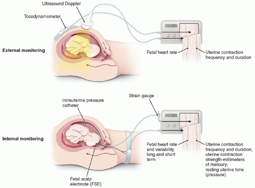 FIGURE 15.1 Cardiotocogram. Modified from Beckman CRB, Ling FW, Barzansky BM, et al. Obstetrics and gynecology, 6th ed. Philadelphia, PA: Wolters Kluwer/Lippincott Williams & Wilkins, 2010:104. |
Regulation of the circulation is a complex fetal behavior, influenced by gestational age and the maternal environment. Under normal circumstances, the reduction in sympathetic tone created by epidural analgesia does not affect Doppler flow characteristics of either the uterine or umbilical artery vessels because the spiral arterioles are maximally dilated and the fetoplacental circulation is stable and tolerant of environmental changes (16,18). In laboring patients with pregnancy-induced hypertension, epidural analgesia has been shown to improve uteroplacental perfusion and effectively reduce maternal blood pressure (19). This offers potential benefits for both the fetus and mother: when uteroplacental perfusion improves, fetal oxygenation and acid-base balance improve, and when blood pressure is restored to normal levels, the risk of vascular accidents and organ damage is reduced.
Fetal Pulse Oximetry
The Apgar Score
The Apgar score is used to record the status of the newborn and the effectiveness of resuscitation. Five physical signs traditionally used by anesthesiologists to monitor patient condition are scored: heart rate, respiratory effort, muscle tone, reflex irritability, and color. Apgar demonstrated that her score was sufficiently sensitive to detect differences among newborns whose mothers had received spinal versus general anesthesia (GA) for cesarean section. The Apgar score is a valuable tool to assess the condition of the infant at birth (20), but it is not specific for the effects of anesthesia on the newborn.
Evaluation of Newborn Neurobehavior
Historically, researchers in obstetric anesthesia have favored the Neurologic and Adaptive Capacity Score (NACS) over other scoring systems to assess the effects of anesthetic medications on the neurobehavior of term, healthy newborns. NACS emphasized evaluation of muscle tone, avoided aversive stimuli, could be completed quickly, and was considered easier to learn. A systematic review of the literature regarding use of NACS in obstetric anesthesia research concluded that reliability and validity evaluations of the tool were lacking (21). Subsequently, it was determined that NACS had poor reliability when used to detect the effects of intrapartum drugs and other interventions on the neonate (22).
Umbilical Cord Blood Gas Analysis
Cord blood gas analysis is the gold standard for assessing fetal acid-base status and uteroplacental function at birth. Umbilical artery pH, base excess, and pCO2 reflect fetal and immediate neonatal condition, whereas umbilical vein values reflect maternal acid-base status and placental function.
“Normal” values vary depending on the definition of normality and the influence of factors (e.g., altitude, parity, breech vaginal delivery, and duration of labor) on the population studied (23). The generally accepted lower limit of normal umbilical artery pH is 7.10, and base excess is -12 mmol/L (23,24).
Values for pH, pCO2, and base excess also vary with differences in sampling technique. Preanalytical error can be introduced if the cord is not clamped immediately, there is an excess quantity of heparin in relation to the amount of blood collected, air is present in the syringe, or the sample is kept at room temperature for longer than 15 minutes. Drawing blood from the “placental end” versus the “umbilical end” of the cord affects pH, pO2, pCO2, and oxygen saturation values owing to the continued influence of gas exchange within the placenta. Current guidelines indicate that blood samples may be obtained from a clamped cord for up to 1 hour; however, the longer the cord is clamped, the more likely it is that base excess and lactate levels will be unreliable (24,25). This observation is relevant in that lactate levels greater than 8 mmol/L support a diagnosis of significant intrapartum asphyxia (26).
The supply of oxygen and the removal of volatile (CO2) and fixed (e.g., lactate) acids by the placenta for excretion by the maternal lungs and kidneys, respectively, allow the fetus to maintain acid-base balance within a narrow range. Interruption of these processes can lead to acidemia in the fetus. In general, respiratory acidosis alone is not associated with newborn complications; rather, it reflects a sudden decrease in uteroplacental or umbilical perfusion such as placental abruption or cord prolapse immediately preceding delivery. Base excess values have greater usefulness than do pH values because base excess does not change significantly with respiratory acidosis and demonstrates linear, rather than logarithmic, correlation with the degree of metabolic acidosis. Umbilical artery base excess is the most direct measure of fetal metabolic acidosis. The process of normal labor and delivery without anesthetic intervention stresses the fetus such that mild acidosis develops in almost all labors.
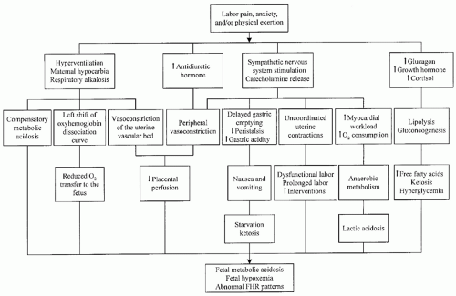 FIGURE 15.2 Potential adverse effects of untreated maternal pain on the fetus. Modified from Brownridge P, Cohen SE, Ward ME. Neural blockade for obstetrics and gynecologic surgery. In: Cousins MJ, Bridenbaugh PO, eds. Neural blockade in clinical anesthesia and management of pain, 3rd ed. Philadelphia, PA: Lippincott Williams & Wilkins, 1998:557-604. |
Reynolds et al. (27) completed a meta-analysis comparing epidural with systemic opioid analgesia to determine the effect of these anesthetic interventions on acid-base status at birth. They concluded that epidural analgesia was associated with an improvement in base excess, suggesting that placental exchange is well preserved in association with this technique.
While umbilical artery pH, base excess, and pCO2 are considered sensitive and objective indicators of fetal hypoxia during labor, the results represent a “snapshot” of fetal status and depict the mixed effluent of all fetal tissues. Cord gases do not distinguish between primary fetal pathologic conditions, fetal effects of maternal conditions (e.g., acid-base disorders), or the influence of inadequate placental blood flow. They also do not indicate in which direction the condition of the fetus is moving, or at what rate, nor do they reflect events that occurred remote from delivery.
Summary
To date, there is no one test that clearly separates effects on the fetus/newborn, if any, of maternally administered medication during labor and delivery.
▪ PAIN MANAGEMENT
For most women, childbirth is one of the most painful events in their lifetime. There are both physiologic and psychological aspects to pain and its management (28).
Labor pain evokes a generalized neuroendocrine stress response that has widespread physiologic effects on the parturient and fetus (29). The neuroendocrine model, presented in Figure 15.2, examines the potential detrimental consequences of untreated pain. The sequelae of hyperventilation, secretion of stress-related
hormones, and increased oxygen consumption can be prevented, obtunded, or abolished by central neuraxial blockade (epidural or spinal anesthesia) (30).
hormones, and increased oxygen consumption can be prevented, obtunded, or abolished by central neuraxial blockade (epidural or spinal anesthesia) (30).
Research in humans supports elements of the neuroendocrine model (31), but studies are not necessarily designed to consider the effects of simultaneously occurring care practices on these same physiologic responses. “This critique is needed because it is somewhat counterintuitive that the procreative physiologic process of labor and birth would by nature have detrimental effects on a healthy mother and fetus” (32). An example of a concurrent care practice is the administration of isotonic “sport drinks” versus water only during labor (33). Sports drinks were shown to prevent the development of maternal ketosis without increasing gastric volume, although there was no difference between the groups in neonatal outcome.
Labor Pain: Implications for the Fetus
Neural pathways and neurochemical systems involved in pain perception are functional from midgestation and are well developed by the third trimester. Gitau et al. (34) conducted a parallel study of the fetal and maternal hormonal responses to fetal blood transfusion. They confirmed that the fetus mounts a hypothalamic-pituitary-adrenal response to transfusion via the intrahepatic vein, which involves piercing the fetal trunk, but not to transfusion in the umbilical vein at the placental cord insertion, which has no sensory innervation. The rise in fetal cortisol and endorphin occurred independently of the maternal reaction. Pretreatment of the fetus with fentanyl for this same procedure attenuated the rise in β-endorphin (35).
Hormonal stress responses do not provide a direct index of pain. While it is true that a rise in cortisol and endorphin is seen as a consequence of painful stimuli in children, other nonpainful situations (e.g., exercise) are also associated with an increase in the levels of these hormones. Nonetheless, the editorial review of Fisk’s fentanyl pretreatment study suggests that fetal analgesia should be given during invasive in utero procedures (36).
At present, there is no literature on fetal “pain” during labor or delivery.
Labor and Delivery Pain: Implications for the Mother
Visceral pain predominates during the first stage of labor. Nociceptive information arising from uterine contractions, distention of the lower uterine segment, and cervical dilation is relayed in C afferent fibers to the dorsal horn of the spinal cord at the T10-L1 levels. As labor progresses, a mixture of visceral and somatic (A-delta fibers) pain results from traction on the pelvic floor structures surrounding the vaginal vault, and eventually from distention and stretch of the vagina and perineum (L2-S1). Delivery pain (Stage II) is somatic in nature and transmitted along the pudendal nerve (S2-S4). Synaptic input at the dorsal horn, mediated by neurotransmitters and chemicals (e.g., excitatory amino acids), is relayed via the spinothalamic tract to higher centers including the reticular formation, hypothalamus, and limbic system. Dorsal horn neurons also initiate segmental spinal reflexes. Descending spinal tracts, endogenous opioids, and other inhibitory systems modulate nociception centrally in the spinal cord. The neural mechanism of labor shares features with other forms of acute pain (37).
To view labor pain only as a neuroendocrine, sensory experience is limiting and undermines the complexity of this phenomenon (32). Pain is just one component of the totality of the labor and birth experience. Assisting women to cope with the affective or distress components of labor and birth in a supportive environment has been shown to reduce the need for pain-relieving drugs, decrease the incidence of operative delivery, result in higher Apgar scores, and improve breastfeeding success (28).
The management of pain and anxiety in labor is a worthwhile goal whether the techniques used are nonpharmacologic, pharmacologic, or include a combination of both. The choice depends on patient preferences, medical status of the mother and fetus, progress of labor, and resources available at the facility for pain management and treatment of potential complications.
Practice guidelines, including a section devoted to specific analgesia techniques, have been developed to enhance the quality of anesthetic care for obstetric patients (38). Likewise, a practice bulletin written for obstetricians that outlines analgesia options for patients has the dual purpose of facilitating communication with patients and anesthesia and neonatology colleagues (39).
Analgesic Techniques for Labor: Effects on the Fetus and Newborn
Analgesia refers to pain relief without loss of consciousness. Regional analgesia denotes partial sensory blockade in a specific area of the body, with or without partial motor blockade. The term neuraxial analgesia pertains to the administration of pain-relieving medications using caudal, spinal, and/or epidural techniques. Drugs given to the mother may affect the fetus directly, via placental transfer, or indirectly by altering maternal physiology and biochemistry (30).
Not all methods of pain relief are available or desirable in all centers, and certain methods are more popular in different parts of the world (40).
Nonpharmacologic Methods
Proponents of nonpharmacologic methods claim that these methods reduce requirements for analgesia during the first stage of labor. This does not necessarily imply that women who use these techniques have less pain, rather that they are able to cope with labor using less analgesia. Nonpharmacologic methods may be combined or used sequentially with drug-based methods (41).
In a systematic review of comfort measures, Simkin and O’Hara (42) commented on five methods evaluated scientifically for their effectiveness in reducing indicators of labor pain. This review also mentioned pain-related outcomes such as obstetric interventions and duration of labor. Continuous labor support was associated with a decrease in duration of labor, requests for analgesia, rates of instrumental and cesarean deliveries, and occurrence of lower Apgar scores. The use of baths offered temporary pain relief and was considered safe provided water temperatures were maintained at or below maternal body temperature and that immersion duration was controlled. Perinatal morbidity and mortality did not increase, even if membranes were ruptured. The authors concluded that there had been insufficient study to provide clear conclusions regarding touch/massage, although emotional and physical relief was demonstrated with this intervention. Intradermal water blocks were effective in reducing severe back pain. The reduction in cesarean deliveries originally reported with this technique was refuted recently (43). Lastly, maternal movement and positioning were reported to impact pain relief in labor and impact several variables related to fetal and neonatal well-being.
Jones et al. published a comprehensive Cochrane review entitled: Pain management for women in labor: an overview of systematic reviews looking at the efficacy and safety of both non-pharmacologic and pharmacologic management during labor. Evidence for nonpharmacologic options was generally limited to small trials with overall low quality methods. The authors concluded that interventions with some impact included hydrotherapy (pain relief with improved satisfaction of birth experience), acupuncture (improved satisfaction and pain relief with fewer assisted vaginal and cesarean deliveries), relaxation techniques (improved satisfaction, pain relief, and fewer assisted vaginal deliveries), and massage (reduction in pain intensity). Lack of conclusive results
were noted in studies pertaining to hypnosis, biofeedback, sterile water injections, aromatherapy, and transcutaneous nerve stimulation (44).
were noted in studies pertaining to hypnosis, biofeedback, sterile water injections, aromatherapy, and transcutaneous nerve stimulation (44).
Systemic Opioids
From the maternal perspective, efficacy and incidence of side effects with systemic opioid analgesia is largely dose dependent rather than drug dependent (30). There is little evidence to suggest one agent is intrinsically superior. Most often, the choice is based on institutional tradition or personal preference.
Opioids may affect the fetus directly as a result of placental transfer and/or indirectly, for example, by altering maternal minute ventilation or uterine tone. As a group, these low molecular weight drugs are lipid-soluble weak bases that readily cross the placenta (45). This implies that maternal to fetal concentration gradients are important; only free, not protein-bound, drug is available for transfer. The amount of “free” drug delivered to the placenta depends on placental blood flow (flow-dependent transfer) and the degree of maternal protein binding. The amount of drug available to the fetus depends on the degree of placental uptake, metabolism, and clearance (30). In single-dose drug studies, key factors influencing umbilical vein/maternal drug ratio are lipid solubility and transit time through the placental bed. In multidrug dosing (e.g., patient-controlled narcotic analgesia [PCA] delivery systems), key factors influencing fetal drug levels are the degree of ionization and degree of fetal protein binding (Fig. 15.3).
Fetal pH is lower than maternal pH; consequently, the fraction of opioid (and other basic drugs) existing in the ionized state is higher in the fetus than in the mother. Ionization results in drug trapping. The degree of ionization depends on the pKa of the drug; the effect is greater for meperidine (pKa approximately 8.5) than morphine (pKa approximately 8.0), and more significant when the fetus is acidotic. This is a simplistic, albeit true, application of opioid pharmacokinetics, a complex, difficult to predict, and incompletely evaluated topic.
All opioids have the potential to decrease baseline FHR and reduce variability, making interpretation of fetal CTG recordings potentially problematic. It has been documented from observational studies that parenteral narcotics can be associated with neonatal respiratory depression, decreased neonatal alertness, inhibition of sucking, and delay in effective feeding. When evidence related to the use of parenteral opioids for labor pain relief was subjected to a systematic review (46), it was noted that none of the studies was sufficiently powered to address the primary outcome measure of need for neonatal resuscitation, a measure of safety. IM opioid was compared to placebo, different IM opioid, same IM opioid but different dose, and same opioid given intravenously; IV opioid was compared to different IV opioid and same IV opioid but different modes of administration. There was insufficient pooled information to draw conclusions regarding any of the secondary outcome measures, including fetal distress, administration of naloxone, Apgar score less than 7 at 5 minutes, baby death, admission to a special care setting, feeding problems, and problems with mother-baby interaction.
The controversial concept of genetic imprinting at birth for opiate or amphetamine addiction in later life was linked to systemically administered pain-relieving labor medications (narcotics, barbiturates, or nitrous oxide [N2O]) (46). More recently, a cohort study showed no association between administration of intrapartum pethidine and substance use disorder in later life (47).
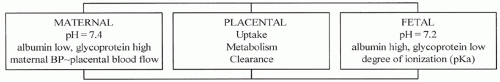 FIGURE 15.3 Factors influencing fetal drug levels with PCA narcotic administration. |
Meperidine is the most commonly used opioid for labor analgesia worldwide. It has been shown that as the time increases from administration of a single-dose IM meperidine 1.5 mg/kg during labor to delivery of the baby, so too does the level of meperidine in the fetus (48). Maximum fetal concentrations reach a plateau between 1 and 5 hours after dosing; therefore, babies born within 1 to 5 hours after meperidine is given to the mother have the greatest chance of narcotic-induced depression. In contrast to single-dose studies, multiple doses of meperidine administered over many hours lead to accumulation of a metabolite, normeperidine, in the mother and fetus (49). Half-lives of 17 to 25 hours for this metabolite are common in the mother; the half-life exceeds 60 hours in the fetus/newborn. Normeperidine is associated with respiratory depression, not reversible by naloxone, and seizures. Because of concerns about meperidine, research has focused on the newer, shorter-acting opioids with no active metabolites such as in the IDiVP trial comparing IM diamorphine with IM meperidine (50).
Fentanyl has been available clinically for more than 30 years. It offers prompt analgesia coupled with a short duration of action and no active metabolites. Both maternal and fetal drug levels decline in a parallel fashion following a single dose of the drug. In the first report of its administration to laboring patients, fentanyl (50 to 100 µg IV q1h) was compared with meperidine (25 to 50 mg IV q2-3h) (51). More mothers were nauseated and sedated and more babies required naloxone in the meperidine group. An advantage of fentanyl is the ability to administer the medication via several routes, including IV, subcutaneous, and oral (52). In a recent multicenter randomized trial comparing patient-controlled epidural analgesia (PCEA) using bupivacaine with PCA fentanyl, there were no differences in mode of delivery, but more neonates in the PCA group required naloxone and had lower 1-minute Apgar scores (30).
Sufentanil is the most lipid soluble (octanol:water partition coefficient 1778) of the commonly used opioids (45). This feature should enhance placental transfer after a single dose, but transfer is impeded by the extent of maternal plasma protein binding (α1-acid glycoprotein) and uptake by the placenta. Sufentanil concentration in the fetus rises slowly, reaching a plateau between 45 and 80 minutes postadministration (53). It is a useful maternal analgesic for pain relief during second stage, when fetal delivery is imminent (<45 minutes).
Remifentanil is an ultra-short-acting opioid. It has the most rapid onset of peak effect (approximately 1 minute), shortest context-sensitive half time (approximately 3 to 5 minutes), and greatest clearance (40 mL/kg/min) of the commonly used opioids (45). Although the maternal cardiovascular and side effect profiles are similar to other fentanyl congeners, remifentanil is chemically distinct because of its ester linkages. This ester structure renders it susceptible to hydrolysis by red cell—and tissue-nonspecific esterases, resulting in rapid metabolism. Remifentanil concentration decreases by 50% within 3 to 5 minutes of stopping drug administration, regardless of the duration of the infusion (45).
Labor pain occurs at intervals and increases in intensity over time. Rapid recovery between contractions and after delivery is desirable. Therefore, the most effective means of administering remifentanil to the laboring patient, taking advantage of the drug’s characteristics, is via patient-controlled analgesia (PCA) with a background infusion. Bolus dose, lockout time, and the rate of infusion can be titrated. The drug rapidly crosses the placenta and is quickly metabolized by fetal esterases. Maternal oxygen desaturation and sedation and reduced FHR variability have been observed. Maternal respiratory arrest with consequent cardiac arrest has been reported (54,55). Remifentanil use should be restricted to highly monitored hospital settings under the supervision of an anesthesia practitioner and with one-on-one nursing care, monitoring for, and intervening to treat maternal sedation, respiratory insufficiency, and desaturation (56,57).
Newborns exposed to remifentanil in utero up until the time of delivery have been vigorous. There have been no reports of lower Apgar scores, unacceptable umbilical cord gas analyses, or respiratory depression necessitating naloxone use (57,58).
Naloxone is used to reverse respiratory depression in narcotic-exposed newborns. It is contraindicated for infants of narcotic-dependent mothers, as administration may precipitate acute withdrawal and seizures. Naloxone has never been shown to reduce the need for assisted mechanical ventilation or reduce admission rates to special care nurseries (59). No studies have evaluated the effect of naloxone on time to spontaneous effective respiration or long-term outcome.
Agonist-Antagonist Opioids
Nalbuphine is used as a systemic analgesic during labor. Reports of severe perinatal cardiovascular and respiratory depression prompted Nicolle et al. (60) to carry out a study designed to delineate placental transfer and disposition of nalbuphine in the neonate. The estimated half-life was 4.1 hours (vs. 0.9 hour in infants and 2 hours in adults). Given that the liver extensively metabolizes nalbuphine, the authors speculated that the slower neonatal plasma disappearance rate compared to the infant or adult could be due in part to immature hepatic function or bypass of the liver via the DV. All 28 babies had 5-minute Apgar scores of 10. Fifty-four percent of the FHR tracings showed reduced variability lasting 10 to 35 minutes and sinusoidal FHRs after maternal injection.
One potential use for this class of drugs is in the treatment of opiate-dependent pregnant women. Babies born to mothers on a buprenorphine maintenance program showed little or no clinically measurable neonatal abstinence syndrome in contrast to findings with methadone, morphine, or heroin maintenance programs (61).
Nitrous Oxide
N2O is an odorless inhalational agent that exerts weak but prompt analgesic activity. It is combined with oxygen in a 50:50 mixture for obstetric use and is self-administered by the patient through a specialized breathing circuit equipped with a demand valve. The negative pressure generated at the onset of inspiration opens the valve, which remains open during inspiration and closes when the patient begins to exhale. Inhalation analgesia with N2O during labor and delivery by itself, as a coanalgesic, or as a temporizing measure pending other forms of pain relief is less common in the United States than in other developed countries.
N2O is a relatively insoluble gas at room temperature, and therefore equilibrates rapidly between the alveoli, blood, and brain. To be fully effective, inhalation needs to be timed with contractions such that the patient begins to breathe the gas about 10 to 15 seconds in advance of the next contraction. This synchronizes the peak effect of N2O with the zenith of pain, assuming the average contraction lasts 60 seconds and peaks at the midpoint.
Any patient at risk of vitamin B12 deficiency (e.g., pernicious anemia or vegetarian) should not use N2O as it irreversibly oxidizes vitamin B12, reducing the activity of methionine synthetase (necessary for myelin formation and DNA synthesis in humans) and other B12-dependent enzymes. Many countries have set maximal environmental limits for N2O (a greenhouse gas), which necessitates the use of ventilation systems that allow exhaled gas to be scavenged (62,63).
N2O readily crosses the placenta. The maternal-fetal concentration ratio reaches 0.8 within 15 minutes of continuous inhalation. It has no effect on uterine contractions or FHR. It is not metabolized and is eliminated quickly and entirely by the lungs with the onset of respiration at birth. This is true whether the mother inhales N2O for 5 minutes or 5 hours. N2O does not affect Apgar scores or sucking behavior (64).
Paracervical Block
This peripheral block provides a therapeutic alternative for first-stage labor pain when central neuraxial blockade is contraindicated or unavailable. The technique involves transvaginal injection of local anesthetics (LAs) on either side of the cervix to interrupt pain transmission at the level of the uterine and cervical plexuses (located at the base of the broad ligament). Paracervical block (PCB) is relatively easy to perform, and, when effective, it provides good to excellent analgesia that lasts 1 to 2 hours.
Since the introduction of PCB in the 1940s, reports of serious adverse sequelae, including injection of LAs directly into the uterine arteries or fetal head, fetal death, and profound bradycardia, have resulted in modification of the injection technique and changes to the concentration and type of LA used. There are statements cautioning against employing this block in situations of uteroplacental insufficiency or nonreassuring FHR tracings. In a review of PCB using dilute bupivacaine solutions, the incidence of fetal bradycardia was 2.2% with an onset between 2 and 10 minutes after injection and a duration of 30 minutes. None of these episodes of bradycardia led to cesarean delivery and Apgar scores, and umbilical arterial and venous pH determinations of the neonates were within the normal range. The exact etiology of the bradycardia is unknown; however, PCB is associated with a small but significant increase in uterine artery impedance, indicating uterine artery vasoconstriction (56).
Neuraxial Analgesia
Spinal, epidural, and combined spinal-epidural (CSE) techniques are commonplace for managing childbirth pain. They are used to administer opioids, LAs, and other pain-modulating adjuvants. Collectively, these methods are considered the most effective forms of pain relief available to laboring women.
Although spinal anesthesia has been in use since 1899, spinal analgesia only became a viable possibility in the 1970s following the discovery of specific opioid receptors in the brain and spinal cord. From a practical standpoint, however, it was not an option for laboring women at that time for two reasons: there was an unacceptably high incidence of postspinal headache (also known as postdural puncture headache) in the young female population, and a single injection technique could not be relied upon to provide analgesia for more than 1 to 2 hours. The advantage of having an epidural catheter in place, either for subsequent bolus dosing or for continuous infusion, is the provision of uninterrupted analgesia between placement of the catheter and delivery of the baby.
The (re-)introduction of fine-gauge, pencil-point, atraumatic (noncutting) spinal needles in the late 1980s fostered a renewed interest in subarachnoid (intrathecal) injection (65). With the advent of CSE equipment and “needle-through-needle” technique, single-level subarachnoid injection, followed immediately by epidural catheter placement at the same site, became possible (66). The CSE procedure has become synonymous with subarachnoid injection of opioid (±a small dose of LA) and simultaneous initiation of a low-dose epidural infusion. The perceived advantages
of CSE compared with more traditional methods continue to be debated, along with the consequences of routine dural puncture (66,67).
of CSE compared with more traditional methods continue to be debated, along with the consequences of routine dural puncture (66,67).
Epidural catheter analgesia alone has been popular for many years. Depending on the choice of agents used, it provides superior pain relief and better overall satisfaction during the first and second stages of labor compared to the modalities mentioned previously. Epidural analgesia can be converted to epidural anesthesia if necessary for cesarean section, instrumented vaginal delivery, manual placental removal, or episiotomy repair.
Neuraxial Opioids
Opioids injected into the lumbar intrathecal space distribute between nerve tissue and cerebrospinal fluid (CSF) on the basis of their partition coefficients (lipid solubility). Opioids injected into the epidural space first diffuse across the dura to reach the subarachnoid space and then behave as their intrathecal counterparts.
Morphine, the least lipid soluble of the commonly used opioids, diffuses slowly from the CSF into the substantia gelatinosa of the dorsal horn to activate opioid receptors. This accounts for its delayed onset and prolonged duration of action. Morphine also spreads rostrally, moving by bulk flow with CSF to reach vasomotor, respiratory, and vomiting centers in the brainstem. In contrast, the highly lipophilic fentanyl and sufentanil penetrate nerve tissue quickly. They have a faster onset of activity coupled with a shorter duration of action. Remifentanil is not approved for use in the intrathecal space because it contains a glycine preservative.
All opioids have some minor intrinsic LA properties, but these effects are marked with meperidine (45), allowing it to be used as the sole agent, even for cesarean section, in the rare event of amide LA allergy. Intrathecal meperidine produces significant sympathetic and motor blockade as well as typical opioid side effects such as nausea and pruritus. It has little value as an adjunct to regional analgesia for labor.
The purported advantages of using opioid drugs alone to induce neuraxial analgesia for labor include the following:
Preservation of motor function, sustaining the ability to ambulate during first stage and to push during second stage.
Avoidance of LA-induced sympathectomy that can be associated with undesirable cardiovascular sequelae such as hypotension.
Reduction in the systemic side effects of opioids themselves, given the receptor-specific route and minute amount of drug needed to exert an effect. Less total opioid means less chance of drug transfer to the fetus and fewer unpleasant maternal side effects such as nausea, vomiting, pruritus, urinary retention, and sedation.
Fetal bradycardia may develop following administration of intrathecal opioid, although its occurrence can follow any type of effective labor analgesia (68


Stay updated, free articles. Join our Telegram channel
Full access? Get Clinical Tree
