Normal and Abnormal Growth and Pubertal Development


Normal and Abnormal Growth and Pubertal Development


image

In many societies throughout history, puberty has been a time of celebration. The changes that accompany puberty announce the transition from childhood to adulthood and the development of fertility. Puberty is the process of cognitive, psychosocial, and biologic maturation. Whereas growth and the development of secondary sexual characteristics are the most visible manifestations of the onset of puberty, changes in body composition and cognitive development are no less significant.1 Puberty can be a difficult transition for many adolescents, even when it progresses normally, and presents substantially greater challenges when its onset is premature, or delayed. The recent trend towards an earlier pubertal maturation and some of its consequences, notably earlier sexuality and the problem of teen pregnancy, make it all the more important to understand the physiology of normal puberty.

This chapter focuses first on the endocrinology and physiology of normal puberty, to provide the foundation for subsequent discussion of the pathophysiology, diagnosis, and management of abnormalities of growth and pubertal development.


The Endocrinology of Normal Puberty

The hypothalamus, anterior pituitary gland, and gonads of the fetus, neonate, infant, and prepubertal child are all capable of secreting hormones in adult concentrations. The key to understanding the endocrinology of puberty lies in first understanding the mechanisms that govern the hypothalamic-pituitary-gonadal axis.


The Ontogeny of the Hypothalamic-Pituitary-Gonadal Axis

The “hypothalamic pulse generator,” the term chosen by Ernst Knobil to describe the rhythmic, pulsatile nature of gonadotropin-releasing hormone (GnRH) secretion,2 consists of approximately 1,500-2,000 specialized neurosecretory cells in the arcuate nucleus, located in the medial basal hypothalamus. The resident GnRH neurons exhibit spontaneous “autorhythmicity” and function as an oscillator in the pulsatile secretion of GnRH.3,4 In response to the pulsatile GnRH signal, pituitary gonadotropes, which contain plasma membrane GnRH receptors, secrete follicle-stimulating hormone (FSH) and luteinizing hormone (LH), also in a pulsatile manner. In turn, the episodic gonadotropin signal stimulates maturation of the germinal elements of the gonads and is transmitted into the pulsatile secretion of sex steroids. Ultimately, the patterns of gonadotropin and gonadal steroid secretion during fetal life, infancy, childhood, adolescence, and adulthood primarily reflect changes in the activity of the hypothalamic pulse generator.


Fetal Life and Infancy

The hypothalamic-pituitary-gonadal axis becomes functional even before birth. Neurons that synthesize GnRH originate in the olfactory placode and migrate to the hypothalamus between 6 and 9 weeks of gestation.5 By 10 weeks, the hypothalamus contains significant amounts of GnRH.6 Development of the hypothalamic-pituitary portal venous system begins between 9 and 10 weeks of gestation and is completed by 19-20 weeks.7 Consequently, FSH and LH concentrations in fetal pituitary glands increase dramatically soon thereafter. The pituitary begins to secrete FSH and LH into the fetal circulation by week 12.8 Fetal serum gonadotropin levels rise progressively, reaching a peak between 20 and 24 weeks,9 then decrease steadily over the last 10 weeks of pregnancy, probably due to a developed sensitivity to the negative feedback effects of high circulating estrogen and progesterone concentrations derived from the placenta.10,11

After birth, steroid levels fall precipitously due to the loss of maternal and placental hormones, allowing the newborn’s hypothalamic-pituitary-gonadal axis to escape their suppressive effects. The characteristic pulsatile pattern of hypothalamic GnRH secretion emerges,12,13 and serum gonadotropin concentrations rise again promptly, with a striking sex difference; FSH rises to a greater extent in females and LH to a greater extent in males.11 In female infants, FSH concentrations occasionally reach levels even greater than those observed in the normal adult menstrual cycle.14,15 Consequently, waves of ovarian follicular development begin and estradiol levels during the first few months of life are comparable to those observed during the midfollicular phase of the menstrual cycle.16 In male infants, elevated LH levels stimulate increased testosterone secretion from the testes. Gonadotropin and gonadal steroid levels peak at about 3-6 months in boys and 12-18 months in girls
and steadily decline thereafter, presumably because normal negative feedback mechanisms become fully functional. By approximately 9-12 months of age in boys and 24-36 months in girls, gonadotropin concentrations fall to typical prepubertal levels, remaining at very low concentrations until the onset of puberty.11 Suppression of hypothalamic pulse generator activity is less intense and shorter in duration in females than in males, probably reflecting the influence of testosterone on hypothalamic programming.17


Childhood and Early Adolescence

During the interval between infancy and puberty, known as the “juvenile pause” in nonhuman primates, the hypothalamic-pituitary-gonadal axis lies dormant. Normal ovulatory menstrual cycles can be induced in prepubertal female monkeys by administering a higher amplitude pulsatile infusion of exogenous GnRH, indicating that neither the anterior pituitary nor the gonads are the limiting factor.18 Although the GnRH pulse generator is active, the frequency and amplitude of pulsatile GnRH secretion generally are irregular and very low.19,20,21 and 22 Low amplitude pulses of gonadotropin secretion can be detected in prepubertal children as young as 5 years of age, primarily during sleep.22,23 and 24 FSH levels rise more than LH, but there is no detectable increase in steroid hormone concentrations.

For a long time, the prevailing theory to explain the juvenile pause that precedes puberty envisioned a hypothalamic “gonadostat” controlling the level of sensitivity to the central negative feedback actions of gonadal steroids. In that context, the changing patterns of gonadotropin secretion were attributed to changes in the “gonadostat” setting. Decreasing gonadotropin levels in late infancy and sustained low concentrations during childhood reflected a rising and ultimately high sensitivity to even very low levels of sex steroid feedback, and increasing gonadotropin concentrations at the onset of puberty reflected a decrease in feedback sensitivity.25,26 The “gonadostat” theory prevailed until cross-sectional and longitudinal studies in children with gonadal dysgenesis revealed a similar, but exaggerated, “diphasic” pattern of gonadotropin secretion. Serum gonadotropin levels in girls with Turner syndrome are markedly elevated in infancy, decline to very low levels during childhood, and rise again to grossly high concentrations at pubertal age, all in the absence of any possible change in the level of negative feedback from gonadal steroids.27 These and similar observations in castrate nonhuman primates demonstrated that steroid hormone feedback affects the amount, but not the pattern, of gonadotropin secretion, contradicting the traditional “gonadostat” theory and establishing a new paradigm. The typical “diphasic” pattern of gonadotropin secretion from infancy to puberty results primarily from changing levels of central inhibition of pulsatile GnRH secretion, and to a lesser extent, from a high sensitivity to low levels of gonadal steroid feedback.


image


Puberty

About 1 year before breast budding in prepubertal girls, the character of nocturnal pulses of gonadotropin secretion changes with LH levels exceeding those of FSH. Breast budding occurs when the nocturnal pulses of gonadotropin secretion become great enough to generate detectable coincident increases in serum estradiol concentrations. At that time, LH peak amplitude increases about 10-fold, whereas FSH pulse amplitude only doubles, resulting in a marked decrease in the serum FSH/LH ratio.22,24 The change reflects an increase in pituitary responsiveness to GnRH, which has a priming effect on pituitary LH secretion and increases the number of GnRH receptors on gonadotropes (up-regulation). Gonadotropes first increase their capacity for response to GnRH by synthesis, and later by secretion of gonadotropins. Pulse frequency also increases, but to a much lesser extent. Gonadotropin pulses become diurnal and the duration of increases in estradiol levels becomes more prolonged. As puberty progresses, the amplitude of pulsatile LH secretion increases further, to levels 20-40 times greater than those detected during prepubertal years, probably reflecting the influence of rising estradiol levels at both the hypothalamic and pituitary levels. Although nocturnal pulse amplitudes are still greatest, significant pulses occur during daytime and basal LH levels become detectable.22 LH bioactivity also increases, due to changes in glycosylation.28

In response to increasing gonadotropin secretion, basal estradiol levels increase progressively.20 Inhibin B levels, which are low or undetectable in prepubertal girls, increase sharply in mid-puberty, then decline in its later stages, first reflecting increasing ovarian stimulation, then the onset of the menstrual cycle and the appearance of a luteal phase, when levels are low.29 Inhibin A concentrations, undetectable or very low through early puberty, increase gradually thereafter but reach adult levels only after menarche, consistent with the corpus luteum being the primary source.29 Menarche occurs in late puberty, after a year-long rise in daily estrogen production,30 probably when estradiol and inhibin B levels become sufficient to exert significant negative feedback on gonadotropin secretion, resulting in cyclic estrogen production. Cycle length and menstrual characteristics vary until the positive feedback relationship between estradiol and gonadotropin secretion matures and ovulation becomes established, often a year or more after menarche.


Central Control Mechanisms

Some of the factors governing the “neuroendocrine switch” for the GnRH pulse generator that is “on” in early infancy, turns “off” during childhood, and switches back “on” again at puberty have now been identified. Most of the important work on the neuroendocrinology of puberty has been performed in nonhuman primate models. The list of factors that modulate the activity of the hypothalamic-pituitary-gonadal axis includes both inhibitory and excitatory neurotransmitters and peptides.


Gamma-Aminobutyric Acid

Gamma-aminobutyric acid (GABA) is an inhibitory neurotransmitter produced by specialized neurons in the hypo thalamus and has an important role in regulating the activity of the GnRH pulse generator. Elegant hypothalamic perfusion studies have revealed that release of GABA into the median eminence decreases as pulsatile GnRH secretion increases at the onset of puberty.31 Conversely, central perfusion with a GABAA receptor antagonist (bicuculline) or the antisense oligodeoxynucleotide for the mRNA coding the GABAA synthesizing enzyme (glutamate acid decarboxylase) stimulates GnRH release.31,32 Chronic administration of bicuculline into the third ventricle induces precocious puberty and menarche in prepubertal female monkeys.33 Evidence suggests that changes in the
subunit composition of GABAA receptors may contribute to the disinhibition of pulsatile GnRH secretion at the onset of puberty.34 These observations suggest that central GABA signaling is one of the factors that restrains GnRH neuronal activity during childhood.


Neuropeptide Y

Neuropeptide NPY (NPY) is a hypothalamic peptide involved in the control of food intake behavior and reproductive function in adults. In castrate adult female monkeys, intracerebroventricular administration of NPY inhibits pulsatile GnRH release.35 In males, the postnatal pattern of GnRH pulse generator activity is inversely related to NPY gene and protein expression in the medial basal hypothalamus and central administration of an NPY receptor antagonist stimulates GnRH release in juveniles.36 These observations suggest that NPY, like GABA, is an important component of the “neurobiologic brake” that restrains the GnRH pulse generator in prepubertal primates. However, others have observed that NPY levels increase in the median eminence at the onset of puberty,37 that infusion of NPY into the median eminence increases GnRH release,38 and that infusion of NPY antiserum into the median eminence did not stimulate GnRH secretion in prepuberal monkeys.37 The varying effects of NPY appear to relate to the site of infusion within the brain and not on the steroid milieu.35 Additional work will be required to clarify the role of NPY in regulation of the hypothalamic pulse generator and the onset of puberty.


Glutamate

Glutamate is an excitatory neurotransmitter in the hypothalamus and stimulates GnRH release via N-methyl-D-aspartate (NMDA) receptors both in vivo and in vitro.39 An intravenous bolus of NMDA stimulates hypothalamic GnRH release,40 and treatment with a specific glutamate receptor antagonist blocks the effect in nonhuman primates. Moreover, prolonged (16-30 weeks) intermittent NMDA stimulation (1 minute every 3 hours) activates the hypothalamic-pituitary-gonadal axis and stimulates precocious puberty and the initiation of spermatogenesis in juvenile males.41 These observations suggest that glutamate signaling may play a role in the resurgence of pulsatile GnRH secretion at the onset of puberty.


Kisspeptins

In just the past few years, kisspeptins have emerged as a critical component of the system that controls the level of GnRH neuronal activity between infancy and puberty. Kisspeptins are neuropeptides (encoded by the KISS1 gene) that signal via the G-protein coupled receptor, GPR54 (encoded by the KISS1R gene).42 Interestingly, the first evidence of their importance in the regulation of the hypothalamic-pituitary-gonadal axis came from observations in humans. Several members of a large consanguineous family with hypogonadotropic hypogonadism and delayed puberty were found to harbor homozygous inactivating mutations for GPR54.43,44 One affected compound heterozygote exhibited an exaggerated pituitary response to exogenous pulsatile GnRH administration, suggesting a hypothalamic locus for the disorder.44 More importantly, the observation also suggested that kisspeptin signaling via GPR54 might play a major role in the resurgence of pulsatile GnRH secretion at puberty in primates. The results of subsequent studies in nonhuman primates and humans strongly support that interpretation.

Neurons expressing KISS1 are located exclusively in the arcuate nucleus,45,46 where GnRH neurons also express GPR54.42 In castrate male and intact female monkeys, the pubertal resurgence of pulsatile GnRH secretion is associated with a nearly 5-fold increase in KISS1 expression and, in females, also with an increase in KISS1R expression.47 Hypothalamic kisspeptin secretion is distinctly pulsatile and highly correlated with that of GnRH.48 An intermittent kisspeptin infusion can sustain pulsatile LH secretion in castrate juvenile animals after discontinuation of a priming pulsatile infusion of exogenous GnRH, but not in the presence of a GnRH receptor antagonist, indicating that the effect of kisspeptin is mediated via pulsatile GnRH secretion.47 The observation that patients with inactivating mutations of GPR54 exhibit pulsatile LH secretion with low amplitude and normal
frequency suggested that kisspeptin might only amplify and not stimulate GnRH pulse generator activity directly.44,49 However, a continuous kisspeptin infusion, which downregulates GPR54, suppresses both LH pulse amplitude and frequency, implying that kisspeptin has similar effects on pulsatile GnRH secretion.50,51 Finally, an activating KISS1R mutation resulting in prolonged activation of the KISS1R signal transduction pathway has been described in a young girl with GnRH-dependent (central) precocious puberty.52 Taken together, these observations indicate that hypothalamic kisspeptin-GPR54 signaling is a key component of the neurobiologic mechanism that triggers the onset of puberty. They further suggest that kisspeptin neurons may provide the fuel for the hypothalamic GnRH pulse generator. The report of undetectable serum gonadotropins in an infant boy bearing a loss-of-function mutation in the KISS1R gene suggests that kisspeptin input to the GnRH neuronal network also is necessary for the increased pulsatile GnRH secretion normally observed during early infancy.53

There is increasing evidence from studies in nonhuman primates that kisspeptin neurons also are involved in mediating the feedback actions of both testicular and ovarian hormones. In males, testosterone negative feedback, which regulates LH secretion by slowing the pace of pulsatile GnRH secretion,54 is associated with a decrease in hypothalamic KISS1 mRNA levels.55 The observation suggests that kisspeptin neurons play an important role in the negative feedback loop that regulates LH secretion in the male, which also involves opioid and GABA neuronal input.56 Gonadal steroids also suppress hypothalamic KISS1 expression in females. In postmenopausal women and ovariectomized monkeys, the density of neurons expressing KISS1 mRNA is significantly higher than in premenopausal women and in intact females monkeys, and treatment with estrogen and progesterone markedly decreases KISS1 expression,46 suggesting that kisspeptin neurons also participate in mediating the hypothalamic negative feedback actions of ovarian steroid hormones. The collective body of evidence thus indicates that kisspeptin neurons are a critical component of the neurobiologic mechanism that regulates the activity of the hypothalamic pulse generator.

In some way, the system that controls the ontogeny of pulsatile GnRH secretion integrates kisspeptin signaling with that of other neuotransmitters (glutamate, GABA) and neuropeptides (NPY). Whether kisspeptin neurons in the arcuate nucleus function as a “pubertal clock,” as a growth-tracking “somatometer,” or simply relay information from such centers to the GnRH neuronal network is unknown.57 Regardless, kisspeptin neurons have emerged as one of the primary transducers of the internal and external environmental cues that regulate the neuroendocrine reproductive axis.


Peripheral Signaling

The age at onset of puberty has been declining steadily as the prevalence of obesity has been increasing, suggesting that a critical body weight58 or body composition59 may be an important factor in determining the timing and progression of puberty.60 Conversely, the suppressive effects of fasting61,62 and chronic malnutrition63 on the neuroendocrine control of reproduction are well known and consistent with the hypothesis. The manner in which such metabolic signals might be communicated and integrated with the reproductive axis in primates is unknown, but studies in ungulates suggest an effect on pulsatile hypothalamic GnRH secretion.64


Leptin

Leptin is produced by adipocytes and serum concentrations are strongly associated with body fat and changes in body fat content. Not surprisingly, leptin has been implicated as one way in which metabolic signals might be communicated to the higher centers controlling the activity of the hypothalamic pulse generator at the onset of puberty.
Leptin-deficient mice and rats fail to enter puberty, and treatment with leptin induces the onset of puberty.65 In humans, serum leptin concentrations in boys and girls diverge at puberty. In males, leptin levels first increase then decrease again to prepubertal concentrations, whereas in females leptin concentrations rise throughout puberty.66,67 One study found that serum leptin levels were directly related to the amount of subcutaneous fat and inversely related to androgen levels.68 Another in girls observed that an increase in the mean serum leptin concentration to 12.2 ng/mL, corresponding to 29.7% body fat and a body mass index (BMI) of 22.3, was associated with a decrease in age at menarche, and that a 1 ng/mL increase in serum leptin lowered the age at menarche by 1 month.69

Evidence from studies in children with congenital leptin deficiency has provided insights into the potential importance of leptin as a somatic stimulus for the onset of puberty. In affected pubertal age children, treatment with recombinant leptin has been associated with endocrine changes consistent with the onset of puberty, whereas all adults with congenital leptin or leptin receptor deficiency described have had severe hypogonadotropic hypogonadism.70 However, similar treatment in younger children has not induced premature puberty.71 These clinical observations suggest that leptin plays an important, but only permissive, role in the onset of puberty. Nonetheless, they are consistent with the idea that a circulating somatic hormone might have the ability to influence or modulate the activity of the hypothalamic GnRH pulse generator.17


Other Candidate Metabolic Signals

Numerous other metabolic signals have been suggested as playing a role in the nutritional regulation of reproduction, such as insulin, ghrelin (the endogenous ligand of the growth hormone secretagogue with a putative role in energy balance)72 galanin-like peptide (a potential neuronal target of leptin),73 and free fatty acids.74 However, the manner in which these signals might interact with inhibitory and excitatory hypothalamic neurotransmitters and peptides and any role they may have in the onset of puberty remain to be established.


The Physiology of Normal Puberty

Although the timing, sequence, and pace of pubertal maturation vary among individuals, the sentinel events of puberty generally follow a predictable pattern. Adrenarche describes the activation of adrenal androgen secretion that begins before puberty and ultimately stimulates pubarche, the appearance of pubic hair. Gonadarche describes the activation of the hypothalamic-pituitary-gonadal axis, which facilitates the pubertal growth spurt, stimulates thelarche, the appearance of breast tissue, and finally menarche, the onset of menses.


Adrenarche

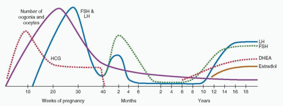
Adrenarche is the term used to describe the increase in adrenal androgen production that begins at approximately 6 years of age in both boys and girls.75,76 Although adrenarche is independent of the maturation of the hypothalamic-pituitary-gonadal axis, the two often are temporally related.77 The increase in adrenal androgen production results from a change in the adrenal response to adrenocorticotropic hormone (ACTH) stimulation, characterized by a shift towards increased production of Δ5-3β-hydroxysteroid intermediates (17α-hydroxpregnenolone; dehydroepiandrostendione, DHEA) and decreased production
of Δ4-ketosteroids (17a-hydroxprogesterone, 17-OHP; androstendione), with no change in cortisol secretion.78 Consequently, an increase in serum DHEA-sulfate (DHEA-S) levels heralds the onset of adrenarche. Generally, the best indicator of adrenarche is a serum DHEA-S concentration greater than 40 μg/dL, which is higher than that normally seen in children 1-5 years of age (5-35 μg/dL).

Adrenal androgens derive from the zona reticularis, the innermost layer of the adrenal cortex,79 which begins to form at approximately 3 years of age and becomes well defined coincident with the increase in DHEA-S production at adrenarche. The molecular mechanisms that govern adrenal cortical differentiation involve a variety of different genes, but little is known about their transcriptional regulation.80 The zona reticularis exhibits a unique enzymatic profile. The activity of 3β-hydroxysteroid dehydrogenase/Δ54 isomerase (3β-HSD), which catalyzes the oxidation and isomerization of Δ5-3β-hydroxysteroid precursors into Δ4-ketosteroids, is low.81 In contrast, the activities of P450c17, including both 17a-hydroxylase (catalyzing the conversion of pregnenolone to 17a-hydroxpregnenolone) and 17,20 lyase (catalyzing the conversion of 17a-hydroxpregnenolone to DHEA), are high, as is steroid sulfotransferase activity.82 Cytochrome b5, which facilitates 17,20 lyase activity, also is preferentially expressed.83 Taken together, the enzyme profile of the zona reticularis favors the formation of DHEA and DHEA-S.

The primary stimulus for adrenarche is unknown. Although ACTH is an obvious candidate, circulating levels of adrenal androgens change without any corresponding changes in ACTH or cortisol during fetal life, puberty, and with aging. In other conditions such as chronic disease, surgical stress, recovery from secondary adrenal insufficiency, and anorexia nervosa, changes in ACTH-induced cortisol secretion are not accompanied by any change in serum adrenal androgen concentrations.84 Although derivatives of proopiomelanocortin (POMC, produced by pituitary corticotropes)85 and other pituitary-dependent factors have been implicated,86 conclusive evidence for an adrenal androgen stimulating hormone is lacking.87,88 Whatever the stimulus might be, it might act to spur the growth and differentiation of the zona reticularis, which may derive from cells originally contained within the “fetal zone” of the adrenal cortex having a unique enzymatic profile (described above). Alternatively, it might act by suppressing 3β-HSD,81 or by stimulating the 17,20 lyase activity of P450c17.82 Interleukin-6 has been implicated as a mediator because it is highly expressed in the zona reticularis and can stimulate DHEA secretion.89 Leptin also has been implicated, because adrenarche coincides with the preadolescent increase in body fat90 and leptin levels91 and leptin stimulates 17,20 lyase activity.92 However, the temporal linkage between adrenarche and increasing body fat also might result from a compensatory hyperinsulinemia or from activation of the growth hormone (GH)/insulin-like growth factor-1 (IGF-1) axis.93

The steady increase in adrenal androgen secretion after adrenarche ultimately stimulates pubarche, the appearance of pubic hair, and also the development and activity of the pilosebacious unit, consisting of a hair follicle and sebaceous gland.94 Adrenal androgen levels correlate with changes in bone density, suggesting they also may contribute to growth in cortical bone.95 If adrenarche has any more fundamental role in the onset of puberty, the mechanism is unknown. Adrenarche generally precedes activation of the hypothalamic-pituitary-gonadal axis, or gonadarche, by approximately 2-3 years. The temporal relationship suggests that adrenal androgen secretion might stimulate the pubertal transition, but several lines of evidence indicate otherwise. First, premature adrenarche generally is not associated with an earlier onset of thelarche or menarche. Second, adrenarche occurs in those with congenital hypergonadotropic hypogonadism (e.g., gonadal dysgenesis) or hypogonadotropic hypogonadism (e.g., Kallmann syndrome). Third, gonadarche occurs in children with Addison disease (hypoadrenalism) treated with glucocorticoids. Finally, in children under age 6 with true precocious puberty, gonadarche precedes adrenarche.


Growth

A substantial proportion of adult height, about 17-18%, is gained during puberty.96 Growth of the limbs precedes that of the trunk, beginning in the distal portions and later also involving the proximal part of the limbs; growth of the trunk occurs primarily during later puberty.
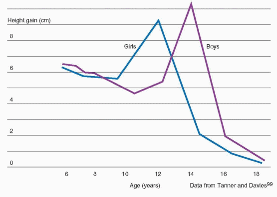
97 The pubertal growth spurt occurs approximately 2 years earlier in girls than in boys and, in general, peak height velocity is reached approximately 6 months before menarche.98

The difference in adult height of men and women relates to the onset and time of the growth spurt in boys and girls. Boys are approximately 2 cm taller than girls when girls reach their peak height velocity, grow 3-4 cm/year for another 1 to 2 years before entering puberty, and reach a greater peak height velocity (10.3 cm/year) than girls (9.0 cm/year).99

Bone mass accumulation during puberty is critical to the development of peak bone mass, which is a major determinant of the risk for developing osteoporosis in later life. Although genetics may be the most important determinant of peak bone mass, other factors such as nutrition and hormone exposure during puberty also contribute. About one-half of total body calcium is accrued during puberty in females, and one-half to two-thirds in males.100,101 In girls, the peak velocity in bone mineral accretion occurs at menarche, approximately 9-12 months after peak height velocity is attained.102,103 The pubertal increase in bone density is greater in black females than in white females.104 Taken together, these data suggest that the window of opportunity to maximize peak bone mass is relatively narrow.101

Weight changes during pubertal maturation reflect changes in body composition and the relative proportions of lean body mass and fat. Skinfold thickness decreases in early puberty and increases after peak height velocity, particularly in girls. Adolescent girls have more body fat than boys, most being deposited in the upper arms, thighs and back; the gender difference increases throughout puberty. The increase in BMI before 16 years of age relates primarily to changes in fat-free mass and thereafter to an increase in fat mass.105 If desired, BMI can be tracked from adolescence to adulthood using published tables for comparison by age and ethnicity.106


image


Growth Hormone

Growth hormone, produced by somatotropes, is the pituitary hormone produced in greatest abundance. The GH gene family includes five distinct genes, all of which are located on chromosome 17 (17q22).107 The pituitary GH gene (GH1) encodes two alternatively spliced mRNAs, yielding a predominant 22 kDa GH molecule and another 20 kDa molecule that accounts for approximately 10% of circulating GH. Placental syncytiotrophblasts express a GH variant in addition to three other genes encoding human chorionic somatotropin, also known as human placental lactogen (discussed in Chapter 8). The regulation of pituitary GH secretion is highly complex. GH secretion is controlled primarily by hypothalamic GHreleasing hormone (GHRH) and by peripheral factors acting on somatotropes that stimulate (e.g., ghrelin),108 or inhibit (e.g., somatostatin)109 GH release.110,111 and 112 Most of the peripheral actions of GH are mediated by insulin-like growth factor I (IGF-I), which inhibits GH release. Nutritional factors also play a role in the regulation of GH secretion; whereas fasting113 and high protein meals114 stimulate GH release, hyperglycemia and leptin inhibit GH secretion.115 Estrogens stimulate, and excess glucocorticoids inhibit GH release. GH secretion peaks during puberty and declines with age, by approximately 50% every 7 years.113

Like the gonadotropins, GH is secreted in a pulsatile fashion, and at the onset of puberty, GH pulse amplitude increases, especially during sleep.116 Consequently, GH concentrations rise progressively. The rate of the increase in circulating GH levels is the most important determinant of the pubertal growth rate; slower growing children exhibit fewer and lower amplitude GH pulses and a more gradual increase in serum GH concentrations.117

GH acts via binding to a specific receptor to stimulate hepatic synthesis and secretion of IGF-I, which promotes both growth and differentiation.107 GH receptor mutations result in GH insensitivity and growth failure (Laron dwarfism);118 in affected individuals, serum GH concentrations are elevated and levels of IGF-I are low. GH stimulates growth via direct and indirect (via IGF-I) actions on the epiphyseal plates of long bones. GH also has a number of metabolic actions, which include increased lipolysis, stimulation of protein synthesis, insulin antagonism, and water and sodium retention.


Insulin-like Growth Factor I

IGF-I is synthesized and secreted by the liver in response to GH stimulation and circulates in serum bound to high affinity IGF binding proteins (IGFBPs). The genes encoding IGF-I, IGF-II, and insulin all belong to the same family. The IGF1 gene has several components and yields several different mRNAs, including the 6 kb form that is regulated by GH. IGF-I acts via its own receptor, which is widely distributed in a variety of tissues and organs.119 IGF-I receptor concentrations are controlled by GH, thyroxine, and other growth factors such as fibroblast growth factor and platelet-derived growth factor. IGF-I acts via a complex signaling cascade to stimulate cell growth and to inhibit apoptosis.

The family of IGFBPs includes six proteins having greater affinities for IGF-I than the IGF-I receptor. IGFBPs are present in all extracellular fluids and serve both to transport IGF-I and to control the amount of IGF-I available to bind to the IGF-I receptor. IGFBP-3 is the most abundant in serum and has the highest affinity for IGF-I, but generally is saturated. Although present in lower concentrations, IGFBP-1 is unsaturated and therefore has greater impact on the levels of free IGF-I. The serum IGFBP-1 concentration is regulated by insulin, increasing during fasting when insulin levels are low, and decreasing after feeding or administration of insulin.120 IGF-I levels are decreased in diseases associated with malnutrition such as inflammatory bowel disease and in hypothyroidism. IGF-I augments the effects of FSH and LH in the ovary, the effect of ACTH on adrenal steroidogenesis, and
the thyroid response to thyroid-stimulating hormone (TSH). IGF-I levels rise 7-fold from very low concentrations at birth to peak values at puberty, fall rapidly by approximately 50% by age 20, then decline slowly with advancing age.121


Gonadal Steroids

The pubertal growth spurt is stimulated primarily by rising levels of GH and IGF-I, but a substantial body of evidence indicates that sex steroids also play an important role. In children with central (gonadotropin-dependent) precocious puberty treated with a longacting GnRH agonist, mean height velocity and nocturnal serum GH and IGF-I levels, initially above the means for chronological age, decrease significantly after 6-12 months and remain suppressed for the duration of treatment.122,123 A study in children with central precocious puberty and GH deficiency (due to an intracranial lesion) observed that bone age was advanced in GH-deficient subjects, but not as much as in control subjects with precocious puberty and normal GH secretion; IGF-I levels were lower in GH-deficient subjects, but greater than in age-matched prepubertal GH-deficient children.124 In a subset of GH-deficient subjects, treatment with a GnRH analog suppressed gonadal sex steroid levels and decreased height velocity, with no appreciable change in GH or IGF-I levels. In girls with Turner syndrome, treatment with exogenous estrogen increases growth velocity and bone age, compared to those observed in placebo-treated controls.125,126 Taken together, these observations indicate that the pubertal growth spurt is mediated, at least in part, by a sex steroid-induced increase in GH secretion. Moreover, they demonstrate that precocious puberty can induce a substantial growth spurt, even in the absence of a normal pubertal increase in circulating GH or IGF-I. However, normal pubertal growth requires the combined actions of sex steroids and GH. Ultimately, sex steroids limit adult height by stimulating epiphyseal fusion.


The Timing of Puberty

What triggers the onset of puberty remains one of the most compelling unanswered questions in reproductive endocrinology. The age at onset of puberty and menarche is influenced by genetics, overall health, social environment, and environmental exposures.

An analysis of two genome-wide association studies including more than 17,000 women from the Nurses’ Health Study and the Women’s Genome Health Study identified 10 common variants or single nucleotide polymorphisms (SNPs) clustered in the regions of chromosomes 6q21 and 9q31.2 that were associated with age at menarche.127,128 Genetic variation in or near the locus (6q21) of the LIN28B gene (encoding a developmentally regulated RNA binding protein)129 has been associated with age at menarche in a number of human populations.128,130,131 Some genetic variants associated with adult height also have been associated with age at menarche, suggesting the association between height and age at menarche has a genetic basis.128,130 Other genes associated with age at menarche include FTO (fat mass and obesity associated gene) and NEGRI (neuronal growth regulator 1), both of which also are associated with childhood obesity.130 Children with a family history of early puberty are more likely to experience an early puberty themselves; age at menarche correlates relatively well between mothers and daughters and between sisters.132

Children who live closer to the equator, at lower altitudes, in urban areas, and mildly obese children generally begin puberty earlier than those who live in northern latitudes, at higher elevations, in rural areas, and those of normal weight. Accumulating evidence suggests that
certain environmental toxicants acting as “endocrine disruptors” also may influence the timing of sexual development.
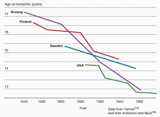
133

The age at onset of puberty has been declining gradually in the general population of the United States over the past century. Although the rate of decrease has slowed considerably more recently, the trend has continued. Overall, the average age at menarche for American girls decreased from approximately 12.75 years in the 1960s to approximately 12.5 years in the early 1990s.134,135 A 1997 study conducted by the Pediatric Research in Office Settings (PROS) network examined the timing of pubertal development in more than 17,000 American girls (90% white, 10% black) and found that the earliest signs of puberty were occurring at ages significantly younger than in the past, with striking racial differences, as follows:136


Pubertal Milestone


Black American Girls


White American Girls


Thelarche


Mean age


Age 6


Age 7


Age 8


Age 9


Age 10


Age 11


Age 12


8.9 yr


6.4 %


15.4%


37.8%


62.6%


80.2 %


96.0 %


98.9 %


10.0 yr


2.9 %


5.0 %


10.5 %


32.1 %


61.5 %


85.4 %


96.0 %


Pubarche


Mean age


Age 6


Age 7


Age 8


Age 9


Age 10


Age 11


Age 12


8.8 yr


9.5 %


17.7 %


34.3 %


62.6 %


85.6 %


95.2 %


98.9 %


10.5 yr


1.4 %


2.8 %


7.7 %


20.0 %


46.4 %


74.3 %


92.2 %


Thelarche and/or Pubarche


Age 6


Age 7


Age 8


Age 9


Age 10


Age 11


Age 12


14.3%


27.2 %


48.3 %


77.4 %


94.6 %


98.4 %


100.0 %


3.7 %


6.7 %


14.7 %


38.2 %


67.9 %


88.0 %


96.6 %


Menarche


Mean age


Age 9


Age 10


Age 11


Age 12


12.2 yr


2.7 %


6.3 %


27.9 %


62.1 %


12.9 yr


0.2 %


1.8 %


13.4 %


35.2 %


These data indicate that a substantial proportion of American girls begin pubertal development 6-12 months earlier than previously observed. On average, black American girls begin puberty between ages 8 and 9, and white American girls by age 10. However, thelarche and/or pubarche can occur normally in black girls as early as age 6 and in white girls as early as age 7.

Subsequent studies analyzing data from the National Health and Nutrition Examination Survey (NHANES) have observed a 2.3 month decrease in the average age of menarche between surveys for the years 1988-1994 (12.53 years) and 1999-2002 (12.34 years), and an overall 4.9 month decrease since 1960.134,137,138 and 139 The decrease in age of menarche has been observed in all ethnic groups, declining from 12.57 to 12.52 years in non-Hispanic white girls, from 12.09 to 12.06 years for non-Hispanic black girls, and from 12.24 to 12.09 for Hispanic American girls.139 Changes in the population distribution of race and ethnicity over time explain the larger change in overall average age compared to those within groups.


image

Historically, the trend to an earlier onset of sexual development has been attributed to improved nutrition and less stressful living conditions.140 The age at menarche has declined as the prevalence of obesity has increased, suggesting that a critical body weight58 or body composition59 is an important factor in determining the onset and progression of puberty.60 Indeed, higher weight and body fat mass are associated with an increased likelihood of early menarche.60,134,141,142 and 143 Data from the U.S. NHANES survey indicated that a girl with a BMI at the 85th percentile is more than twice as likely to have reached menarche as a girl of the same age and race/ethnicity having a BMI at the 50th percentile.139 However, girls reach menarche over a wide range of weight and BMI, and age at menarche cannot be predicted reliably for individuals on that basis.144 Importantly, early pubertal development is associated with slightly decreased adult height and an increased risk for obesity, compared to a late menarche.145,146


Average Ages of Pubertal Milestones in Different Populations147


Country


Year(s)


Subjects


Ages


Thelarche


Pubarche


Menarche


Chile


2000


758


6-16


8.9


10.4


12.7


China


1993


3,749


7-19


9.8


11.6


12.4


Denmark


1991-1993


1,100


6-20


10.9


11.3


13.4


Egypt


2000


1,550


6-18


10.7


10.5


12.4


England


1960-1970


192


3-19


11.2


11.7


13.5


India


1988-1991


9,951


5-18


10.2


12.6


Iran


2003-2004


1,420


6-17


9.7


10.5


12.7


Italy


1998-2001


1,642


6-15


10.5


10.6


11.9


Japan


1990-2000


832


6-14


9.7


12.2


Korea


1993-1995


4,237


14-20


12.5


Lithuania


1999-2000


1,231


7-12


10.2


11.7


Netherlands


1996-1997


3,028


8-20


10.7


11.0


12.9


Spain


2000


266


8-10


10.7


12.4


Thailand


1997-1999


300


9-19


9.4


11.1


11.2


Turkey


2005


1,562


6-16


10.2


10.6


12.4


U.S.


1992-1993


15,439 white


3-12


9.9


10.5


12.9


1,638 black


8.9


8.8


12.2


Stages of Pubertal Development

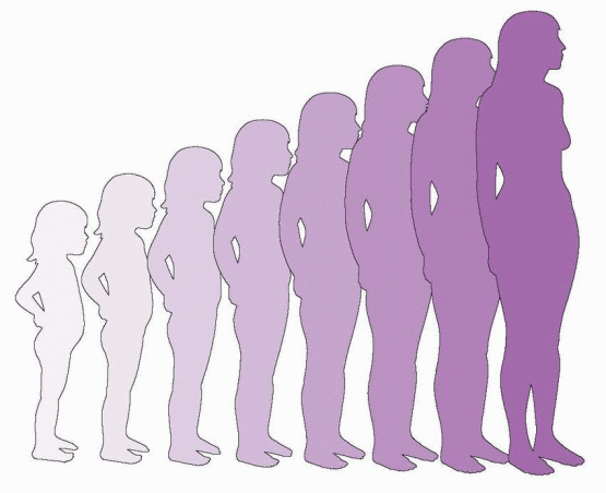
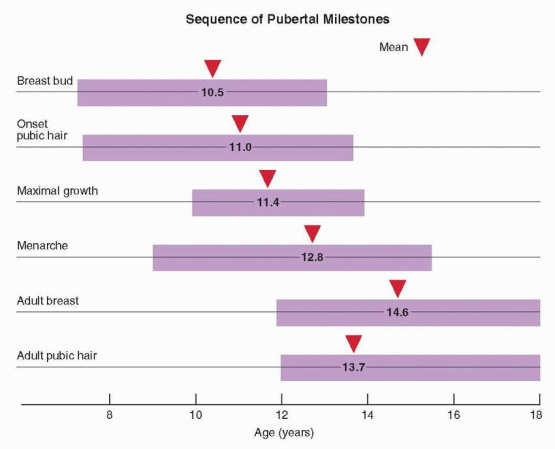
Puberty includes a series of predictable events that vary in timing, sequence, and pace. In general, the first sign of puberty in most adolescent girls is an acceleration of growth, followed by breast budding (thelarche), the appearance of pubic hair (pubarche), and finally, the onset of menses (menarche).

The staging systems used most frequently to describe the physical changes of puberty were first described by Marshall and Tanner in 1969 (girls)148 and 1970 (boys).
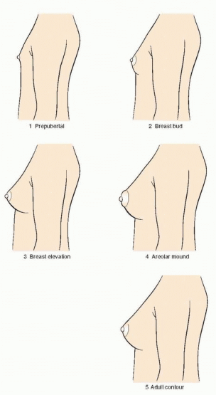
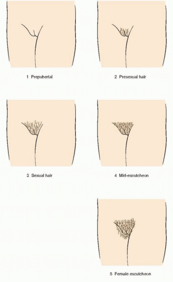
149 The Tanner stages describe secondary sexual characteristics, including breast development in girls, pubic hair growth in both sexes, and genital development in boys. As shown in the diagrams, there are five Tanner stages of breast and pubic hair development in girls, with stage 1 representing the prepubertal state and stage 5 representing adult development.

Breast development follows a recognized sequence of events. Breast budding (Tanner stage 2) is distinguished by enlargement and by widening of the areolae. The breast then enlarges, becoming elevated beyond the areolae (Tanner stage 3). The breast enlarges further and the areolae and nipple form secondary mounds (Tanner stage 4), just before the breast achieves an adult contour (Tanner stage 5).


In the majority of adolescents, pubarche closely follows thelarche, but in a substantial minority the sequence is reversed and pubarche precedes thelarche. In either case, the two are closely linked and progress in parallel. Pubarche (Tanner stage 2) is distinguished by the emergence of a small amount of long, relatively straight hair on the labia majora. The hair then becomes curly, coarser, and extends outward (Tanner stage 3). Hair extends further to cover the labia (Tanner stage 4) before assuming an adult pattern with extension onto the medial thigh (Tanner stage 5).

Menarche occurs an average of 2.6 years after the onset of puberty and after the peak of growth has passed.98,102,148 On average, the pubertal sequence of accelerated growth, thelarche, pubarche, and menarche requires a period of 4.5 years (range, 1-6 years). The relationship between menarche and the growth spurt is relatively fixed. After menarche, growth slows and generally does not increase more than about 6 cm (2.4 inches). The menses immediately following menarche usually are anovulatory, irregular, and occasionally heavy. Anovulatory cycles frequently persist for as much as 12-18 months and are not
uncommon even up to 4 years after menarche.150,151 However, the frequency of menses generally increases rapidly over the first year after menarche; 65% of adolescent girls report having 10 or more periods per year at the end of the first postmenarcheal year, and 90% after 3 years.30 The hallmark of the maturation of the hypothalamic-pituitary-ovarian axis and of the completion of puberty is the development of estrogen positive feedback, which stimulates the midcycle LH surge and ovulation. In general, ovulatory cycles become progressively more frequent. The time required to establish ovulatory cycles relates to the age at menarche; when menarche occurs after the age of 13, only one-half have ovulatory cycles within 4.5 years.152


image


image


image


Serum Hormone Concentrations During Female Puberty153,154,155,156,157,158 and 159


Tanner


FSH


LH


Estradiol


DHEA-S


Stage


IU/L


IU/L


pg/mL


ng/dL


Stage 1


0.9-5.1


1.8-9.2


<10


19-302


Stage 2


1.4-7.0


2.0-16.6


7-37


45-1,904


Stage 3


2.4-7.7


5.6-13.6


9-59


125-1,730


Stage 4


1.5-11.2


7.0-14.4


10-156


153-1,321


Adult Follicular


3.0-20.0


5.0-25.0


30-100


162-1,620


Common Problems Associated with Puberty

Some of the common physical manifestations of pubertal maturation may be viewed by patients or their parents as abnormal, including anemia, acne, psychosocial problems, myopia, and dysfunctional uterine bleeding. Adolescents also are at high risk for sexually transmitted infections.

Girls tend to eat less of foods high in iron content, such as meat, and a low heme iron intake increases the risk of low iron stores.160 The third National Health and Nutrition Examination Survey (1988-1994) observed a 9% prevalence of anemia among American girls between the ages of 12 and 15 years.161

Acne is a disorder of the pilosebaceous unit caused by androgen stimulation characterized by follicular occlusion and inflammation. During puberty, the number of acneiform lesions increases, at all stages.162 In girls, acne tends to be more severe in the late stages of puberty, which are associated with higher serum concentrations of DHEA-S.163

The psychosocial changes during puberty predispose to a increased incidence of depression, which is twice as common in girls as in boys.164 Many girls become unhappy with their physical appearance, resulting in a decrease in self-esteem that is more common in white girls than in black girls.165 The problem is most common when pubertal development is not synchronous with that of peers.166 Early maturing girls are more likely to develop psychopathology,167 to have older friends,168 and to be vulnerable to peer pressures.169

The prevalence of myopia (nearsightedness), caused by growth in the axial diameter of the eye, is greatest during puberty. Dysfunctional uterine bleeding is a consequence of anovulatory cycles and is common in adolescent girls during the first year or two after menarche.

Adolescents represent the highest-risk age group for nearly all sexually transmitted infections (STIs).170 The risk for acquiring an STI relates to age at first intercourse, the number of partners, the perceived risk, and attitudes about acquiring an STL.171 The persistence of a columnar epithelium on the exocervix (ectropion) also may predispose to infection with Chlamydia172 and human papillomavirus.173,174


Precocious Puberty

Precocious puberty describes pubertal development that begins at an earlier age than expected, based on established normal standards. Its causes are many, ranging from variants
of normal development, such as premature adrenarche, to serious pathology, including malignant intracranial neoplasms. Children with precocious puberty warrant careful evaluation to define the cause and, when indicated, prompt treatment to avoid the psychosocial and growth consequences of abnormally early sexual development.


Indications for Evaluation

Abnormally early or precocious puberty generally is defined as pubertal development occurring more than 2.5 standard deviations earlier than the average age. Traditionally, using 10 years as the average age of onset of puberty in girls, precocious puberty has been defined as secondary sexual development before the age of 8 years. However, as discussed in an earlier section of this chapter,134,135 the age at onset of puberty has been declining over the past few decades, raising questions about when pubertal development should be considered precocious and warrants clinical evaluation.

The 1997 study conducted by the Pediatric Research in Office Settings (PROS) network observed that 6.7% of American white girls and 27.2% of black girls had breast or pubic hair development before the age of 8 years.136 These observations suggested that continued application of the traditional definition of precocious puberty would result in a large number of potentially normal girls having extensive, costly, and unnecessary testing. Consequently, new guidelines were proposed, lowering the age at which evaluation is indicated to age 7 in white girls and age 6 in black girls,175 sparking a vigorous debate.

Some authorities questioned the new recommendations because the PROS study population was not a random sample drawn from the general population, because the study focused on premature thelarche and premature adrenarche and not on “true” precocious puberty (typically characterized by early breast and pubic hair development), and because no cause was determined in those having precocious puberty. Criticism centered on concerns that the recommended lower ages in the newly proposed guidelines might increase significantly the risk for under-diagnosis of important endocrine pathology.176,177,178 and 179 Indeed, a subsequent large European study involving 443 girls with central precocious puberty identified 35 with an occult intracranial lesion (8%) and reported that application of the revised American guidelines lowering the age for evaluation for precocious puberty would have missed 4/35 girls (11%) with cranial pathology.180,181 Others favoring the new recommendations emphasized that fewer than 2% of girls with precocious puberty over age 6 had an intracranial lesion and that unnecessary imaging has high financial and emotional costs.182

In another American study involving 223 patients (white girls ages 7-8 years and black girls ages 6-8 years) referred to a single tertiary center solely for evaluation of precocious puberty over a 5-year period, 105 (47%) exhibited both breast and pubic hair development, 83 (37%) had only pubic hair, 24 (11%) had only breast development, and 11 (5%) had no signs of early sexual development.179 Ultimately, 186/212 (88%) with signs of early puberty had a diagnosis of idiopathic gonadotropin-dependent precocious puberty and 26 (12%) had a treatable endocrinopathy amenable to early intervention, including acanthosis nigricans/hyperinsulinemia, hypothyroidism, neurofibromatosis, GH deficiency, pituitary adenoma, McCune-Albright syndrome, and congenital adrenal hyperplasia. More importantly, more than one-third of the girls with only breast development or with breast and pubic hair development had bone ages that were advanced significantly and were, therefore, at risk for diminished growth potential.179

Clearly, more and larger prospective studies are needed because questions regarding the indications for evaluation for precocious puberty remain unsettled. However, taking all of the available data into consideration, we believe that the following guidelines offer a good balance between safety and cost-effectiveness:


Classification of Precocious Puberty

Traditionally, precocious puberty has been classified according to the underlying pathophysiology. However, the classification has limited practical utility in clinical practice because it reflects the final diagnosis, after evaluation is completed.

Gonadotropin-dependent precocious puberty, also known as “central precocious puberty” or “true precocious puberty,” describes early maturation and activation of the hypothalamic-pituitary-gonadal axis and is characterized by both breast and pubic hair development in girls, and by pubic hair development and testicular enlargement (>4 mL in volume or 2.5 cm in diameter) in boys. The early developing sexual characteristics are “isosexual,” meaning they are consistent with the child’s gender.

Gonadotropin-independent precocious puberty, also known as “peripheral precocious puberty” or “pseudo-precocious puberty,” describes early sexual development that is independent of GnRH and gonadotropins and generally results from exposure to sex steroid hormones that derive from the gonads, the adrenals, or the environment. Gonadotropinindependent precocious puberty is further sub-classified as isosexual when sexual characteristics are consistent with gender, and as “contrasexual” when inconsistent with gender (virilization in girls, or feminization in boys).

Incomplete precocious puberty describes children with isolated premature thelarche or premature adrenarche. Both usually represent variants of normal pubertal development, but some will progress to complete precocious puberty that may be gonadotropin-dependent or independent.


Gonadotropin-Dependent Precocious Puberty

Gonadotropin-dependent precocious puberty results from early maturation of the hypothalamic-pituitary-gonadal axis and is much more common in girls than in boys.183 Although puberty begins earlier than normal, the sequence of pubertal events generally is normal and proceeds at the normal pace.


Up to 90% of children with gonadotropin-dependent precocious puberty have no identifiable cause (idiopathic), a diagnosis made by exclusion.184,185 However, the disorder can be associated with a variety of central nervous system lesions, including tumors, irradiation, hydrocephalus, cysts, trauma, inflammatory diseases, and midline developmental defects such as septo-optic dysplasia. Consequently, head magnetic resonance imaging (MRI) is indicated even when there are no neurological signs or symptoms.180,185,186

Tumors associated with gonadotropin-dependent precocious puberty include hamartomas, astrocytomas, ependymomas, pineal tumors, and optic and hypothalamic gliomas. Hamartomas are heterotopic neuronal masses containing GnRH neurons that typically attach to the tuber cinereum or floor of the third ventricle where they can function as an ectopic hypothalamic GnRH pulse generator, divorced from the central inhibitory mechanisms that normally restrain activity during childhood; they are the most common tumor associated with precocious puberty and can be associated with gelastic seizures (laughing, giggling)187,188 some produce transforming growth factor alpha, which mediates release of GnRH.189 The precocious puberty that can be observed in children with neurofibromatosis usually relates to an optic glioma.190

As described in an earlier section of this chapter, activating mutations in the gene encoding the GPR54 receptor (KISS1R), which mediates the actions of kisspeptin (an excitatory neuroregulator of GnRH secretion) can cause gonadotropin-dependent precocious puberty.52

Children exposed to high circulating androgen or estrogen concentrations, as may occur with congenital adrenal hyperplasia, virilizing tumors, and the McCune Albright syndrome, often exhibit early maturation of the hypothalamic-pituitary-gonadalaxis, which then results in gonadotropin-dependent precocious puberty.191,192 and 193

Although quite rare, girls with severe primary hypothyroidism can present with precocious puberty, exhibiting breast development, galactorrhea, and episodic menstrual bleeding. In most cases, the very high serum levels of TSH, which has structural similarity to FSH, appear to activate the FSH receptor.194

Rarely, gonadotropin-dependent precocious pubertal development has resulted from an autonomous pituitary gonadotropin-secreting tumor rather than from early maturation of the hypothalamic-pituitary-gonadal axis.195,196


Gonadotropin-Independent Precocious Puberty

Gonadotropin-independent precocious puberty can result from excess sex steroids secretion from the gonads or adrenals or from exposure to exogenous estrogens.

Autonomous functional ovarian follicular cysts are the most common cause of gonadotropinindependent precocious puberty in girls. Transient breast development and vaginal bleeding are the most common presentation, which can be an isolated event or recur at unpredictable intervals.197,198 and 199 Serum estrogen levels typically are elevated, but not always (due to regression of the cyst), and both basal and GnRH-stimulated gonadotropin concentrations are low. In most cases, bone age is not advanced. Ovarian ultrasonography usually demonstrates one or more unilateral or bilateral ovarian cysts greater than 15 mm in diameter.200 The disorder is self-limited in most and requires no treatment. However, recurrent cysts resulting in prolonged or repeated estrogen exposure can precipitate early maturation of the hypothalamic-pituitary-gonadal axis, resulting in gonadotropin-dependent precocious puberty.198 Autonomous ovarian cysts also can be an early manifestation of McCune-Albright syndrome, arising before emergence of the characteristic skin (“café-au-lait spots”) or bone lesions; affected patients therefore require careful longer-term follow-up.197,199


Ovarian tumors are rare causes of gonadotropin-independent precocious puberty in girls and include granulosa cell tumors, Leydig cell tumors and gonadoblastomas.201,202 and 203

McCune-Albright syndrome is a rare disorder characterized classically by precocious puberty, café-au-lait skin pigmentation, and polyostotic fibrous dysplasia of bone, all caused by a somatic mutation of the alpha subunit of the G-protein (encoded by the GNAS1 gene), which results in a mosaic distribution of cells bearing constitutively active adenylate cyclase.204,205 and 206 The mutation results in continuous stimulation of endocrine function and, in addition to precocious puberty, also can cause gigantism, Cushing syndrome, adrenal hyperplasia, and thyrotoxicosis, in varying combinations. Although precocious puberty is the most common clinical manifestation,207 the phenotype varies with the tissues that are affected by the mutation and can include hepatitis, intestinal polylps, and cardiac arrhythmias. As in other forms of gonadotropin-independent precocious puberty, the sequence of pubertal development may be abnormal; for example, vaginal bleeding frequently precedes breast development.208 The skin and bone lesions can increase over time and may not be present at the initital presentation. Early and repeated exposure to elevated sex steroid levels can result in accelerated growth, advanced bone age, and reduced adult height; it also may induce early maturation of the hypothalamic-pituitary-gonadal axis, resulting in secondary gonadotropin-dependent precocious puberty. McCune-Albright syndrome is more common in girls than in boys. The diagnosis merits consideration in girls presenting with recurrent functional ovarian follicular cysts and episodic menses.209 Partial forms of the syndrome also have been described.206

Adrenal pathology, such as androgen-secreting tumors and congenital adrenal hyperplasia, is another cause of gonadotropin-independent precocious pubertal development.

Exposure to exogenous estrogens or environmental pollutants having estrogenic activity (xenoestrogens) can result in premature sexual development in infants or toddlers.210,211 and 212 Examples include accidental exposure to estrogens, xenoestrogens, or placental extracts contained in cosmetics or personal hair and skin care products and environmental pollutants that can act as endocrine disruptors by mimicking estradiol, such as polychlorinated biphenyls, herbicides, pesticides, and plasticizers, which may be found in water contaminated with industrial products.213 Serum hormone levels in affected children typically are in the normal range, but can vary widely depending on the nature, time, and frequency of use or exposure. Children are extremely sensitive to the effects of estrogen and may respond with increased growth or breast development even at serum levels below the limits of detection.214


Incomplete Precocious Puberty

Incomplete precocious puberty includes premature adrenarche or premature thelarche and usually is a variant of normal puberty. Such cases present a clinical dilemma due to uncertainty regarding whether the condition is entirely benign, as usual, or might be the first indication of true precocious puberty.


Premature Adrenarche

Premature adrenarche is the most common cause of premature pubarche, describing other-wise unexplained early growth of genital hair associated with increased levels of adrenal androgens.215 Generally, the best indicator of adrenarche is a serum DHEA-S concentration greater than 40 μg/dL, which is higher than that normally seen in children 1-5 years of age (5-35 μg/dL). In children with premature adrenarche, the growth rate and bone age often are above average but still within normal ranges. Exaggerated adrenarche is the term used to describe the clinical extreme of premature adrenarche, wherein the serum DHEA-S
level exceeds that typical of adrenarche or for age and usually, but not always, is associated with a somewhat early onset of true puberty.216

The cause of premature adrenarche is unknown. The condition traditionally has been considered an early variant of normal development and, as such, generally has no serious consequences. However, up to 20% of girls with premature adrenarche may subsequently develop gonadotropin-dependent precocious puberty and close follow-up therefore is recommended.77,217 Other evidence indicates that girls with premature adrenarche also are at increased risk for developing polycystic ovary syndrome, suggesting that premature adrenarche may be an early manifestation of the disorder.218,219,220,221,222 and 223 In many, premature pubarche is preceded by low birth weight and is followed by hyperandrogenism, hirsutism, and oligomenorrhea in adolescence, often accompanied by hyperinsulinemia and dyslipidemia. These observations suggest that insulin resistance may be the underlying metabolic disorder, causing decreased growth during fetal life, premature pubarche, and hyperandrogenism that worsens during late puberty or the early postmenarcheal years.224 In those affected, metformin treatment can decrease insulin resistance and hyperandrogenism, improve the lipid profile, often restore cyclic menses, and may help to prevent later development of diabetes and cardiovascular disease.225

Premature pubarche usually results from a premature adrenarche but also has other causes. Idiopathic premature pubarche, unassociated with any demonstrable increase in adrenal androgen production, probably reflects an increased sensitivity of hair follicles to normal androgen concentrations. Premature pubarche sometimes can be the only clinical manifestation of a mild form of congenital adrenal hyperplasia (CAH).226,227 Other rare causes of ACTH-dependent childhood virilization include Cushing syndrome, glucocoticoid resistance, cortisone reductase deficiency, and androgen-producing neoplasms of the adrenal gland or ovary.

The evaluation of premature pubarche should focus first on determining whether the growth of pubic hair is an isolated phenomenon or may be associated with other signs and symptoms suggesting another of the diagnoses mentioned above. The single most important and useful test is an x-ray of the left hand and wrist for bone age. If sexual hair is small in amount and slow-growing and bone age is normal, precocious puberty is unlikely and expectant management is appropriate, with re-evaluation at 6 months and periodically thereafter. A limited endocrine evaluation should include measurements of serum testosterone and DHEA-S, for comparison to age-adjusted normal values. The presumptive diagnosis of premature adrenarche can be made when both are appropriate for pubarche, bone age is normal, and predicted adult height is within the range expected for the family.228 More extensive endocrine evaluation can be reserved for those children having other signs suggesting true precocious puberty or a virilizing disorder.

An ACTH stimulation test to exclude the diagnosis of CAH is indicated when bone age is advanced abnormally, the predicted adult height is abnormally low, or when the serum testosterone and DHEA-S concentrations are elevated above the ranges typical of premature adrenarche. The test is performed by obtaining blood samples before and 60 minutes after administering cosyntropin (synthetic ACTH 1-24; 1 μg/m2 or 0.25 mg). A stimulated serum 17-OHP concentration greater than 1,000 ng/dL generally indicates 21-hydroxylase deficiency.229 In children with premature pubarche, diagnosis of the rare 3β-HSD deficiency requires a stimulated 17a-hydroxpregnenolone level greater than 9,790 ng/dL.227

Premature adrenarche is a benign condition and requires no specific treatment. Parents can be reassured that the condition is a normal variant relating to increased sensitivity of hair follicles to low levels of androgen, or an early occurring incomplete form of puberty. However, children with a diagnosis of premature adrenarche merit periodic re-evaluation for evidence of progressive virilization.


Premature Thelarche

Premature thelarche generally is defined as isolated breast development in girls before the

age of 8 years. In girls, premature thelarche usually is a benign condition considered a variant of normal puberty. Early breast development is particularly common during the first year of life when the hypothalamic-pituitary-gonadal axis is still active.230,231 Studies using ultrasensitive bioassays for estrogen have detected higher estrogen levels in many, but not all, girls with premature thelarche than in normal controls.232 The breast also may be more sensitive to estradiol than normal in some girls.233 Although most affected children subsequently experience normal puberty and growth,234,235 and 236 a significant proportion experiences an earlier than average menarche.237

Physical examination typically reveals a light pink areola with an infantile appearance, with Tanner stage 2 or 3 breast development; frequently, the change may be unilateral or asymmetrical. There is no sign of androgen exposure.

Exaggerated thelarche describes those with premature thelarche who also exhibit increased growth velocity and/or advanced bone age and may represent an intermediate between premature thelarache and precocious puberty.232,238 Even girls with exaggerated thelarche exhibit a prepubertal pattern of response to acute stimulation with GnRH or a GnRH agonist; FSH levels rise more than LH, which remains below 5 IU/L.239 Some cases have been associated with the presence of functional ovarian cysts.197 Genetic studies in girls with exaggerated thelarche have revealed that some harbor a mutation in the GNAS1 gene, suggesting that the disorder can be an early or the only sign of McCune-Albright syndrome.232,240

Premature thelarche also has been related to exposure to exogenous estrogen, including environmental chemicals that degrade slowly in the environment and can accumulate in the food chain, but no clear relationship with premature thelarche has been established.

The evaluation of premature thelarche, like that of premature adrenarche, should focus on determining whether breast development is an isolated phenomenon or associated with other signs of precocious puberty; here again, the most important initial test is an evaluation of bone age. In children with Tanner stage 2 breast development and normal bone age, precocious puberty is unlikely and expectant management is appropriate, with reevaluation at 6 months and periodically thereafter.

Approximately 15-20% of girls with premature thelarche subsequently develop gonadotropin-dependentprecocious puberty, at a mean age of 7.1 ± 0.7years and mean bone age of 9.0 ± 1.1 years.241,242 A longitudinal study involving more than 150 girls with premature thelarche observed that 69% had complete regression of breast development (13% of these later developing true precocious puberty), 21% had recurrent episodes of breast development (32% later developing true precocious puberty), and 10% had persistent breast development (57% later developing true precocious puberty).242


Evaluation of Precocious Pubertal Development

The evaluation of early sexual development begins with a careful history and physical examination and measurement of bone age to determine whether there is any corresponding increase in linear growth. Subsequent evaluation is limited to those with precocious puberty and is aimed at determining the cause and at directing treatment.

The medical history should determine when the physical change(s) were first noticed, in the siblings and parents as well as in the patient, seek evidence of growth acceleration, exclude previous history of neurological disease or trauma or exposure to sex steroids, and identify any associated symptoms of headache, seizures, or abdominal pain.


The physical examination should include height, weight, and calculation of growth velocity (cm/year), which often is an early indication of evolving precocious puberty.243 A fundoscopic examination should be performed to detect papilledema, a sign of increased intracranial pressure. Evaluation of visual fields may reveal evidence to suggest a sellar mass lesion. A careful examination of the skin should be performed to identify any café-aulait spots, which suggest the diagnosis of McCune-Albright syndrome.

Tanner staging of pubic hair and/or breast development should be performed. The diameter of the glandular breast tissue should be measured, taking care to distinguish it from adipose. Accurate assessments are important for determining whether additional evaluation is warranted.

A measurement of bone age is indicated when examination demonstrates signs of early sexual development.


Tanner Staging


Breast


Public Hair


Stage 1 (prepubertal)


Elevation of papilla only


No public hair


Stage 2


Elevation of breast and papilla as small mound, increased areola diameter


Sparse, long, pigmented hair, primarily on labia majora


Stage 3


Further enlargement without separation of breast and areola


Dark, coarse, curled hair sparsely distributed over mons


Stage 4


Secondary mound of areola and papilla above the breast


Adult-type hair, abundant but limited to the mons


Stage 5


Recession of areola to contour of the breast


Adult-type hair, extending onto the medial thigh


Endocrine Evaluation and Imaging

Children with advanced bone age and those having normal bone age accompanied by both breast and pubic hair development, or normal bone age with evidence of accelerated growth and breast or pubic hair development, warrant further endocrine evaluation and imaging.

Basal and GnRH-stimulated serum gonadotropin levels differentiate gonadotropin-dependent from gonadotropin-independent precocious puberty, which then guides further evaluation. Serum gonadotropin concentrations should be measured using ultra-sensitive assays having low detection limits for pediatric patients (approximately 0.1 IU/L).244,245 and 246

The GnRH stimulation test is performed by obtaining blood samples before and 30-40 minutes after a single dose of GnRH (100 μg), administered intravenously. Because synthetic GnRH currently is not available in the United States, a GnRH agonist can be used instead,247,248 and 249 obtaining blood samples before and 60 minutes after a single dose of leuprolide acetate (20 μg/kg), administered subcutaneously.249 The stimulated serum LH concentration is the most useful diagnostic parameter; although a normal threshold value has not been firmly established, due to differences in assay methods and the limited amount of normative data, a stimulated LH value of 3.3-5.0 IU/L defines the upper limit of normal for prepubertal children (Tanner stage 1, T1) with most assays.249 Both basal and stimulated serum LH concentrations have high specificity and positive predictive value for diagnosis of gonadotropin-dependent precocious puberty. In a study comparing the results of GnRH stimulation tests performed in normal children (T1) with those obtained in
children with gonadotropin-dependent and gonadotropin-independent precocious puberty, the mean basal serum LH concentration was 1.6 IU/L in the group with gonadotropindependent precocious puberty and less than 0.6 IU/L in the other two groups. The mean stimulated LH value in the group of children with gonadotropin-dependent precocious puberty was 21.6 IU/L, compared to 3.2 IU/L in normal children (T1) and 1.4 IU/L in the group with gonadotropin-independent precocious puberty.250

In children with gonadotropin-dependent precocious puberty (as identified by elevated basal or stimulated serum LH levels), a head MRI is indicated to exclude an intracranial mass lesion.249,251 Thyroid function tests (TSH and free T4) should be obtained if there is any clinical evidence of hypothyroidism.

In children with gonadotropin-independent precocious puberty (as identified by normal basal and stimulated serum LH levels), serum concentrations of estradiol, testosterone and hCG (functional ovarian cysts and tumors, functional adrenal tumors), late afternoon Cortisol (Cushing syndrome), DHEA-S (premature adrenarche), and 170HP (congenital adrenal hyperplasia) should be obtained to determine the peripheral source of sex steroid production and the cause of early sexual development.


image


Treatment of Precocious Puberty

The treatment of precocious puberty differs according to whether it is gonadotropin-dependent or gonadotropin-independent and on the underlying cause, when that can be determined. The principal goals of treatment are to stop or slow development until normal pubertal age, to maximize adult height, and to reduce the risk of psychosocial problems associated with early sexual maturation.


Treatment of Gonadotropin-Dependent Precocious Puberty

The decision to treat gonadotropin-dependent precocious puberty depends on the underlying pathology and on the speed of sexual development. In those having an identified intracranial lesion, treatment should be directed to the lesion, if that is possible. In those having no intracranial lesion, the decision to treat should be based primarily on the pace of progression and on the estimated adult height.

Treatment for gonadotropin-dependent precocious puberty generally is indicated when sexual maturation progresses to the next stage within 3-6 months, when growth velocity is accelerated to greater than 6 cm/year (unless peak height velocity has already passed), when bone age is advanced by 1 year or more, or when predicted adult height is below the target range or is decreasing on serial determinations.251 Conversely, those with stable or regressing pubertal signs, normal growth velocity (for age), bone age within 1 year of chronological age, and a predicted adult height within the target range may not require treatment. In most cases, growth velocity should be monitored for 3-6 months before making the decision to treat.251 Treatment aimed only at avoiding the potential psychosocial consequences of precocious puberty should be carefully considered because there are few data regarding outcomes and effectiveness.


GnRH Agonist Treatment

Long-acting GnRH agonists have proven both safe and effective for the treatment of idiopathic gonadotropin-dependent precocious puberty.252,253,254,255,256,257,258 and 259 GnRH agonist treatment causes a brief initial “flare” of gonadotropin release, followed by pituitary desensitization (exhaustion of available stores of releasable gonadotropins), and down-regulation (decrease in GnRH receptors). By suppressing the pituitary-gonadal axis, GnRH agonist therapy can prevent progressive pubertal development, and increase final adult height, compared to pretreatment predictions. Young children and those who exhibit rapidly progressive development can be expected to have early epiphyseal fusion, are at greatest risk for compromised adult height, and can benefit most from treatment.249

In girls under 6 years of age with idiopathic gonadotropin-dependent precocious puberty, treatment with a GnRH agonist can be expected to add 9-10 cm to adult height. In older children already past their peak with slowing growth velocity, treatment can be expected to slow it further, to delay epiphyseal fusion, and to yield slow but steady increases in predicted adult height. In girls between 6 and 8 years of age, GnRH agonist treatment typically results in a gain of 4-7 cm in height, less if bone age is significantly advanced.249 Girls already close to the age of normal puberty, those with slowly progressive maturation, and girls with a predicted height above 150 cm have less to gain and may not benefit significantly from treatment.260,261 and 262

The choice among the available GnRH agonist formulations depends mostly on physician preference and availability. Depot preparations generally are preferred because of improved compliance. Direct comparisons in randomized trials have not been made,
but any of the following treatment regimens generally can be expected to suppress the pituitary-gonadal axis:263,264 and 265


  • Buserelin 6.3 mg every 2 months


  • Goserelin 3.6 mg every month, or 10.8 mg every 3 months


  • Histrelin 50 mg implant every year


  • Leuprolide 3.75-7.5 mg monthly, or 11.25 mg every 3 months


  • Triptorelin 3.0-3.75 mg monthly, or 11.25 mg every 3 months

Nonetheless, the dose of GnRH agonist treatment required can vary significantly.266 Inadequate treatment can permit progressive sexual development and bone maturation. Conversely, over-treatment can suppress endogenous GH and decrease growth velocity and bone mineral accumulation to levels below those normally expected during the prepubertal years.267 The adequacy of GnRH agonist treatment can be monitored simply by measuring the serum LH concentration 30-60 minutes after each repeated injection of the agonist; the LH level should be less than 3.0 IU/L, consistent with prepubertal norms after acute GnRH agonist stimulation.268

GnRH agonist treatment should be monitored at 3-6 month intervals with serial physical examinations to detect any progressive pubertal development; bone age also should be evaluated periodically.249 Breast development should cease and growth velocity and the pace of advancing bone age should decrease. Pubic hair development may continue due to normal adrenarche.269 Although bone density may decline during longer durations of treatment, bone mass is regained after treatment ends and peak bone mass is normal; consequently, there is no reason or need to monitor bone density.249

Treatment with GnRH agonists does not appear to have any significant long-term adverse effects on function of the hypothalamic-pituitary-gonadal axis.270 It can be continued until the epiphyses are fused or until the pubertal and chronological ages are appropriately matched. Prompt reactivation of the pituitary-gonadal axis and pubertal development, in a pattern similar to that in normal adolescents, generally follows the discontinuation of treatment.271

GnRH agonist therapy also is recommended for treatment of GnRH-secreting hypothalamic hamartomas187,272; the tumor can be monitored by serial imaging and risky surgery can be avoided. Treatment for other hypothalamic, pituitary, cerebral, or pineal tumors must be individualized. Many that are small and do not extend around or into vital structures can be excised successfully.

Only gold members can continue reading. Log In or Register to continue

Stay updated, free articles. Join our Telegram channel

Jul 5, 2016 | Posted by in GYNECOLOGY | Comments Off on Normal and Abnormal Growth and Pubertal Development

Full access? Get Clinical Tree

Get Clinical Tree app for offline access