Liver Tumors
Anthony D. Sandler
John J. Meehan Jr.
Department of Surgery, University of Iowa Health Care, University of Iowa, Iowa City, Iowa 52242.
The management of hepatic malignancies in the pediatric patient presents a substantial diagnostic and therapeutic challenge. A comprehensive understanding of the disease is critical for optimizing surgical and chemotherapeutic interventions. The type, size, and location of the tumor can dramatically change the course of therapy. Complete surgical excision plays a pivotal role in determining patient survival, whereas preoperative chemotherapy is often crucial for shrinking the tumor and enabling complete resection. Advances in chemotherapy, liver resection techniques, and perioperative patient care since the mid-1980s have markedly improved the overall survival of pediatric patients with liver tumors. However, despite these advances, there is still much room for improvement.
EPIDEMIOLOGY
Primary tumors of the liver constitute between 0.5% and 2% of childhood tumors (1). About 1.9 per 1 million children a year will develop malignant hepatic tumors in the United States (2). Nearly 70% of all primary pediatric liver tumors are malignant (Table 38-1) (3). Hepatoblastomas are the most common pediatric liver malignancy followed by hepatocellular carcinoma (HCC).
Hepatoblastomas are embryonal tumors that generally occur in young children younger than 3 years of age. Conditions associated with an increased risk of hepatoblastoma include Beckwith-Wiedemann syndrome, Fanconi’s anemia, cirrhosis, renal or adrenal agenesis, and hemihypertrophy (4). Chromosomal abnormalities are documented in some cases of hepatoblastoma (5). Particular genetic alterations include trisomy 2, trisomy 20, and mutations in 11p. Interestingly, Beckwith-Wiedemann syndrome is also mapped to 11p15 locus, suggesting the presence of a tumor suppressor gene in this location (6). Hepatoblastomas occurs twice as frequently in males than in females (7), and there is a report of hepatoblastoma in a set of twins in a family with familial adenomatous polyposis (8).
Hepatocellular carcinoma is an aggressive tumor that tends to occur in older children with an average age of 11 years (9). Like hepatoblastoma, males are more commonly affected than females. Unlike hepatoblastoma, HCC often develops in the presence of underlying liver disease. Predisposing factors include hepatitis B (10,11), alpha-1 antitrypsin deficiency, tyrosinosis, glycogen storage disease, cirrhosis (3), and cerebral giantism due to Soto’s syndrome (12). Biliary atresia is also considered a potential predisposing condition (13). In addition, environmental factors such as aflatoxin and nitrosamines have been linked to pediatric hepatocellular carcinoma (14). Although hepatoblastoma is more common in the United States, hepatocellular carcinoma far exceeds hepatoblastoma in many parts of Africa and Asia where hepatitis B is prevalent. Improved worldwide implementation of immunizations for hepatitis B may help reduce the number of cases (15).
PATHOLOGY
Hepatoblastoma and hepatocellular carcinoma are epithelial malignancies. They each tend to occur more commonly in the right lobe of the liver. Hepatoblastomas tend to be solitary lesions that spread within the liver by direct extension and although multicentricity occurs, it is less common.
Hepatoblastoma may present with either of two histologic types (epithelial and mixed variant), and there are six histologic subtypes listed in Table 38-2. Of the epithelial subtypes, the fetal subtype is well differentiated, whereas the embryonal variant is immature and poorly differentiated. Both of these variants have cells arranged in cords. The mixed epithelial-mesenchymal variants may have spindle-cell characteristics. Early studies suggested that the purely fetal variant had a better prognosis with survival as high as 92% at 2 years in stage I patients. This figure appeared to be a significant advantage over all other subtypes that had a 2-year survival of only 57% for stage I patients (9). However, subsequent studies from the Armed Forces Institute of Pathology suggested there may be no improvement in survival for the pure fetal variant (16).
TABLE 38-1 Primary Pediatric Liver Tumors. | ||||||||||||||||||||||
|---|---|---|---|---|---|---|---|---|---|---|---|---|---|---|---|---|---|---|---|---|---|---|
| ||||||||||||||||||||||
Hepatocellular carcinoma consists of large pleomorphic tumor cells that may resemble mature hepatocytes and bilobar involvement is fairly common. Extension of the tumor occurs by intrahepatic dissemination that often leads to multicentricity. Fibrolamellar carcinoma is a variant of hepatocellular carcinoma that may offer an improved prognosis when compared with other hepatocellular carcinomas. Fibrolamellar carcinoma appears grossly nodular, and histologically the lesions are eosinophilic hepatocytes encircled by fibrous bands that give the tumor its lamellar appearance (17).
CLINICAL PRESENTATION
Hepatoblastoma most frequently presents as a right upper quadrant mass, which brings the patient to the attention of the medical practitioner (18). Unfortunately, by this time, most lesions may have grown to considerable size. In fact, more than one-half of hepatoblastomas are believed to be unresectable on initial presentation prior to the induction of chemotherapy (19). Other signs and symptoms include abdominal distention, weight loss, abdominal pain, nausea, and vomiting. Anemia is often present. Occasionally, patients will present with precocious puberty secondary to human chorionic gonadotropin production from the tumor. Bilirubin levels are usually normal, and transaminases may be only minimally elevated if at all. The serum alpha-fetoprotein (AFP) level is elevated in more than 90% of patients (20). Elevation of AFP alone, however, is not diagnostic for hepatoblastoma. Increased serum AFP is also seen in malignant teratomas, such as yolk sac tumors, as well as sarcomas and mesenchymal hamartomas. AFP levels can also be used as a marker for recurrence of tumor postoperatively (21).
TABLE 38-2 Histologic Subtypes of Hepatoblastoma. | |
|---|---|
|
Hepatocellular carcinoma has the same clinical features in children as seen in adults. It occurs in patients between 5 and 25 years of age and, like hepatoblastoma, may demonstrate only a minimal elevation in liver function tests. Many patients with hepatocellular carcinoma have an underlying primary liver disease such as cirrhosis; thus, elevated liver function tests may be detected. The serum ferritin level is elevated in most patients. As noted previously, extension of the tumor occurs by intrahepatic dissemination that often leads to multicentricity. This characteristic, especially in the setting of underlying liver disease, makes resection for hepatocellular carcinoma difficult, if not impossible.
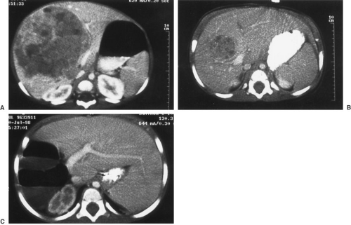
Clinical suspicion of a liver mass warrants a thorough diagnostic imaging investigation. A plain film radiograph of the abdomen may reveal a mass effect containing calcifications. Chest films are recommended to assist in ruling out metastatic disease. Ultrasound will demonstrate the size and location of the mass and will also help differentiate other potential sources of upper abdominal masses. In addition, Doppler studies during the ultrasound can yield vital information to plan operative strategy, including hepatic vein or vena cava involvement as well as proximity of the tumor to the hepatic artery, biliary ductal system, and the portal vein. A computed tomography (CT) scan will demonstrate the size of the mass and help determine resectability (Figs. 38-1A and 38-1B). A CT of the chest is also indicated to better evaluate for potential metastatic disease. Magnetic resonance imaging (MRI) is a reasonable alternative to a CT scan and can be used as a roadmap for planning resection. A CT multiplanar reformation, CT angiogram, or MR angiogram is helpful for reconstructing images of the venous and arterial anatomy to determine tumor proximity to the vessels (Figs. 38-2A and 38-2B). Arteriography has little value in the diagnostic workup and evaluation of these tumors, but may have a role in either chemoembolization or intraarterial infusion therapy in select patients.


