Hemodynamics


Hemodynamics


Patrick J. McNamara

Dany E. Weisz

Regan E. Giesinger

Amish Jain


▪ INTRODUCTION

The care of the premature neonate has improved dramatically over the past 100 years, during which time neonatal mortality has fallen from 40 deaths per 1,000 live births at the turn of the 20th century to the current rates of 4 deaths per 1,000 live births in the developed world. These improvements are a direct result of enhanced maternal health, and access to quality obstetric and neonatal intensive care. Although overall survival for premature infants in general has improved, mortality remains high, particularly at the limits of viability. The contribution of impaired cardiovascular performance and/or systemic hemodynamics to ongoing neonatal morbidity is poorly understood. The approach to hemodynamic monitoring and cardiovascular decision making in critically preterm and term infants has traditionally been based on limited information and remains a poorly understood aspect of neonatal intensive care. The challenges stem from a lack of readily available information that provides insights regarding cardiovascular health, its influence on endorgan performance, and the underlying physiologic processes. The dynamic nature of cardiovascular physiology and its impact on cellular and metabolic mechanisms further complicates the problem. Enhanced cardiovascular monitoring, and earlier therapeutic intervention, may prove to be a necessary step toward improving survival further and minimizing adverse neurodevelopmental sequelae.


▪ PHYSIOLOGY OF THE POSTNATAL TRANSITION

The condition of the infant at birth is dependent on intrauterine well-being and growth in addition to birth- or delivery-associated complications. Both term and preterm infants undergo dramatic cardiorespiratory changes at birth, which coincide with improved lung compliance and termination of the placental circulation. These critical adaptive changes include the following:


  • i. Increased pulmonary blood flow, to about 10 times fetal levels. In utero pulmonary blood flow is low as a function of the vasoconstricted state of pulmonary blood vessels. This occurs in part due to the exposure of the pulmonary vascular bed to higher alveolar concentrations of oxygen as compared to the relatively hypoxic intrauterine environment. Other metabolically active substances, such as metabolites of prostaglandin, bradykinins, or histamine, may play some role through inducing pulmonary vasodilation.


  • ii. Alteration in flow through fetal channels such as the ductus arteriosus and foramen ovale that may last for many days. The major change in flow through fetal channels is either a direct result of increased flow to the lungs or higher arterial oxygen tension. The augmentation of pulmonary venous return leads to an increase in left atrial pressure, which causes displacement of the flap of the foramen ovale over the rims of the fossa, thus abolishing any right-to-left atrial flow. There is oftentimes some residual left-to-right transatrial flow for a period of time as the circulation readjusts. The pattern of flow through the ductus arteriosus is influenced by both systemic and pulmonary vascular resistance (PVR). As lung compliance improves, PVR decreases leading to a reversal in the directionality of the shunt from right to left to bidirectional and eventually left to right. The increase in systemic vascular resistance (SVR), upon removal of the compliant placenta from the systemic circuit and as a result of increased systemic arterial oxygen tension, further exaggerates this change. Recent normative data from term human cohorts suggest that the transductal shunt is left to right by 24 hours, after which a persistent right-to-left shunt should be considered pathologic (1). In premature infants, the administration of surfactant can alter transductal flow significantly through reduced PVR. Finally, the processes leading to ductal closure differ between term and premature infants. The architecture of the immature arterial duct differs such that ductal tone is less responsive to oxygen, thus delaying closure and potentially contributing to excessive flow to the lungs and compromised systemic flow. There is evidence of functional closure by 6 hours in some immature patients, although this is rare (2).


  • iii. Increased left ventricular output (LVO) and right ventricular output (RVO) are necessary to meet the metabolic needs of immature neonate with insufficient thermoregulatory mechanisms and increased work of breathing. The increase in RVO relates to improved systemic blood flow and lower right ventricular (RV) afterload secondary to pulmonary vasodilation. The transition from right-to-left ventricular dominance occurs over hours and is secondary to increased left atrial preload and left ventricular (LV) afterload. In total, there is a threefold increase in LVO, which is necessary to meet the increased demands of the body. The enhanced ability of the left ventricle to increase its output is related, in part, to elimination of constraint by the pressure-loaded right ventricle.


Regulation of Myocardial Performance


Architecture of the Myocyte

Fetal myocardial tissue differs in many respects from its mature adult counterpart, which may explain, at least in part, the differential responsiveness of the immature myocardium to stressors. First, only 30% of the immature myocardium is contractile tissue, compared to 60% in the mature adult heart, rendering it less compliant. Histologic studies have shown that LV myocytes are aligned circumferentially in the midwall and longitudinally in the subepicardial and subendocardial layers of the walls (5). The immature sarcomere and contractile apparatus are relatively disorganized. Intrauterine and early postnatal cardiac growth is a combination of both hyperplasia and hypertrophy. Exposure of the developing rodent heart to dexamethasone led to cardiac hypertrophy, characterized by myocytes that were longer and wider, with increased volume (6). In humans, neonates born to mothers who had received a single antenatal course of steroids had higher systolic blood pressures (SBPs) and increased myocardial thickness (7).


Control of Myocytic Activation

The control mechanisms governing contraction and relaxation in the immature heart are poorly understood but thought to be substantially different from the fully mature heart. In the mature heart, graded control of release of calcium is related to so-called L-type activity, which triggers release from the sarcoplasmic reticulum. Graded control of release in immature myocytes is thought to be related to factors influencing the activity of the sodium-calcium channel. Recently, isoproterenol-induced β-adrenergic stimulation of sodium-calcium exchanger was identified in guinea pig ventricular myocytes. An improved understanding of factors that govern myocardial contractility and relaxation may facilitate more physiologically appropriate choice of therapeutic interventions in premature infants.


Performance and Physiology of the Immature Myocardium

At physiologic heart rates, the immature myocardium shows a positive relationship; contractility falls with extreme or sustained tachycardia.

Cardiac function is dependent on the following factors: preload (residual blood present in the ventricle at end diastole), which is dependent on the intravascular volume status of the infant and diastolic compliance of the ventricle; afterload (resistance against which the myocardium must contract), which depends on vascular resistance, blood viscosity; myocardial performance (the intrinsic ability of the myocardium to contract); and heart rate. Both the force-rate trajectory and the “optimal” heart rate reflect myocytic function and global myocardial contractile behavior. Developmentally, the immature myocardium has been shown to exhibit a higher basal contractile state and a greater sensitivity to changes in afterload (8). The intolerance of the immature myocardium to increased afterload may be attributable to differences in myofibrillar architecture, or immaturity of receptor development or regulation (9). The Frank-Starling law appears less applicable to the immature myocardium. The immediate postnatal period following the loss of the low-pressure system of the placenta and postoperative period following patent ductus arteriosus (PDA) ligation represent two clinical situations in which the neonatal myocardium is subjected to afterload stress. The net effect is impaired myocardial systolic performance and consequential poor systemic blood flow due to low CO, oftentimes despite a normal systemic blood pressure. This problem is further compounded by any potential stressors such as hypoxia, anemia, and mechanical ventilation, which reduces venous return and causes pressure on the myocardium preventing effective contraction.


Control of Vascular Tone

After the transitional period, vascular tone is modulated by a balance between vasoconstrictors, for example, thromboxane, vasopressin, and vasodilators, for example, nitric oxide (NO) and prostaglandins. Immaturity of the central nervous system may also impact on transitional vascular changes. NO is produced by actions of nitric oxide synthase (NOS), present in abundance in smooth muscle tissue; it acts via cyclic guanosine monophosphate (cGMP) on calcium-sensitive potassium channels and myosin light-chain phosphatases to cause smooth muscle relaxation. Endotoxins and cytokines such as tumor necrosis factor-alpha (TNF-α) and a variety of interleukins can induce NOS and NO synthesis leading to profound dilatation and a reduced systemic blood flow in the presence of sepsis. In addition, excess NO leads to formation of free oxygen radicals leading to vascular wall damage. Vasopressin also plays an important role in regulating vascular tone postnatally; its vasoconstrictor effects are modulated through V1 receptors in many organs (excluding the lung, brain, and heart), which in turn increase calcium release from the sarcoplasmic reticulum, up-regulate adrenaline receptors on smooth muscle walls, and reduce NO synthesis. Its implication in shock has been studied in adults. Initially, vasopressin levels increase in response to shock to maintain vascular tone; however, as shock progresses, vasopressin stores are depleted and vascular tone is therefore compromised. Prostaglandins are eicosanoids derived from cell membrane arachidonic acid by the actions of cyclooxygenase (COX) enzymes and play an important role of regulation of vascular tone. Prostaglandin E2 (PGE2), a vasodilator, and thromboxane A2, a vasoconstrictor, are both implicated in the early regulation of vascular tone and may have a role in the pathogenesis of hypovolemia associated with shock.


▪ ASSESSMENT OF THE CARDIOVASCULAR SYSTEM


Clinical Assessment of the Cardiovascular System

A healthy cardiovascular system is defined as one that ensures sufficient oxygen delivery to tissues to meet the metabolic needs of the cells. Assessment of the cardiovascular system begins with a comprehensive clinical history to identify relevant maternal, deliveryrelated, or postnatal factors that increase the risk of cardiovascular ill health. Continuous monitoring of heart rate and blood pressure, which are commonly used as surrogate markers of CO, provides an objective and longitudinal assessment of cardiovascular wellbeing. Physical exam and investigations should be targeted toward assessing end-organ health and function as well as surrogate markers of the adequacy of oxygen delivery. For example, in the absence of primary central nervous system pathology, general observation of level of consciousness and tone can act as surrogate markers of cerebral perfusion. However, there is considerable variability in the normal range for many of the aforementioned hemodynamic measurements. In addition, many of the measurement tools are subject to operator-dependent variability and multiple confounding factors making single measurements unreliable (Table 29.1). The recommended approach is to perform a comprehensive appraisal of the cardiovascular system, which facilitates more holistic insights into illness severity and the pathophysiologic nature of disease, enabling a more targeted approach to therapeutic intervention.


Heart Rate

Tachycardia is often cited as a sensitive marker of low CO based on the concept that stroke volume is relatively fixed due to limitations in the myocardial reserve. While there is some weak evidence of a direct correlation, many neonatal and animal studies have concluded that heart rate is not a major determinant of CO in neonates. In a fetal ovine experimental paradigm, LVO remained stable despite wide variations in heart rate, suggesting that stroke volume may vary significantly to maintain an adequate CO (10). In human preterm neonates, stroke volume also varies with changing heart rate. Echocardiography-based evaluations of CO reaffirm this relationship; specifically, there is no difference in heart rate between patients with low systemic blood flow and those in whom
the systemic blood flow is normal. Elevation in heart rate beyond 160 beats per minute or a sustained elevation from baseline may indicate hypovolemia. However, many noncardiovascular factors can also influence heart rate. Elevated heart rate can be caused by pain, hyperthermia, and commonly used medications such as caffeine and atropine. Sinus bradycardia occurs commonly in sleep, and the neonate should respond with increased heart rate when awakened. Other factors that should be considered if the heart rate remains low include hypothermia, hypothyroidism, electrolyte imbalance, and some medications such as β antagonists (e.g., propranolol).


TABLE 29.1 Clinical Indicators of Cardiovascular Health


Clinical Indicator


Pathophysiology


Confounding Factors


Tachycardia


Increasing HR may increase CO if stroke volume is unchanged


Medications, pain, temperature, agitation


Systolic hypotension


Marker of decreased CO


Transitional circulation, left-to-right shunts


Diastolic hypotension


Marker of SVR and preload


Transitional circulation, left-to-right shunts


Increased CRT


Vasoconstriction of the skin


Wide range of normal


Pallor/acrocyanosis


Vasoconstriction of the skin


Lighting, temperature, skin tone, anemia


Decreased level of consciousness


Decreased cerebral perfusion pressure


Sedative medications, meningitis, seizures


Decreased urinary output


Decreased renal perfusion pressure


Renal pathology, transitional changes


Elevated lactate


Anaerobic metabolism


Some IEM, hemolysed samples, gluconeogenesis


Metabolic acidosis


Anaerobic metabolism


Bicarbonate loss


Low central venous saturation


Increased oxygen consumption


Catheter placement, peripheral shunts, necrotic tissue


HR, heart rate; CO, cardiac output; IEM, inborn error of metabolism.


Blood Pressure

Blood pressure is used as a surrogate marker for blood flow because it is noninvasive and reproducible. In the absence of left-to-right shunt, outside of the transitional period low blood pressure represents either low CO or low SVR. Both of these conditions can lead to inadequate organ perfusion. Typically, a cutoff of mean blood pressure (MBP) less than gestational age (GA) in completed weeks is used; however, most neonates after the first 72 hours of life will maintain their MBP greater than 30 mm Hg in the absence of a hemodynamically significant PDA. In the transitional period, blood pressure and systemic blood flow are poorly correlated. At this time, using an MBP less than GA in completed weeks has been shown to correctly capture only 71% of neonates with low systemic blood flow and falsely identify 12% of neonates as hypotensive (see “Hypotension” section for more detail).


Capillary Refill Time

Studies suggest that capillary refill time (CRT) in healthy neonates varies considerably. Central CRT less than 4 seconds and peripheral CRT less than 10 seconds have been documented in normal neonates. Comparative evaluations using Doppler echocardiography are inconsistent and have shown variable correlation between markers of systemic blood flow and central CRT (11). However, the combination of central CRT of greater than 4 seconds and arterial lactate greater than 4 has been shown to correlate with low CO.


Skin Color

The color of the skin is modified by many factors including temperature, hemoglobin, skin translucency, oxygenation, jaundice, underlying pigmentation, and ambient factors such as environmental light. Interobserver variability of color assessment is high (12). When combined with other markers of low CO, pallor and/or acrocyanosis may suggest peripheral vasoconstriction in skin vessels.


Urinary Output

In the absence of renal parenchymal disease and urinary retention, low urinary output can be a marker of cardiovascular compromise. Glomerular filtration is driven by the pressure gradient across the wall of the glomerular capillary. Renal perfusion pressure is generated by renal blood flow, which is dependent on CO, systemic-to-pulmonary shunts, and SVR. Like the adult kidney, the preterm kidney has the ability to autoregulate blood flow above an unknown threshold of renal perfusion pressure. Below that critical threshold, glomerular filtration (hence urinary output) decreases proportionally to prerenal blood flow.

In the preterm neonate, this relationship is obscured in the first week of life, and caution must be used in relying on urinary output as a marker of renal perfusion. Urine output in preterm neonates follows a predictable pattern. There is minimal urine output in the prediuretic phase (birth to day 2) followed by an abrupt increase during the diuretic phase (days 1 to 5) and a gradual leveling out during the homeostatic phase. During this time, high urine output is not a reliable marker of adequate renal perfusion, though a significant decrease from baseline may be concerning (13).


Laboratory Markers

An increase in serum lactate may occur in clinical situations where anaerobic metabolism occurs when cellular oxygen delivery is compromised. This may occur due to inadequate oxygen content in the blood as occurs in significant anemia or hypoxemia, inadequate tissue blood flow, or a combination of the two. Hence, elevated arterial lactate may be a marker of low CO particularly when used in combination with other clinical or echo markers of low systemic blood flow. Metabolic acidosis will also occur in conditions leading to tissue hypoxia and anaerobic metabolism; however, base excess is significantly affected by bicarbonate loss and has been shown to correlate poorly with tissue perfusion.

In isolation of other clinical indices of cardiovascular health or CO, elevated lactate has poor specificity (12). Lactate is produced as a result of increased gluconeogenesis, and in some inborn errors of metabolism. It is spuriously increased with sample hemolysis, and caution must be interpreted when catecholamines are being administered as sympathomimetic medications increase lactate via perfusion-independent mechanisms. Finally, lactate may be sequestered in damaged tissue, and only when perfusion is improved will serum levels rise.

Mixed venous oxygen saturation (svO2) is widely used in adult intensive care to measure tissue extraction of oxygen. Low svO2
(<70%) is associated with negative outcomes in children, but neonatal studies are lacking (12). Poorly perfused organs in situations of low systemic blood flow extract as much oxygen as possible to optimize aerobic metabolism; hence svO2 will be low. Caution should be used interpreting svO2 in disorders such as necrotizing enterocolitis (NEC) as ischemic tissue does not extract oxygen and therefore high svO2 may be misleading. In neonates, catheter positioning is difficult, and acquiring a mixed svO2, for which the optimal catheter position is the main pulmonary artery is not typically possible. Central venous saturation, which is optimally acquired from the right atrium at the junction of the superior vena cava (SVC) or inferior vena cava (IVC), can be measured and changes in parallel with mixed svO2. The absolute values of mixed and central svO2 are not interchangeable, and central svO2 is affected by catheter position. Due to differential blood flow to vital organs in conditions of shock, a catheter placed in the SVC will overestimate mixed svO2, while one in the IVC will underestimate it. The presence of a high-volume peripheral shunt decreases systemic organ perfusion and increases pulmonary blood flow and hence leads to overestimation of svO2. If catheter position is appropriate, changes in central svO2 over time can be helpful in assessment of neonates with low CO.

In general, no individual marker should be used in isolation. Longitudinal monitoring of arterial pressure provides an objective and reliable surrogate of cardiovascular well-being; however, it should not be used as the sole basis to inform decision making independent of other markers. The components of blood pressure, systolic and diastolic thresholds, may provide insights regarding the specific pathophysiologic process that may be useful in guiding treatment choices. Clinical and laboratory assessment should be combined with a compatible history before treatment decisions are made. Echocardiography or other measures of organ blood flow should be considered whenever possible.


Role of Targeted Neonatal Echocardiography

The approach to cardiovascular care in neonates was previously limited by the lack of reliable clinical tools that provided longitudinal information regarding cardiovascular well-being. The consequences include diagnostic assumptions, incorrect treatment choices, and limited ability for longitudinal evaluation. Targeted neonatal echocardiography (TNE) refers to the use of ultrasound by trained neonatologists to assess cardiovascular health, obtain physiologic information relevant to the clinical situation, formulate a diagnostic impression, make a therapeutic recommendation, and evaluate the response to treatment. It has now become the standard of care within many neonatal intensive care units (NICUs) throughout the world, and there is increasing evidence of its benefit to neonates. Within the immediate postnatal period, it is difficult to determine whether hemodynamic instability is a result of intracardiac and extracardiac shunting, alterations in SVR and PVR, or a developmentally immature myocardium. The goals of TNE include longitudinal assessment of myocardial function, systemic blood flow and pulmonary blood flow, shunts, organ and tissue blood flow. TNE is usually performed by a neonatologist, is directed by a specific clinical question, and may provide hemodynamic information that either complements clinical findings or provides novel physiologic insights. The availability of real-time physiologic data is thought to help the attending physician provide more focused and targeted cardiovascular care. Combination of clinical examination and bedside echocardiography has been shown to improve clinical diagnosis and patient management in the adult population. There is some evidence that routine use of TNE in the neonatal unit may lead to identification of cardiovascular compromise and changes in management and potentially improve short-term outcomes. TNE has enabled more targeted cardiovascular management, determining the type of inotropic agent most likely to be of benefit, and monitoring treatment response. The provision of real-time information on cardiovascular performance and systemic hemodynamics, noninvasive nature of the technique, rapidity of data acquisition and report generation, and ability to perform longitudinal functional assessments have all contributed to the increased utilization of functional echocardiography by neonatologists in the NICU. There is growing evidence of the value of TNE in aiding diagnosis and guiding hemodynamic therapy in the NICU. Carmo et al. (14) showed the benefit of serial echocardiography in directing the duration of indomethacin treatment in infants with a PDA . Jain et al. (15) showed the benefits of TNE in preventing postoperative cardiorespiratory instability following PDA ligation. There are many case reports and cohort studies showing benefits for select patients. Future research should continue to investigate “diseasespecific” impact of TNE on relevant clinical outcomes. Neonatologists who wish to develop expertise in TNE must undergo formalized and structured training and evaluation to ensure competency. Specifically, it is imperative that trained personnel are able to obtain high-quality images and have the necessary knowledge of cardiovascular physiology and therapeutics to ensure that their medical recommendations are rational and scientifically valid.


Novel Methods for Assessment of the Cardiovascular System

There is increasing emphasis, in contemporary neonatal practice, on the importance and need to monitor minute-by-minute indices of physiologic stability in an attempt to understand neonatal disease and enhance patient outcomes. Most of these newer devices are not used in routine clinical practice, but they provide novel insights that will improve our understanding of neonatal hemodynamics, thus informing clinical practice.


Noninvasive Cardiac Output Monitoring

Continuous CO monitoring is a potentially valuable tool in the NICU in the management of a wide range of neonatal illnesses. In adults and older children, continuous CO measurement is frequently achieved by invasive methods, including thermodilution using a pulmonary artery catheter, an intraesophageal probe for continuous Doppler velocity flow assessment, or an arterial catheter for pulse contour analysis. Size constraints or suboptimal reliability preclude the use of these methods in preterm and term infants, and in lieu, several noninvasive and semiinvasive modalities have been evaluated.

Detectable alterations in the electrical properties of the thorax have been used to estimate CO. In children, bioimpedance estimates of CO are unreliable when compared with magnetic resonance imaging and direct Fick methods (16,17). Two newer electrical approaches, based on extensions of the theory of bioimpedance, are currently under evaluation. Electrical velocimetry is based on the principle that the conductivity of the blood in the aorta varies during the cardiac cycle. During systole, red blood cells align in the direction of flow, and the electrical current applied from external electrodes is easily conducted. In diastole, red blood cells assume a random orientation, which results in lower conductivity of the injected electrical current. Electrical velocimetry measures the peak rate of change in conductivity across the cardiac cycle and uses it to derive the mean aortic blood velocity index, from which LV stroke volume is estimated using calculated aortic cross-sectional area and LV ejection times (Fig. 29.1). Studies in small children have compared the CO estimates of electrical velocimetry with transthoracic echocardiography (TTE). In a mixed population of children with repaired/unrepaired congenital heart disease, electrical velocimetry estimates of CO had unacceptably high absolute and percentage bias compared with thermodilution and subxiphoid Doppler measurements (18,19). Noori et al. (20) compared CO estimates of electrical velocimetry and TTE in healthy term neonates and found a mean difference of 4 mL/min (limits of agreement -234 to 242 mL/min) and an adjusted percentage bias of 31.6%. Grollmus et al. (21) reported a similar 29% relative TTE-electrical velocimetry bias in a cohort of infants postoperatively from the arterial switch operation. Transthoracic bioreactance is another newer method of noninvasive CO monitoring (NICOM, Cheetah Medical, MA). In contrast
to bioimpedance, which aims to detect changes in the amplitude of an applied electrical current, bioreactance estimates changes in the frequency of the current (the relative phase shift) between the input and output signals, which is induced by blood ejected into the aorta from the left ventricle. Stroke volume is estimated using the measured peak rate of change of the phase shift, ventricular ejection time, and a constant of proportionality that accounts for patient age, gender, and body size. Studies have reported variable reliability of NICOM-measured CO. NICOM was compared with invasive measurements of CO using an aortic root catheter in anaesthetized beagles and demonstrated a bias of 63 ± 38 mL/min, a percent bias of 6.1%, and high responsiveness to pharmacologically induced changes in CO (22). In contrast, NICOM demonstrated poor reliability and responsiveness compared with pulmonary artery thermodilution-measured CO in critically ill adults being treated with volume expansion (mean bias 0.9 L/min/m2 and limits of agreement -2.2 to 4.1) (23). In a heterogeneous group of term and moderately preterm neonates, NICOM estimates of CO were strongly correlated with TTE (r = 0.95) but NICOM systematically underestimated CO by 31 ± 8% (24). In a cohort of extremely preterm infants undergoing PDA ligation, NICOM similarly underestimated CO relative to echocardiography (mean bias 39%, limits of agreement 8% to 69%), with increasing bias over time (25). Collectively, these studies suggest that NICOM is able to trend CO over time, but it is not interchangeable with invasive or noninvasive measures. Use as a trending tool of CO in neonates likely requires initial and periodic calibration with echocardiography. Additional studies demonstrating adequate reliability are needed prior to independent use.


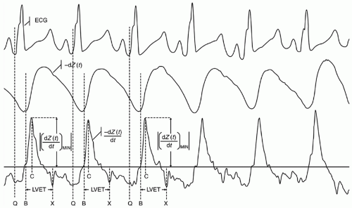
FIGURE 29.1 Surface ECG, impedance waveform [-dZ(t), also known as ΔZ(t)], and the electronically differentiated first time derivative of -dZ(t), -dZ(t)/dt, obtained from electrical velocimetry monitoring on a 25-day-old male (HR = 142 beats/min, SV = 3.3 mL, CO = 0.47 L/min). The marker labeled “Q” on the ECG marks the beginning of ventricular depolarization and thus the onset of electromechanical systole. Shortly after aortic valve opening (“B”), the -dZ(t) waveform exhibits a significant upslope and, consequently, its time derivative -dZ(t)/dt exhibits a nadir (“C”). The amplitude at the point that in the traditional presentation is depicted as a positive deflection is the maximum slope or peak rate of change of the transthoracic electrical impedance during a particular cardiac cycle and measured beat to beat. The time to peak (rise time) of -dZ(t)/dt is concordant with the time to peak of -dv(t)/dt of the aortic blood velocity waveform. The magnitude at the peak of -dZ (t)/dt, that is | (dZ (t)/dt) MIN|, is analogous to the magnitude |dv (t)/dt) MIN| of this waveform. The first-time derivative of the impedance waveform, -dZ (t)/dt, exhibits a deflection at the time of aortic valve closure (“X”). The temporal interval between points B and X is defined as the left ventricular ejection time. Reproduced from Norozi K, et al. Electrical velocimetry for measuring cardiac output in children with congenital heart disease. Br J Anaesthesia 2008;100:88, Copyright 2008, with permission from the author and Oxford University Press.


Noninvasive Cerebral Perfusion Imaging: Near-Infrared Spectroscopy and Arterial Spin-Labeled Perfusion Magnetic Resonance Imaging

Near-infrared spectroscopy (NIRS) is a diffuse optical technique that measures variations of cerebral absorption and scattering within the spectral window of the near-infrared range. It is sensitive to tissue chromophore (oxy- and deoxyhemoglobin) concentrations and thus can be used to estimate regional cerebral oxygen saturation (rcSO2), cerebral fractional tissue oxygen extraction (cFTOE), and cerebral blood volume. Neonates are ideal candidates for NIRS monitoring because the decreased thickness of the neonatal skull permits deeper penetration of near-infrared light. NIRS measurements are performed continuously and noninvasively at the bedside, without the need for general anesthesia.

The surveillance and prevention of early cerebral injury in neonates is one area where the clinical utility of NIRS in the NICU may be justified. Normative ranges of rcSO2 and cFTOE during normal neonatal transition for term (26) and preterm (27) infants have been established. Lower rcSO2 and higher cFTOE heralds the subsequent development of severe peri- and intraventricular hemorrhage (IVH) in very preterm infants (28), which potentially facilitates earlier identification of high-risk infants who may benefit from intervention to prevent cerebral injury.

Arterial spin-labeled perfusion magnetic resonance imaging (ASL-pMRI) is a newer noninvasive method of cerebral blood flow assessment that uses electromagnetically labeled arterial blood water as an endogenous contrast agent, in lieu of intravascular contrast agents (e.g., gadolinium), or radioactive-labeled
tracers. ASL-pMRI-estimated cerebral blood flow measurements in neonates undergoing hypothermia for perinatal hypoxic-ischemic encephalopathy correlate strongly with NIRS (r = 0.88) (29). ASLpMRI may be used to identify and quantify the severity of cerebral hyperperfusion after global cerebral anoxic brain injury, though at this stage, the technique is not useful as a bedside clinical tool to guide decision making.


▪ PATENT DUCTUS ARTERIOSUS


Pathophysiologic Continuum of the Ductal Shunt in Neonates

During normal transition at birth, clamping of the umbilical cord and the initiation of ventilation in air result in an increase in SVR and a decrease in PVR. The ductal flow pattern, previously right to left in utero, becomes bidirectional and eventually left to right as PVR decreases below systemic arterial pressure. In unwell neonates, the ductal shunt is variable in direction, reflecting disordered pulmonary and/or systemic blood flow or perturbations in the programmed postnatal decrease in PVR. Consideration of the role of the ductus arteriosus (DA) should not be binary. The PDA may play a supportive or neutral role in infants with persistent pulmonary hypertension of the newborn (PPHN), in whom there is a postnatal failure of vasorelaxation of the pulmonary arterioles (due to either abnormal fetal pulmonary development or maladaptive neonatal transition), resulting in persistently elevated PVR. In severe cases, PVR remains suprasystemic, and the ductal shunt is from the pulmonary artery to the aorta, supporting the postductal systemic blood flow, albeit with deoxygenated blood that results in a difference in oxygen saturation between preductal and postductal circulations. Here, the PDA also results in reduced RV afterload and may help preserve RV function. In mild cases of PPHN, the ductal shunt is bidirectional and may not contribute significantly to either pulmonary overcirculation or RV afterload reduction. Instead, the PDA may be an innocuous bystander that provides a measure of the pressure gradient between the pulmonary and systemic circulations.

In preterm and a small minority of term infants whose PVR decreases at birth but in whom the ductus remains patent, a continuous left-to-right shunt develops. Shunt volume is determined by the pressure gradient between the pulmonary artery and aorta and by the resistance to transductal flow, which is primarily influenced by ductal diameter and length. Determinants of PVR such as hypocapnemia, hyperoxemia, or alkalosis may augment the shunt volume. A large shunt results in volume overload of the pulmonary artery, and subsequent alveolar edema, reduced pulmonary compliance, and increased need for mechanical ventilation. Increased blood flow to the left heart results in increased end-diastolic volume of the left atrium and ventricle. LV dilatation occurs, and it compensates by increasing stroke volume. Impaired LV diastolic compliance results in pressure loading of the left atrium and contributes to left atrial dilatation. Diastolic flow reversal from the descending aorta to the pulmonary artery via the PDA is common, as is absent or reverse end-diastolic flow in systemic arteries such as the celiac and superior mesenteric arteries. Diastolic “steal,” combined with shorter diastolic times due to tachycardia and increased myocardial oxygen demand from LV dilatation, may result in subendocardial ischemia.


Clinical Importance of Patent Ductus Arteriosus in Preterm Infants: Severe Morbidities of Prematurity

One-third of very-low-birth-weight (VLBW) infants and up to 65% of infants born at GA less than 28 weeks have PDA on the 3rd day of life (30). Infants with persistent PDA have increased mortality, IVH, bronchopulmonary dysplasia (BPD), retinopathy of prematurity (ROP), and NEC compared with infants without a PDA. Perinatal hemodynamic instability, cerebral ischemia, and subsequent reperfusion injury may contribute to the development of germinal matrix hemorrhage and subsequent extension into the cerebral ventricles (IVH) or periventricular hemorrhagic venous infarction. Most IVH occurs in the first week of life, coincident with the emergence of a left-to-right ductal shunt, which increases cerebral (preductal) blood flow and may contribute to reperfusion injury. Both the targeted and indiscriminate administration of prophylactic indomethacin reduce the incidence of all grades of IVH, possibly by mitigating the emergence of a significant ductal shunt (31).

PDA-induced pulmonary overcirculation and systemic hypoperfusion may contribute to increased BPD and NEC. Increased pulmonary blood flow results in pulmonary interstitial edema and greater need for invasive mechanical ventilation. Ventilator-induced lung injury promotes alveolar inflammation and ongoing need for respiratory support, a major risk factor for the development of BPD. Diastolic flow reversal in the abdominal aorta, celiac artery, and superior mesenteric artery (SMA) is common in infants with PDA, and this diastolic “steal” may contribute to intestinal hypoperfusion and an increased risk of NEC. While a causal relationship among PDA and IVH, BPD, NEC, and ROP has not been definitively established, a number of physiologic, observational, and randomized trials strongly support this association (30,32). The association of these morbidities with increased mortality and adverse neurodevelopmental outcome is the impetus behind the need to identify and potentially mitigate the multisystem effects of a pathologic ductal shunt.


Determining the Hemodynamic Significance of the Patent Ductus Arteriosus

For term neonates with PDA, treatment in infancy is considered if left heart volume loading and pulmonary overcirculation lead to feeding difficulty, inadequate weight gain, or pulmonary insufficiency. In the absence of these clinical findings, infants are permitted to grow to allow later catheter-device closure in lieu of an open surgical technique, and PDA closure is performed to prevent irreversible changes to the pulmonary arteriolar musculature and pulmonary hypertension (PHT).

The variability in timing of ductal closure in term infants highlights the pathophysiologic spectrum associated with PDA-related shunting. In contrast, the clinical evaluation of the hemodynamic significance of PDA in preterm infants is more challenging due to the common coexistence of pulmonary disorders and immature oral feeding ability. Respiratory distress syndrome (RDS) and ventilator-induced lung injury result in pulmonary insufficiency that can be exacerbated by a significant ductal shunt. Myocardial diastolic dysfunction is also common in preterm infants (33) and reduces the infant’s tolerance of left heart volume loading associated with a PDA. The clinical determination of the relative pathologic effect of the PDA and primary pulmonary disorders is also influenced by GA-based expectations and the evolution of an infant’s respiratory and feeding physiology as he or she matures. The clinical hemodynamic significance of a PDA may therefore be considered to fall along a continuum between innocent bystander and prime pathologic contributor. The diagnosis of PDA, using clinical examination, electrocardiography, and chest radiography, is explored in detail in a subsequent chapter. Echocardiography is the primary method of PDA evaluation in preterm infants and requires assessments of the following: ductus arteriosus size and flow pattern, pulmonary overcirculation and left heart loading, and systemic arterial diastolic flow reversal (“systemic steal”).


Assessment of the Ductus Arteriosus Size and Transductal Flow Pattern

Shunt volume is positively correlated with PDA radius; vessel size ≥1.5 mm on the first day of life predicts a subsequent symptomatic PDA (34) and correlates well with Doppler flow pattern in assessments of hemodynamic significance (35). For infants with birth weight near 0.5 kg, the 1.5-mm threshold may be insensitive, and in lieu, PDA diameter may be indexed to weight (>1.5 mm/kg) or left pulmonary artery (LPA) diameter (PDA:LPA ratio >0.5). The spectrum of PDA pulsewave Doppler flow patterns reflects the varied and evolving status of a PDA as a barometer of pulmonary arteriolar pressure versus primary pathologic contributor to left heart volume loading and pulmonary overcirculation. In severe neonatal PHT, the ductal shunt is right to left
when pulmonary artery pressure is suprasystemic and bidirectional (right to left in systole, left to right in diastole) when pulmonary artery pressure is approximately systemic. While a bidirectional ductal shunt is common in preterm infants on the first day of life, a persistent bidirectional ductal shunt has been associated with increased mortality, likely a surrogate marker of persistent PHT due to severe pulmonary disease. The restricted or “closing” pattern of a PDA depicts a veryhigh-velocity ductal shunt (peak systolic velocity >2.0 m/s) and high peak systolic-to-minimum diastolic velocity ratio (<2.0) (Fig. 29.2). A hemodynamically significant PDA is characterized by an unrestrictive or “pulsatile” left-to-right flow pattern, with the highest peak velocity at the end of systole and very low diastolic velocity. While peak systolic velocity less than 1.5 m/s has been traditionally described as “unrestrictive,” higher peak systolic velocities may be seen in infants with large unrestrictive ductal shunts, owing to very large shunt volumes or in the situation of a chronic funnel-shaped PDA, posttreatment, where there may be partial restriction at the pulmonary end.


Assessment of Pulmonary Overcirculation—Left Heart Loading

A large left-to-right ductal shunt is associated with increased pulmonary arterial blood flow, pulmonary venous return, LV end-diastolic volume, and LVO. Pulmonary artery diastolic velocity, left atrial and LV chamber sizes, mitral valve Doppler velocities, and LVO provide surrogate estimates of pulmonary overcirculation and left heart volume and pressure loading, though they may be of reduced value in the presence of a large transatrial shunt. A large left-to-right ductal shunt delivers blood to the pulmonary artery throughout the cardiac cycle, resulting in more turbulent flow and increased antegrade diastolic flow in the main and branch pulmonary arteries. Left pulmonary artery diastolic velocity correlates with increased ductal shunting estimated by cardiac catheterization and a prolonged need for mechanical ventilation (36). Maximum left pulmonary artery diastolic velocity less than 0.2 m/s is suggestive of a small ductal shunt, while greater than 0.5 m/s is associated with a large shunt.


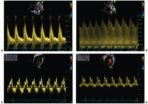
FIGURE 29.2 Pulse-wave Doppler interrogation of a PDA (A and B) and left ventricular inflow across the mitral valve (C and D). An unrestrictive left-to-right ductal shunt (A) has the Doppler profile of an arterial pulsation, with low diastolic velocity, while a restrictive shunt demonstrates high peak systolic and diastolic velocities and low systolic-to-diastolic peak velocity ratio. Left ventricular filling in diastole consists of early (E) and late (A, during atrial contraction) phases, with normal E:A ratio greater than 1. Preterm infants with no PDA demonstrate normal inflow and an E:A ratio less than 1 due to prematurity-related decreased myocardial compliance and impaired early filling (C). Large PDA is associated with left atrial pressure loading and increased early diastolic ventricular filling, resulting in a “pseudonormalized” E:A ratio greater than 1.

Infants with PDA have increased LVO and LV end-diastolic dimension (LVEDD), which are surrogates for increased LV enddiastolic volume. LVEDD can be compared to previously published normative values for LV chamber size for VLBW infants. LVO greater than 300 mL/kg/min is highly specific in predicting a symptomatic PDA (34). Left atrial dilatation occurs due to volume and pressure loading from increased pulmonary venous return and LV diastolic dysfunction. The relatively fixed transaortic diameter permits indexing of LA chamber size for comparison of the LA:Ao ratio between infants. LA:Ao greater than 1.4 has a high sensitivity for ductal significance (37); however, higher LA:Ao ratios (≥1.6) are more specific for a significant ductal shunt (38).

Mitral valve inflow Doppler indices and isovolumic relaxation time (IVRT) are affected by left atrium volume and pressure loading associated with a large ductal shunt. In healthy term infants, the mitral valve E (“early”) wave to A (“atrial”) wave ratio (E:A) is greater than 1,
signifying a predominance of early LV diastolic filling. The myocardium in preterm infants is less compliant resulting in impaired passive diastolic filling, reliance on atrial contraction for ventricular filling, and a resultant mitral valve E:A ratio less than 1. In the presence of a large ductal shunt, increased end-systolic left atrial pressure results in earlier mitral valve opening and a shortened IVRT (often <45 milliseconds) (33) and increased early passive filling velocity and flow, with E:A greater than 1, termed “pseudonormalization” (Fig. 29.2).


Systemic Steal

Diastolic flow reversal in the abdominal aorta and splanchnic circulation due to a ductal shunt occurs when the aortopulmonary pressure gradient is greater than the diastolic pressure in the abdominal aorta or the specific end-organ arterial resistance. This diastolic “steal” in the abdominal aorta is one of the well-recognized and reliable indicators of a hemodynamically significant shunt in preterm infants, both clinically and on cardiac MRI (39). Celiac artery Doppler flow (CAF) to LVO ratio (CAF:LVO) less than 0.1 is highly sensitive for the presence of a significant PDA (40). Infants with PDA may have reduced SMA blood flow, which may impair postprandial increases in intestinal perfusion. Although the middle cerebral artery (MCA) is supplied by preductal CO, a large ductal shunt may result in reduced, absent, or reverse MCA diastolic flow, though the clinical sequelae of this are unknown. Reductions in diastolic flow associated with a PDA may be compensated for by increases in systolic blood flow. Echocardiography may assess arterial blood flow but not vascular resistance. Reduced blood flow velocity and/or diastolic flow reversal in the splanchnic circulation improves immediately after PDA ligation.


Echocardiographic PDA Score

While individual echocardiographic parameters are variably sensitive for PDA hemodynamic significance, their aggregation into a comprehensive PDA score permits practical application in infants with variable echocardiographic indices (Table 29.2). Several PDA scores have been reported and demonstrated strong predictive ability for neonatal outcomes. El-Khuffash et al. (41) combined six echocardiographic markers in a PDA score that predicted severe neurologic disability or death in 2-day-old VLBW infants (Table 29.3). Sehgal et al. (42) reported that a comprehensive echocardiographic PDA score, assessed just prior to ibuprofen treatment, predicted the development of chronic lung disease in VLBW infants with a PDA. While these scoring systems identify infants with a PDA at risk of adverse outcome, it remains to be demonstrated whether treatments aimed at ductal closure or shunt management may improve outcomes.


TABLE 29.2 Echocardiography Parameters of Ductal Hemodynamic Significance


Hemodynamic Significance


Parameter


Mild


Moderate


Severe


Ductus arteriosus size and flow pattern


PDA diameter


2D diameter


<1.5 mm/kg


1.5-3 mm/kg


>3 mm/kg


PDA:LPA ratio


<0.5


0.5-1


>1


PDA Doppler


Peak systolic velocitya


>2.5


1.5-2.5


<1.5


Peak systolic velocity: minimum diastolic velocity


<2


2-4


>4


Pulmonary overcirculation/left heart loading


LV chamber size


No dilatation


Moderate dilatation


Marked dilatation


LA hypertension


LA:Ao


<1.5


1.5-2.0


>2.0


Mitral valve E:A ratio


<1


>1


IVRT


>45 msec


30-45 msec


<30 msec


LPA Vmax diastole


<0.3 m/s


0.3-0.5 m/s


>0.5 m/s


Systemic steal


Abdominal aorta


No diastolic reversal


Diastolic reversal


Diastolic reversal


Celiac:aorta VTI ratio


<0.10


aVery large left-to-right ductal shunts may have higher peak systolic velocities (>1.5 m/s), indicating high shunt volume rather than flow restriction. Ao, aorta; IVRT, isovolumic relaxation time; LA, left atrium; LPA, left pulmonary artery; LV, left ventricle; PDA, patent ductus arteriosus; VTI, velocity-time integral.


TABLE 29.3 Echocardiographic PDA Score at 48 Hours of Life in Preterm Infants Less Than 32 Weeks of GA and the Outcome of Death or Neurodevelopmental Impairment at 2 Years


Score


Parameter


Not Significant (0 Points)


Significant (1 Point)


Ductal diameter


<1.5 mm


≥1.5 mm


Left atrium:aorta


<1.5


≥1.5


Descending aorta end-diastolic velocity


Present


Absent or reversed


Left ventricular output


<300 mL/kg/min


≥300 mL/kg/min


Mitral valve E:A


<1


>1


Celiac artery flow: left ventricular output


>0.15


<0.15


From El-Khuffash AF, Slevin M, McNamara PJ, et al. Troponin T, N-terminal pro natriuretic peptide and a patent ductus arteriosus scoring system predict death before discharge or neurodevelopmental outcome at 2 years in preterm infants. Arch Dis Child Fetal Neonatal Ed 2011;96(2):F133.


Biomarkers

In response to left heart pressure and volume loading, cardiac myocytes cleave pro-brain natriuretic peptide (BNP) into biologically active BNP and the inactive fragment amino-terminal pro-BNP (NTpBNP). BNP inhibits the renin-angiotensin-aldosterone axis, vasodilates the pulmonary and systemic circulation, and promotes natriuresis and diuresis. While modalities to measure plasma BNP and NTpBNP concentrations are widely available, NTpBNP has the advantage of having a longer half-life (60 minutes vs. 20 minutes). Their widespread use in the NICU has been hampered by
the evaluation of many different testing kits, each with its own reference value range, rendering the interpretation of results more challenging. The early identification of infants at high risk for developing a symptomatic PDA may be clinically useful in centers seeking to administer targeted treatment aimed at early ductal closure. In very preterm infants, elevations in plasma BNP and NTpBNP concentrations at birth and on the first day of life correlate with lower gestational and birth weight, but not the development of PDA. After the 2nd day of life, elevated concentrations predict hemodynamically significant PDA (Table 29.4), though the interpretation of widely varying cutoff values of BNP and NTpBNP is hampered by the use of different testing kits and diagnostic criteria for symptomatic PDA. Martinovici et al. (43) found that plasma NTpBNP level less than 10,000 pg/mL measured on the 2nd day of life had 89% sensitivity and 100% specificity for spontaneous ductal closure.


TABLE 29.4 Relationship Between Plasma BNP/NTpBNP Concentration in the First 3 Days of Life and a Diagnosis of hsPDA in Preterm Infants


Author (Year)


Biomarker


Mean or Median GA (Weeks)


No. of Infants (with hsPDA)


Age (Days)


Area Under ROC (95% CI)


Threshold


Sensitivity (%)


Specificity (%)


Choi (2005)a


BNP


29


66 (23)


3


0.997 (0.99-1.00)


>1,100


100


95


Flynn (2005)a


BNP


28


20 (N/A)


3


N/A


<300


52


100


Czernik (2008)b


BNP


25


67 (24)


2


0.86 (0.75-0.96)


>550


83


86


El-Khuffash (2007)c


NTpBNP


27


48 (25)


3


0.87 (0.76-0.97)


>5,000


70


87


Farombi-Oghuvbu (2008)c


NTpBNP


30


49 (18)


3


0.98 (0.93-1.03)


>1,347


100


95


Nuntnarumit (2009)c


NTpBNP


29


35 (12)


3


0.96 (0.91-1.02)


>1,204


100


91


Ramakrishnan (2009)c


NTpBNP


28


56 (20)


3


0.90 (0.81-0.99)


>1,280


>5,160


95


60


58


95


Martinovici (2011)c


NTpBNP


28


27 (12)


2


0.92 (0.67-0.99)


0.98(0.84-1.00)


>1,182


>591


89


91


100


100


Buddhe (2012)d


NTpBNP


27


69 (22)


3-5


0.98 (p < 0.001)


>697


96


90


aTriage BNP test, Biosite Diagnostics


bADVIA Centaur analyzer, Siemens/Bayer


cElecsys proBNP, Roche Diagnostics


dVITROS NTpBNP reagent pack, Ortho Clinical Diagnostics


BNP, brain natriuretic peptide; GA, gestational age; hsPDA, hemodynamically significant patent ductus arteriosus; NTpBNP, amino-terminal pro-brain natriuretic peptide; ROC, receiver operating curve.


BNP thresholds expressed in pg/mL.


NTpBNP thresholds expressed in pmol/L. Studies reporting NTpBNP concentrations in pg/mL were converted to pmol/L. 1 pmol/L = 8.457 pg/mL.


After the onset of clinical symptoms of suspected ductal shunting, plasma BNP/NTpBNP discriminate between infants with and without a hemodynamically significant PDA, as diagnosed by echocardiography. Chen et al. (44) reported that a plasma BNP (Triage BNP assay, Biosite Diagnostics) cutoff value of 40 pg/mL had a 92% sensitivity, 46% specificity, 1.70 positive likelihood ratio (LR), and 0.17 negative LR for predicting a moderate or large PDA shunt as assessed by echocardiography. The low specificity and positive LR reflect the large overlap in values among infants with and without a hemodynamically significant PDA. Higher cutoff values have lower sensitivity but higher specificity. A plasma BNP cutoff value of 200 pg/mL had a 59% sensitivity, 91% specificity, 6.91 positive LR, and 0.46 negative LR for predicting a moderate or large echocardiographic PDA shunt. These findings suggest that plasma BNP less than 40 pg/mL or greater than 200 pg/mL indicate a moderate likelihood of not having or having a hemodynamically significant PDA, respectively. In centers where access to echocardiography is limited, these cutoffs may be helpful for guiding administration of empiric pharmacologic therapy aimed at ductal closure. However, plasma BNP concentrations in the range of 40 to 200 pg/mL are poorly discriminatory, and echocardiography is needed.


Management of a Hemodynamically Significant PDA


Supportive Management—Strategies to Limit Shunt Volume

Conservative measures comprise treatment strategies aimed at reducing ductal shunt volume or improving an infant’s physiologic tolerance of the ductal shunt without medical or surgical interventions to close it. Positive end-expiratory pressure (PEEP), target oxygen saturations, diuretics, fluid restriction, and targeting high blood hematocrit are commonly used. Higher PEEP, administered via invasive or noninvasive mechanical ventilation, decreases systemic and pulmonary venous return, reducing pulmonary alveolar edema and LV end-diastolic volume. An increase in PEEP from 5 to 8 cm H2O has been demonstrated to reduce echocardiographic indices of left-to-right ductal shunting (45). This may be due, in part, to mitigation of ductal shunting by mean airway pressure (MAP)-associated increases in PVR. Diuretics may be used to reduce pulmonary edema and work of breathing in infants with large leftto-right ductal shunts. Furosemide is the most commonly prescribed diuretic, and its use in preterm infants with PDA-associated volume overload has largely been extrapolated from older studies in infants with edema of varying etiologies, including congestive heart failure (CHF). However, furosemide increases renal prostaglandin production and may mitigate ductal constriction (46). The administration of furosemide to prevent fluid retention in preterm infants with PDA during treatment with indomethacin may result in excessive weight loss without a demonstrated benefit on PDA closure rates (47).

The rationale for the administration of other conservative treatments is based primarily on physiologic principles and is generally unsupported. Higher oxygen saturations may improve spontaneous ductal closure due to vasoconstrictive effects of higher arterial oxygen tension. However, recent large randomized trials comparing high versus low oxygen saturation targets in extremely preterm infants found no difference in the development of PDA (48). Similarly, while a higher hematocrit may increase PVR and mitigate left-to-right shunting across ventricular septal defects, randomized trials have found no difference in PDA development or management using strategies that increase hematocrit, such as liberal red cell transfusion practices or delayed cord clamping at birth (49). Fluid restriction has been reportedly used to reduce left heart volume loading in infants with PDA. While excessive fluid administration in the first days of life has been associated with higher rates of developing PDA, moderate fluid restriction does not improve pulmonary or systemic hemodynamics in infants with a PDA (50). Most importantly, to achieve a reduction in intravascular volume, daily fluid intake would have to be restricted beyond renal concentrating ability (typically <100 mL/kg/d), and this is not recommended due to reduced nutritional intake and somatic growth.


Ductal Closure Strategies


Pharmacotherapy: Prostaglandin Synthase Inhibitors

Ductal patency is promoted by the production of circulating prostaglandins catalyzed by the enzyme prostaglandin H2 (PGH2) synthase. PGH2 synthase has both peroxidase (POX) and COX moieties that work in series to produce PGH2, the precursor to PGE2. Arachidonic acid is converted to PGH2 by sequential reactions catalyzed by COX and POX. Inhibitors of COX, and more recently POX, which decrease circulating prostaglandins, constitute the dominant pharmacologic therapies targeting ductal closure.

Ibuprofen and indomethacin are the most commonly used COX inhibitors. Their efficacy in ductal closure wanes with decreasing GA, due to an immaturity-related absence of ductal intimal thickening, which is a necessary precursor for neointimal mound formation and subsequent anatomic closure after ductal vasoconstriction. The optimal timing of administration of COX inhibitor treatment for PDA remains uncertain. Prophylactic indomethacin reduces the risk of severe IVH, periventricular leukomalacia, pulmonary hemorrhage, symptomatic PDA, and PDA ligation in extremely low-birth-weight (ELBW) infants, compared with later symptomatic treatment only. The use of prophylactic indomethacin declined after the publication of a large randomized trial that found no improvement in 18- to 24-month neurodevelopmental outcome (31). However, improved neurodevelopmental outcome has been demonstrated in later childhood, when psychological assessments are more reliable (51). The clear short-term benefits coupled with evidence of a lack of harm have led to its continued use in some centers. Targeted indomethacin prophylaxis, administered to infants with PDA greater than 1.5 mm in the first 6 hours of life, reduces symptomatic PDA and pulmonary hemorrhage and avoids the indiscriminate administration to infants who may have never developed PDA. Screening echocardiography also facilitates the identification of the small minority of infants with an entirely right-to-left shunt (indicating suprasystemic RV pressures), in whom the PDA reduces RV afterload and indomethacin prophylaxis is contraindicated (52). The cardiovascular consequences of prophylaxis in the setting of a bidirectional shunt remain unknown.

For the treatment of symptomatic PDA, pre-surfactant era trials suggested that early treatment (in the first week of life) improved PDA closure rates and reduced pulmonary morbidity, such as the number of days of oxygen therapy and a more rapid wean of mechanical ventilation, but without demonstrating improvement in mortality, BPD, or other morbidities (53). Recent trials reported no difference in mortality or severe neonatal morbidities in infants treated early for mildly symptomatic PDA compared with expectant management followed by late treatment (54,55). However, studies to date have relied on oversimplified clinical and echocardiographic criteria to identify a hemodynamically significant PDA. Failure to define the spectrum of PDA-related hemodynamic disturbance may have resulted in diluted study populations underpowered for the target outcomes. The contemporary clinical decision to administer pharmacologic treatment should integrate a comprehensive echocardiographic assessment of the hemodynamic significance of the PDA, rather than a simplified approach based on size. Therapy should target infants in whom the ductal shunt is estimated to be a primary pathologic contributor to current physiologic instability, compared with other concurrent pathologies (most commonly pulmonary immaturity and severe RDS related to prematurity).

COX inhibitor use in preterm infants is associated with reductions in cerebral and splanchnic blood flow. Adverse effects include oliguria, weight gain, gastrointestinal injury, transient dysfunction of platelet aggregation, and increased serum hyperbilirubinemia due to competitive binding of albumin. Their use is contraindicated in the setting of renal failure, NEC, intracranial (but not intraventricular) hemorrhage, and severe jaundice. Indomethacin should not be administered concurrently with systemic corticosteroids due to an increased risk of spontaneous intestinal perforation. Ibuprofen is as effective as indomethacin in achieving PDA closure, and ibuprofen-treated infants have lower risks of NEC and transient renal insufficiency (56). While early studies comparing ibuprofen and indomethacin identified an increased risk of BPD in ibuprofen-treated infants, a recent meta-analysis revealed no difference in BPD and earlier weaning from respiratory support with ibuprofen (56). Both oral and intravenous forms of ibuprofen are widely used, and intravenous formulations of indomethacin predominate. Indomethacin dosing of 0.2 mg/kg every 12 to 24 hours for three doses is a commonly used treatment regimen. Prolonged or higher total doses of indomethacin (more than 0.6 mg/kg) have been associated with increased risk of NEC without improvement in PDA closure rates (57). Oral and intravenous ibuprofen have similar rates of PDA closure and adverse effects and are typically administered in a 10 mg/kg initial dose, followed by two once-daily 5 mg/kg doses.

Only gold members can continue reading. Log In or Register to continue

Stay updated, free articles. Join our Telegram channel

May 30, 2016 | Posted by in PEDIATRICS | Comments Off on Hemodynamics

Full access? Get Clinical Tree

Get Clinical Tree app for offline access