Hematology


Hematology


Yigal Dror

Anthony K. C. Chan

Jillian M. Baker

Maria Laura Avila


▪ RED BLOOD CELL DISORDERS


Red Blood Cell Development

Early or primitive hematopoiesis appears in the extraembyonic yolk sac during gastrulation at about day 16 to 18 in humans and forms macrophages, nucleated erythrocytes, and some megakaryocytes (1). At about day 28, a different pathway called definitive hematopoiesis arises and gradually replaces the primitive hematopoiesis. Definitive hematopoiesis is characterized by formation of hematopoietic stem cells that give rise to various types of leukocytes (granulocytes, basophils, and eosinophils), lymphocytes, megakaryocytes, and anucleated erythrocytes. The appearance of definitive hematopoiesis is most evident in the aorta-gonad-mesonephros region (2,3). By the 8th week of gestation, fetal hematopoiesis is migrated to the liver. The liver remains the primary site of blood cell production throughout the early fetal period. By 6 months of gestation, the bone marrow becomes the principal site of blood cell development. During gestation, a switch occurs in the type of hemoglobin being formed. Hemoglobin is a tetramer of four globin proteins; two are encoded from genes on chromosome 16 and two from genes on chromosome 11. On chromosome 16, there is a switch from epsilon (ε) to gamma (γ) and then to delta (δ) and beta (β). On chromosome 11, there is a switch from zeta (ζ) to alpha (α). At birth, about 60% to 90% of the hemoglobin comprises of two alpha and two gamma globins and is termed fetal hemoglobin. During the first 6 to 10 months of life, most of the fetal hemoglobin is gradually replaced by the adult hemoglobin (HbA) that is comprised of alpha and beta globins. The site of production of erythropoietin (EPO) switches from the less sensitive hepatic to the more sensitive renal site (4).

Erythroid precursors are identified by their cell surface antigen expression and growth characteristics in culture. The earliest characterized precursor is the burst-forming unit (BFU-E), which gives rise to colony-forming units (CFU-E). More than 40 times the number of BFU-E can be cultured from fetal blood as from an equivalent volume of adult blood; however, total body erythropoietic potential of the fetus might be comparable to that of an adult (4). BFU-Es respond to stem cell factor and interleukin-3 by proliferation, and their number declines along the series of fetal blood, cord blood, postdelivery bone marrow, and postdelivery blood. Neonatal BFU-E and CFU-E are as sensitive as their adult counterparts to EPO stimulation (5). However, there are differences between fetal and adult erythropoiesis in EPO levels and in mounting EPO levels in response to various cues. EPO controls erythropoiesis by a feedback loop according to erythrocyte mass and central venous oxygen tension. The same feedback loop involving EPO levels and measures of oxygen delivery (such as hemoglobin level and oxygen tension) exists in premature neonates (6); however, the measured levels of EPO in the preterm baby are much lower than those of older children and adults with corresponding degrees of anemia (7,8). The magnitude of the EPO response is lowest in the least mature infant (27 to 31 weeks of gestation) (8). EPO values in cordocentesis samples from infants between 18 and 37 weeks of gestation were found to be low (9), but there was no correlation between gestational age and EPO level. The poor EPO response persists through the neonatal period, resulting in a reduced erythropoietic stimulus and lower hemoglobin levels in premature infants (10). The poor EPO response derives from various factors, including a high oxygen level after birth, incomplete transition of EPO production from a low EPO-responsive organ (liver) to a high EPO-responsive organ (kidney) until the first 3 to 4 months of life, and a high rate of EPO clearance in neonates.


Normal Hemoglobin Levels

In the newborn period, the hemoglobin concentration is undergoing constant physiologic change. A clear definition of the normal hemoglobin range is therefore important for proper evaluation and management.

Normal hemoglobin values at birth have been determined through measurement of levels in cord blood. The normal mean hemoglobin level is 16.9 ± 1.6 g/dL in term neonates and 15.9 ± 2.4 g/dL in premature infants (11). Definitive values for premature infants have been elucidated through cordocentesis sampling (Table 43.1). Based on these data, cord hemoglobin levels less than 13.0 g/dL should be considered abnormal in term and premature (<36 weeks of gestation) neonates. In the very premature infant (<26 weeks of gestation), values as low as 12.0 g/dL may be acceptable. If anemia is confirmed, a prompt and careful search for the cause should be initiated.

The hemoglobin level in newborn infants is significantly influenced by the amount of placental transfusion. At birth, blood is rapidly transferred from the placenta to the infant, with one-fourth of the placental transfusion occurring within 15 seconds of birth and one-half by the end of the first minute (12). The placental vessels contain 75 to 125 mL of blood at birth (13). In a randomized study of delayed cord clamping in preterm infants (24 to 32 weeks of gestational age), the mean blood volume (74.4 mL/kg) was significantly greater in the delayed cord clamping group compared to the group in whom the cord was clamped immediately after birth (62.7 mL/kg) (14). Several studies have reported an association between delayed cord clamping and decreased requirement for blood transfusions, decreased hypotension and intraventricular hemorrhage in preterm babies (15). A meta-analysis of 15 controlled trials comparing late (at least 2 minutes) versus early (at birth) cord clamping in term infants showed an association between late cord clamping and improved hematocrit, stored iron, and a clinically important reduction in risk of anemia (RR 0.53; 95% CI, 0.40 to 0.70) in a period 2 to 6 months after birth (16). Asymptomatic polycythemia was found to be associated with late cord clamping in term infants; however, jaundice and respiratory distress were not (16).


Fetal Hemoglobin, Neonatal Erythrocytes, and 2,3-Diphosphoglycerate

Human tissue metabolism depends critically on an adequate supply of oxygen. The oxygen transport system in humans is the erythrocyte, which contains the iron-protein conjugate hemoglobin. The erythrocyte’s primary function is to deliver oxygen to the tissues at a partial pressure sufficient to permit its rapid diffusion from the blood to the cells (see Chapter 28, “Oxygen Delivery” section for detailed description).


Anemia in the Newborn Period

Anemia at birth or appearing during the first few weeks of life can be broadly categorized as resulting from blood loss, hemolysis, or underproduction of erythrocytes. It is rarely due to sequestration of blood in a large spleen.


Physiologic Anemia and the Anemia of Prematurity

The hemoglobin concentration of healthy term and premature infants undergoes typical changes during the first weeks of life. After birth, there is a transient increase in hemoglobin concentration as plasma moves extravascularly (17). Thereafter, the hemoglobin concentration gradually falls, to reach a nadir of 11.4 ± 0.9 g/dL in term infants by 8 to 12 weeks of age and 7.0 to 10.0 g/dL in premature infants by 6 weeks of age (Fig. 43.1) (18).


TABLE 43.1 Normal Erythrocyte Values during Gestationa


Weeks of Gestation


Erythrocytes (×1012/L)


Hemoglobin (g/dL)


Hematocrit (%)


Mean Corpuscular Volume (fL)


18-21


2.85 ± 0.36


11.7 ± 1.3


37.3 ± 4.3


131.11 ± 10.97


22-25


3.09 ± 0.34


12.2 ± 1.6


38.6 ± 3.9


125.1 ± 7.84


26-29


3.46 ± 0.41


12.9 ± 1.4


40.9 ± 4.4


118.5 ± 7.96


>36


4.7 ± 0.4


16.5 ± 1.5


51.0 ± 4.5


108 ± 5


aValues are means ± 1 standard deviation.


There are several reasons for the fall in hemoglobin. The first is the decline in erythrocyte production in the first few postnatal days as evidenced by a fall in reticulocyte count (Fig. 43.2). Normally, reticulocyte counts may be elevated during the first 1 or 2 days of life (200 to 300 × 109/L) but then fall to low levels (in the order of 50 × 109/L) through the remainder of the neonatal period. This diminution of erythropoiesis is probably related to negative feedback caused by the increased oxygen delivery after birth and consequently decreased EPO production (19). The reduced EPO response persists until approximately 6 weeks of age, at which time erythrocyte production increases, as evidenced by a sharp rise in reticulocyte numbers in the blood and an increase in total body hemoglobin (Fig. 43.2). Other factors that contribute to the physiologic anemia in newborns, particularly the more profound anemia in premature infants, are the shortened survival of neonatal erythrocytes (20) and rapid body growth (Fig. 43.2).


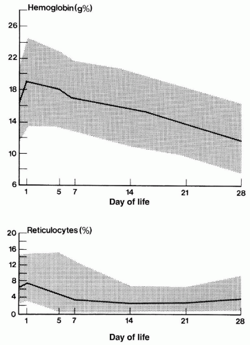
FIGURE 43.1 Hemoglobin values of 178 normal premature infants ≤36 weeks of gestation. Data at the first point, day 0, are cord blood values. Subsequent points represent data from capillary blood samples on 1, 5, 7, 14, and 28 days of life. The dark line represents the mean value, and the shaded area includes 95% of all values.

The effect of rapid body growth on hemoglobin levels is unique to neonates. Healthy premature infants rapidly grow while active erythropoiesis, as evidenced by a mild reticulocytosis, resumes at 6 to 8 weeks of age. Associated with this rapid weight gain is an obligatory increase in the total circulating blood volume. The resultant hemodilution may cause a peripheral hemoglobin concentration that is static, or even falls slightly. The apparent paradox of a stable or falling hemoglobin concentration despite active erythropoiesis (i.e., mild reticulocytosis and an increasing erythrocyte mass) gradually corrects, and the peripheral hemoglobin concentration increases (see Fig. 43.2). Failure to recognize the important effect of rapid body growth on the peripheral hemoglobin concentration may lead to inappropriate investigation and treatment of apparent anemia (21).

In preterm infants, there is a faster and more pronounced decline in hemoglobin after birth than in term babies. This anemia of prematurity typically occurs at 4 to 6 weeks of age, and the hemoglobin may fall to 70 or 80 g/L in infants of less than 1.0 or
1.0 to 1.5 kg, respectively (19,22). Anemia of prematurity largely results from relatively low production of EPO, reduced response to EPO, and blood sampling (23). The signs and symptoms of this early anemia in premature infants are nonspecific and reflect changes in metabolic rate or cardiorespiratory function and perfusion.


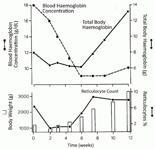
FIGURE 43.2 Changes in total body hemoglobin, blood hemoglobin concentration, reticulocyte count, and body weight in a representative premature infant. The vertical bars represent the infant’s body weight. During the first 6 weeks of life, the blood hemoglobin concentration and total body hemoglobin fall as a result of decreased erythrocyte production, as evidenced by the low reticulocyte count. The more rapid decline in blood hemoglobin concentration from the 3rd to the 6th week is the result of the increasing body size and dilution of the hemoglobin mass. After 6 weeks of age, hemoglobin production increases, as evidenced by the increased reticulocyte count and the rapid increase in total body hemoglobin. The blood hemoglobin concentration during that period may rise slightly, or not at all, because the total body size increases at approximately the same rate as the total hemoglobin mass.

Trials of folate, iron, and vitamin E have not shown any evidence of benefit in preventing the physiologic anemia of infancy (24); however, their supplementation may improve the efficacy of EPO administration (25). Optimal protein supplementation is important for maintaining good hematopoiesis (26,27) and may similarly optimize the effect of EPO in the preterm neonate (26).

Surveys of blood product use in neonatal units show that most transfusion practices are based on either a hemoglobin value or symptoms and signs that the clinician interprets as indicators of anemia (apnea, poor weight gain, etc.) (28). A recent international survey of transfusion practices for extremely premature infants found that 44% of the 1,018 neonatologists considered the degree of oxygen requirement and the need for respiratory support to be very important factors when considering transfusion (29). Similar clinical criteria have been incorporated into guidelines for the transfusion of premature infants (30,31,32).

Transfusion therapy may expose infants to infectious agents (e.g., cytomegalovirus [CMV], hepatitis, human immunodeficiency virus [HIV]) (33,34) and other risks from blood products (e.g., graft versus host disease). Transfusions in preterm infants have unique risks. Many studies have described an association between transfusions and necrotizing enterocolitis (NEC) particularly in very-low-birthweight (VLBW) infants (35,36). A recent meta-analysis of observational data in 12 studies of transfusion-associated NEC also reported this association. These authors also found patients with transfusion-associated NEC to be at higher risk of mortality but acknowledge the need for further studies to adjust for possible confounders (37). Others have reported the association between red blood cell (RBC) transfusions and severe intraventricular hemorrhage (36,38,39). Prevention of anemia by limiting blood work and delayed cord clamping among other practices remains an important strategy.

Two main trials have studied the optimal hemoglobin trigger for RBC transfusion in preterm neonates by comparing transfusion to maintain higher (153 or 135 g/L) versus lower (73 or 77 g/L) hemoglobin levels, respectively (40,41). Both trials show that a restrictive practice reduces the number of transfusions; however, the authors of the studies differed in their conclusions as to whether liberal use of transfusions may help to prevent complications such as apnea and brain injury (40,41,42). A study, which evaluates the long-term impact of transfusions on neurologic outcome, is in progress (42,43,44).

The desire to avoid the use of blood products coupled with the need to treat symptomatic anemia has led to trials of recombinant EPO in premature newborns. Low plasma EPO levels as described above are a major contributing factor in the pathogenesis of anemia of prematurity (10). In clinical trials of EPO administration to prevent or treat anemia of prematurity, EPO has been started either early (within the first week of life) or late (about 3 weeks of age). In general, infants receiving larger EPO doses at both time points have shown improvement in reticulocyte counts and hemoglobin levels and a decrease in the number of transfusions per infant. Of importance, however, early initiation of EPO therapy was not associated with a significant reduction in the number of infants transfused, and in groups treated later, most RBC exposures occurred before the start of EPO therapy.

Cochrane Database Systematic Reviews were performed on early or late EPO administration in preterm and/or low-birth-weight (LBW) infants, respectively (45,46,47). These Cochrane reviews, as well as older meta-analyses (48,49), highlight the heterogeneous design of studies, for example, with regard to the inclusion criteria (birth weight) and treatment (EPO doses).

The Cochrane reviews of early EPO utilization found that administration of EPO reduced the number and volume of RBC transfusions only minimally and to a degree that is of questionable clinical importance. The authors concluded that this practice is not recommended (45). The review of late EPO use found that the reduction in number and volume of transfusions was also of marginal clinical significance and that exposure to blood donors was not likely to be substantially reduced, as most studies included infants who had received transfusions prior to enrollment. The authors recommended that future study should focus on limiting donor exposure in the first few days of life (46).

A concerning association has been reported between treatment of premature babies with EPO and retinopathy of prematurity (50). A Cochrane review of early EPO use found a significant increase in the risk of stage III retinopathy of prematurity (45). Another concern regarding treatment with EPO is the development of neutralizing antierythropoietin antibodies. This has been demonstrated in adult patients with chronic renal failure who developed pure red cell aplasia following treatment with EPO for 3 to 67 months. The complication resolved several months after cessation of EPO treatment; however, the patients required multiple RBC transfusions until then. Whether neonates who are treated with EPO for less than 3 months can develop neutralizing antibodies remains to be determined (51); however, this potential complication of EPO therapy should be taken into consideration when assessing the risk:benefit of EPO therapy in newborn infants (52).

The long-acting erythropoiesis-stimulating agent darbepoetin has also been studied. A recently published randomized, masked placebo-controlled study of 102 preterm infants given either weekly darbepoetin, or EPO or placebo demonstrated fewer transfusion rates and fewer donor exposures in those treated with darbepoetin or EPO compared to those receiving placebo. The incidence of adverse events was equal in all the study groups, including the incidence of retinopathy of prematurity (53).

In summary, the use of EPO for anemia of prematurity has generally fallen out of favor in North America. The overall incremental benefit of EPO or related therapy in neonates, who are managed with rigorous transfusion guidelines, strategies to minimize iatrogenic blood losses from phlebotomy, and appropriate iron and protein supplementation, is not clear and requires further study.


Anemia Caused by Blood Loss

Blood loss resulting in anemia may occur prenatally, at the time of delivery, or postnatally. Blood loss may be the result of occult hemorrhage before birth, obstetric accidents, internal hemorrhages, or excessive blood sampling for diagnostic studies (Table 43.2). Faxelius et al. (54) associated a low erythrocyte volume with a maternal history of bleeding in the late third trimester, placenta previa, abruptio placentae, nonelective cesarean section (CS), deliveries associated with cord compression, Apgar scores less than 6, an early central venous hematocrit less than 45%, and a mean arterial pressure less than 30 mm Hg.


Occult Hemorrhage Before Birth

Occult hemorrhage before birth may be caused by fetomaternal hemorrhage or by the bleeding of one fetus into another in multiple pregnancies. In approximately 50% of all pregnancies, some fetal cells can be demonstrated in the maternal circulation (55). In about 8% of pregnancies, from 0.5 to 40.0 mL of blood is transferred from the fetus to the mother at birth, and in 1% of pregnancies, the blood loss exceeds 40 mL. Fetomaternal hemorrhage is more common after traumatic diagnostic amniocentesis or external cephalic version.


Fetomaternal Hemorrhage

The clinical manifestations of a fetomaternal hemorrhage depend on the volume of the hemorrhage and the rapidity with which it has occurred. A sudden and unexpected decrease in fetal movements may be a warning sign of an acute, massive fetomaternal hemorrhage. The prognosis for such cases is poor and may be improved by prompt delivery and a neonatal transfusion or if the fetus is premature by cord
sampling and an intrauterine transfusion (56). If the hemorrhage has been prolonged or repeated during the course of the pregnancy, anemia develops slowly giving the fetus an opportunity to hemodynamically compensate. These infants may manifest only pallor at birth. After acute hemorrhage just before delivery, the infant may be pale and sluggish, with gasping respirations and signs of circulatory shock.


TABLE 43.2 Types of Hemorrhage in the Neonate


Occult hemorrhage before birth


Fetomaternal


Traumatic amniocentesis


Spontaneous


After external cephalic version


Twin-to-twin


Obstetric accidents, malformations of the placenta and cord


Nuchal cord with placental blood trapping


Rupture of a normal umbilical cord


Precipitous delivery


Entanglement


Hematoma of the cord or placenta


Rupture of an abnormal umbilical cord


Varices


Aneurysm


Rupture of anomalous vessels


Aberrant vessel


Velamentous insertion


Communicating vessels in multilobed placenta


Incision of the placenta during cesarean section


Placenta previa


Abruptio placentae


Internal hemorrhage


Intracranial hemorrhage


Giant cephalohematoma


Subgaleal hemorrhage


Retroperitoneal hemorrhage


Laceration of the liver


Ruptured spleen


Pulmonary hemorrhage


The degree of anemia varies. Usually, the hemoglobin is less than 12.0 g/dL before the physician is able to recognize signs and symptoms of anemia. Hemoglobin values as low as 3.0 to 4.0 g/dL have been recorded in infants who were born alive and survived. If the hemorrhage has been acute, and particularly in hypovolemic shock, the hemoglobin value may not reflect the magnitude of the blood loss. Several hours may elapse before hemodilution occurs, and the magnitude of the hemorrhage is appreciated. In general, a loss of 20% of the blood volume acutely is sufficient to produce signs of shock and is reflected in a fall in hemoglobin concentration within 3 hours of the event. A study of long-term outcome of 48 infants who had massive fetomaternal hemorrhage found that blood loss greater than 20 mL/kg was associated with severe prenatal and neonatal complications (57). Huissoud et al. (58) found that detection of over 2.5% of the RBCs with fetal hemoglobin in the maternal blood by the Kleihauer-Betke test is predictive of adverse outcomes.

After acute hemorrhage, the erythrocytes usually appear normochromic and normocytic. In chronic hemorrhage, the cells appear hypochromic and microcytic, indicating fetal iron deficiency anemia (59).

If anemia is a direct result of a fetomaternal hemorrhage, the Coombs test is negative, and the infant is not jaundiced. Infants with anemia secondary to blood loss generally have lower than average bilirubin values throughout the neonatal period as a consequence of their reduced erythrocyte mass.

The diagnosis of a fetomaternal hemorrhage great enough to result in anemia at birth can be made with certainty only by the demonstration of fetal cells in the maternal circulation. Traditionally, the Kleihauer technique of acid elution was the simplest and most commonly employed method for the detection of fetal cells (60). The test is based on the property of HbF to resist elution from the cell in an acid medium; therefore, it may provide false results when other conditions capable of producing elevations in maternal HbF levels are present. These include maternal thalassemia minor, sickle cell anemia, hereditary persistence of HbF, bone marrow failure, and a pregnancy-induced rise in HbF production (61). In these conditions, however, the appearance of the Betke-Kleihauer test, with many cells containing variable amounts of HbF, is different from that of a true transplacental hemorrhage, in which the fetal cells containing high concentrations of HbF are readily differentiated from the maternal cells containing no HbF. The diagnosis of a fetomaternal hemorrhage may be missed in situations in which the mother and infant are incompatible in the ABO blood group system. In such instances, the infant’s A or B cells are rapidly cleared from the maternal circulation by the maternal anti-A or anti-B and may not be seen in the Kleihauer preparation.

More recently, flow cytometry has replaced the Kleihauer technique in some centers. The flow cytometry using fluorescent-labeled antihemoglobin F antibody was found to be more sensitive and is shorter to perform than the traditional Betke-Kleihauer test (62).


Twin-to-Twin Transfusion (See Chapter 24)

Twin-to-twin transfusion syndrome (TTTS) has a reported prevalence of 1 in 2,000 pregnancies or in 10% to 15% of monochorionic twin gestations (63). In these cases, blood exchange between twins may cause anemia in the donor and polycythemia in the recipient. In 5.5% to 17.5% of cases with TTTS, the anemia is severe (64). If a significant hemorrhage has occurred, the difference in hemoglobin between the twins may exceed 5.0 g/dL. The anemic twin may develop congestive heart failure and hydrops, and the plethoric twin may manifest symptoms and signs of the hyperviscosity syndrome, disseminated intravascular coagulation (DIC), and hyperbilirubinemia.

The hemorrhage may be acute or chronic. Tan et al. (65), on the basis of a review of 482 twin pairs in which 35 were found to have TTTS, pointed out how the difference in weight of the twins could be used to establish the timing of the hemorrhage. If the weight difference exceeded 20% of the weight of the larger twin, the transfusion was chronic, and the smaller infant was invariably the donor. The anemic, smaller twin displayed reticulocytosis. If the difference in the weight of the twins did not exceed 20% of the weight of the larger twin, the larger twin was the donor in almost 50% of cases. In these presumably acute transfusions around the time of birth, significant reticulocytosis was not observed in the anemic donor. The diagnostic criteria that are based on discrepancy in hemoglobin levels and weight between the twins have a low specificity. Recently, the World Association of Perinatal Medicine published revised diagnostic criteria for TTTS. The criteria that must be met include (a) confirmation of monochorionic pregnancy, (b) polyhydramnios in one twin (recipient) and oligohydramnios in the other (donor), and (c) markedly enlarged bladder in one twin (recipient) and markedly small bladder in the other (donor) (63).

If TTTS is suspected, attempts to confirm it by placental examination should be made. The placentas of all multiple pregnancies should be routinely examined for purposes of genetic counseling. If hematologic evidence has not been obtained, and the infants have died, other findings may suggest the diagnosis, including polyhydramnios of the recipient’s amniotic sac and oligohydramnios of the donor and marked differences in the size and organ weights of the twins.

With the advent of accurate ultrasound assessment of the fetus, the diagnosis of twin-to-twin transfusion in utero has become
possible, and a careful assessment of chorionicity in twins undergoing first trimester ultrasound scanning is recommended (66). The early detection of monochorionic twins identifies a high-risk pregnancy that should be managed in obstetric centers experienced in dealing with such cases. In cases of severe TTTS, the donor (anemic) twin is smaller, and there is associated oligohydramnios; the recipient (polycythemic, hypervolemic) twin is larger, and there is associated polyhydramnios. Intrauterine diagnosis is therefore dependent on identification of same sex, size difference, oligohydramnios/polyhydramnios, and a monochorionic placenta. When diagnosed in utero, TTTS can be classified into five discreet stages, which correlate with probability of survival (67). Stage I is defined by the finding of isolated discrepancy in amniotic fluid volumes between fetuses; absence of a urine-filled bladder in the donor fetus defines stage II, absent or reversed end-diastolic flow in the umbilical artery of the donor fetus or abnormal venous Doppler pattern in the recipient, such as reversed flow in the ductus venosus or pulsatile umbilical venous flow stage III, hydrops fetalis stage IV, and demise of one or both fetuses stage V. Perinatal outcomes correlate with disease severity as assessed by the stage at presentation and gestational age at delivery; overall the perinatal mortality rate for the TTTS is 30% to 50% (68). Therapy has included repeated amniocentesis to reduce polyhydramnios, laser photocoagulation of placental vascular anastomoses, amniotic septostomy, and selective feticide by cord occlusion (66).


Internal Hemorrhage

Anemia that appears in the first 24 to 72 hours of life and is not associated with significant jaundice is commonly caused by hemorrhage at the time of birth or by a postnatal internal hemorrhage. Traumatic deliveries may result in subdural or subarachnoid hemorrhages or cephalohematomas of sufficient magnitude to produce anemia. Subaponeurotic or subgaleal hemorrhages are relatively common after vacuum extraction and may lead to significant neonatal anemia and can be life threatening.

Breech deliveries may be associated with hemorrhage into the adrenals, kidney, spleen, or retroperitoneal area and can present with abdominal mass and anemia. Rupture of the liver or subcapsular hemorrhage into the liver may occur more commonly than is clinically recognized (75). An infant with a ruptured liver may appear well for the first 24 to 48 hours of life and then suddenly go into shock. The abdomen may appear distended, and a mass contiguous with the liver is often palpable. Shifting dullness on abdominal percussion can often be demonstrated, and an elevation of the right hemidiaphragm may be seen on the radiograph. Splenic rupture may occur after a difficult delivery or as a result of the extreme distension of the spleen that is often seen in babies with severe erythroblastosis fetalis. The physician should always suspect a rupture of the spleen when an anemic, and often hydropic, infant with erythroblastosis is found to have a low initial venous pressure at the time of exchange transfusion. The diagnosis of intra-abdominal hemorrhage is readily made with ultrasonography.

In infants with birth weights less than 1,500 g, bleeding into the cerebral ventricles, subarachnoid space, and parenchyma can also produce significant decreases in hemoglobin concentration.


Iatrogenic Anemia due to Blood Sampling

Anemia appearing during the first week of life is often caused by blood removal for diagnostic studies required for the frequent monitoring of critically ill infants. Removal of more than 20% of a subject’s blood volume produces anemia. In an infant of 1,500 g, this represents a blood loss of 25 mL. If frequent blood sampling is necessary, a flow sheet should be used to record the amount removed at any given time. This simple technique often converts a diagnosis of idiopathic anemia to one of iatrogenic anemia.

Despite the use of methods to analyze small volumes of blood by most laboratories, cumulative blood losses through sampling for laboratory monitoring are often surprisingly large in small infants. Blanchette and Zipursky (76) reported an average blood loss of 22.9 mL from 59 premature infants studied through the first 6 weeks of life. Forty-six percent (26 of 57) of the infants studied had cumulative losses that exceeded their circulating erythrocyte mass at birth (Fig. 43.3); in a few cases, losses were equivalent to two or three times the infants’ initial circulating erythrocyte masses. Approximately 10% of all blood loss during sampling for laboratory
monitoring was hidden and represented blood on cotton swabs or in the dead space of syringes or tubing of butterfly sets used to collect blood samples (77).


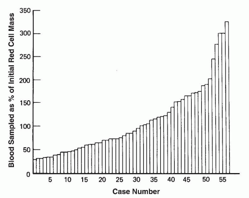
FIGURE 43.3 Cumulative blood losses through sampling in premature infants, expressed as a percentage of their erythrocyte mass at birth. Infants were studied during the first 6 weeks of life, and each vertical bar represents a single infant.


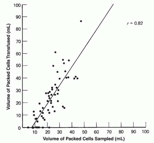
FIGURE 43.4 Relation during the first 6 weeks of life between the cumulative volumes of blood sampled from and transfused into 57 premature infants who had birth weights less than 1,500 g. Volumes represent milliliters of packed erythrocytes (r, correlation coefficient).

There is a strong correlation between the volume of blood sampled and that transfused (Fig. 43.4), suggesting that much of the erythrocyte transfusion requirements of ill, premature infants is a direct consequence of blood loss for essential laboratory monitoring (76). Autologous cord blood collection, fractionation, and reinfusion gained increasing interest (78). However, the clinical significance is unclear due to insufficient volumes to cover multiple transfusions (79) as well as other problems such as clotting, hemolysis, bacterial contamination, and high costs. Widness et al. reported a clinically meaningful (46%) reduction in packed RBC transfusions in a cohort of extremely LBW with the use of a bedside point-of-care blood gas and chemistry monitor (80) and with the use of in-line blood monitoring (81).


Treatment of Anemia Secondary to Blood Loss

The treatment of anemia secondary to blood loss depends on the degree of anemia and the acuteness of the hemorrhage. For acute hemorrhage, the following measures must be employed:


  • If the infant is pale and limp at birth, clear the airway and assist ventilation.


  • Obtain venous access immediately usually by insertion of a low umbilical venous line. Blood specimens for complete blood count and crossmatching should be drawn. If an umbilical line is placed, it may be possible to measure a central venous pressure that will be low.


  • As soon as it is apparent that pallor is a result of hypovolemic shock or profound anemia and not a consequence of asphyxia, administer 15 to 20 mL/kg, depending on ready availability, of O Rh-negative packed RBCs or an isotonic crystalloid solution such as normal saline in the interim. Albumin is no longer recommended for volume replacement (82). Infants with acute external blood loss usually demonstrate dramatic improvement after such a procedure. Infants with massive internal hemorrhages show less evidence of response.


  • A further infusion of 10 to 20 mL/kg of whole blood or reconstituted whole blood (packed RBCs plus fresh frozen plasma [FFP]) may be given if clinically indicated.

After resuscitation and stabilization, the cause of the blood loss should be sought. Examine the placenta and cord for evidence of abnormalities. Obtain a blood sample from the mother for the detection of a fetomaternal hemorrhage. The infant who is mildly anemic at birth as a consequence of chronic blood loss but hemodynamically stable generally does not require a transfusion.

For anemic infants still requiring intensive support, especially mechanical ventilation, it is probably appropriate to treat anemia with blood transfusion. The decision to transfuse should be based on the hemoglobin level and on the clinical condition of the baby.


Hemolytic Anemia

Anemia as a consequence of a hemolytic process is common in the newborn period and has multiple causes that may be classified as immune (alloimmune or autoimmune) or nonimmune (membranopathies, enzymopathies, and hemoglobinopathies). Hemolysis is almost always associated with elevation of the serum indirect bilirubin value to 170 µmol/L (10 mg/dL) or greater. Most commonly, a hemolytic process is first detected during the investigation of jaundice occurring during the first week of life.


Alloimmune Hemolytic Disease

Hemolytic disease in the newborn as a consequence of alloimmunization of the mother is caused by the passage of fetal erythrocytes into the maternal circulation, where they stimulate the production of antibody. Antibodies of the IgG class cross the placenta to the fetal circulation, attach to antigenic sites on the surface of the erythrocyte, and cause its rapid removal by the fetal reticuloendothelial system. The incidence and clinical manifestations of alloimmunization depend on the type of blood group incompatibility between the mother and fetus. Hemolytic disease due to Rh incompatibility became less frequent than ABO incompatibility since prevention by anti-D immune globulin injections to women at risk; however, it is still considered the prototype of alloimmune hemolytic anemia of the newborn.

Rh Hemolytic Disease The rhesus (Rh) blood group system includes the C/c, E/e, and D antigens. Among the various Rh antigens, Rh(D) is the most common cause of hemolytic disease of the newborn and is the focus of this discussion. The incidence of Rh incompatibility in a population depends, in large part, on the prevalence of the Rh-negative antigens. The prevalence of the Rh-negative genotype ranges from approximately zero in Japanese, Chinese, and North American Indian populations to 5.5% among African Americans and 15% among American Caucasians (83,84,85). Among Caucasian women, it has been estimated that in approximately 9% of all pregnancies, an Rh-negative woman carries an Rh-positive fetus. In 6% of pregnancies at risk, alloimmunization of the mother occurs if there is no immunoprophylaxis.

The severity of Rh hemolytic disease varies greatly from infant to infant. It is estimated that, without antenatal diagnosis and treatment, the perinatal mortality in this disease would be approximately 17.5%, with stillbirths accounting for about 14% of deaths (86). The degree of hemolytic disease tends to be more severe in subsequent pregnancies than in the initial one in which sensitization occurred.

Pathogenesis Entry of as little as 0.05 to 0.1 mL of fetal blood into the maternal circulation will produce immunization. Transplacental hemorrhage, and subsequently Rh immunization, tends to occur more frequently in pregnancies that have been complicated by toxemia, CS, or manual removal of the placenta. It is estimated that 1% of Rh-negative women develop antibodies as a consequence of these transplacental hemorrhages before the delivery of their first child. An additional 7.5% manifest evidence of sensitization within
6 months of the delivery of their first child, and another 7.5% show no evidence of immunization 6 months after delivery but develop antibodies during their next pregnancy if their fetus is Rh positive, presumably as a consequence of a sensitization during the first pregnancy.

Destruction of Fetal Erythrocytes by Anti-D The transfer of antibody from the mother into the fetal circulation is responsible for the clinical manifestations of the hemolytic process. The erythrocyte, coated with an antibody of the IgG class, is removed primarily in the spleen of the fetus. The rate of destruction is generally proportional to the amount of antibody on the cell. At very high levels of antibody, the cell may be destroyed by intravascular hemolysis and splenic sequestration.

Before birth, the chief danger of excess erythrocyte destruction is profound anemia. After birth, the infant is primarily at risk from the products of erythrocyte breakdown, such as bilirubin. In utero, the infant responds to the increased breakdown of cells by increasing the rate of erythrocyte production. This is reflected by an elevated reticulocyte count and the presence of nucleated erythrocytes in the peripheral circulation. This accelerated demand for erythrocytes results in active erythropoiesis in nonmarrow sites such as the liver, spleen, and lung. A major portion of the hepatosplenomegaly observed in infants with hemolytic disease is a result of this extramedullary erythropoiesis. In infants with severe Rh incompatibility, the liver and pancreas exhibit pathologic changes. Islet cell hyperplasia can be observed in the pancreas, and focal cellular necrosis with cholestasis may be seen in the liver.

The most severely affected infants manifest anasarca with life-threatening pleural effusions and ascites, with resulting hydrops fetalis. In addition to anemia, intrauterine hypoxia, hypoproteinemia, and a low oncotic pressure of the plasma play a role in the development of hydrops. Hydrops fetalis has been observed in a variety of other conditions (Table 43.3).

Clinical Manifestations The main signs of hemolytic disease in the newborn are jaundice, pallor, and hepatosplenomegaly. Jaundice usually becomes evident during the first 24 hours after birth (frequently within the first 4 to 5 hours), and peaks by the 3rd or 4th day. Jaundice and the metabolism of bilirubin are extensively discussed in Chapter 32.

The degree of anemia reflects the severity of the hemolytic process and the infant’s capacity to respond to it with increased erythrocyte production. Late anemia may develop in infants with Rh alloimmunization. This is observed in two clinical settings. In one, the infant does not become sufficiently jaundiced in the initial newborn period to require exchange transfusion. This is more common since the advent of phototherapy, which may control the jaundice even though the hemolytic process continues. Continued erythrocyte destruction occurs, and the infant can develop severe or fatal anemia between 7 and 21 days of life. The other, more common situation occurs in infants who have had exchange transfusions. In these infants, a gradual fall in hemoglobin may be observed, with hemoglobin values of 5 to 6 g/dL being reached by 4 to 6 weeks of life. This results from the continued presence of IgG anti-D in the neonatal circulation with destruction of residual and newly formed Rh-positive cells. Spontaneous correction can be expected by 6 to 8 weeks of age.

Petechiae and purpura may be observed in infants with severe anemia as a result of thrombocytopenia and a disturbance in the intrinsic system of coagulation. This disturbance may result from DIC or from hepatic dysfunction with consequent inability to synthesize the vitamin K-dependent factors (87).

Laboratory Findings Decreased hemoglobin concentration, increased reticulocyte count, and increased numbers of nucleated erythrocytes in the peripheral blood reflect the presence of the hemolytic process. Hemoglobin values that are less than 13 g/dL in the cord blood should be regarded as abnormal. The reticulocyte count is usually greater than 6% and may reach 30% to 40%. In the peripheral blood, nucleated erythrocytes may be observed in addition to some degree of polychromasia and anisocytosis. Spherocytes are not abundant (compared to healthy infants) in patients with Rh hemolytic disease.


TABLE 43.3 Some Causes of Hydrops Fetalis


Severe chronic anemia in utero


Parvovirus infection


Erythroblastosis fetalis


Homozygous alpha-thalassemia


Chronic fetomaternal transfusion or twin-to-twin transfusion


Inherited bone marrow failure syndromes with severe anemia (e.g., Diamond-Blackfan anemia and CDA) (see Table 43.6)


Glucose-6-phosphate dehydrogenase deficiency (rarely)


Cardiac failure


Severe congenital cardiomyopathy or myocarditis


Premature closure of the foramen ovale


Large arteriovenous malformation (e.g., hemangioma)


Intrauterine arrhythmias


Hypoproteinemia


Renal disease


Congenital nephrosis


Renal vein thrombosis


Congenital hepatitis


Intrauterine infections


Syphilis


Toxoplasmosis


Cytomegalovirus


Miscellaneous


Maternal diabetes mellitus


Parabiotic syndrome of multiple pregnancies


Sublethal umbilical or chorionic vein thrombosis


Fetal neuroblastoma


Cystic adenomatoid malformation of the lung


Pulmonary lymphangiectasia


Chorioangioma of the placenta


Transient leukemia of Down syndrome


The erythrocytes of infants with Rh hemolytic disease test positive on the direct antibody test (also called Coombs test), indicating the presence of maternal IgG on the infant’s erythrocyte surface. An eluate obtained from cord RBCs, if available, should confirm the presence of anti-D. Of note, affected infants may type as Rh negative at birth as a result of maternal anti-D blocking the Rh antigen on cord or neonatal RBCs reacting with the Rh typing reagent.

Prevention The prevention of Rh hemolytic disease focuses primarily on the administration of anti-D immune globulin (such as the human immunoglobulin concentrate of anti-D, WinRho) to the mother in the antenatal period, usually at the 28th week of gestation (or in some countries at the 34th week of gestation), and after delivery, abortion, and invasive procedures. For immunized women, the focus is on the prevention of fetal hydrops and death by intrauterine transfusion until safe delivery can be assured, usually at the 36th week of gestation (88,89).

Prevention of Rh sensitization with anti-D administration is both effective and cost saving (90,91) (see Chapter 32).

Pregnant women at risk for delivery of an infant with Rh(D) hemolytic disease (i.e., Rh(D)-negative mothers with Rh(D)-positive partners and anti-D antibodies in the serum) must be followed carefully during pregnancy. Stillbirths in this setting may be
prevented by intrauterine transfusions or by the early termination of pregnancy.

Intrauterine Diagnosis and Treatment The severity of hemolysis in the at-risk fetus can be estimated by measuring the amniotic fluid bilirubin levels by spectrophotometry, which is more accurate than the maternal antibody titer (92,93). Women who should be considered for amniocentesis are those with a history of hemolytic disease in previous infants and those whose anti-D titers are greater than 0.125 by the indirect antibody test (also called indirect Coombs test), remembering that titers may vary from laboratory to laboratory. Peak velocity of the middle cerebral artery flow by Doppler ultrasound is a less invasive method of estimating fetal anemia and has now become a standard practice (94). Fetal Rh(D) typing using DNA extracted from amniotic fluid cells can identify the fetus at risk (95) and thus avoid the need for fetal blood sampling to determine the Rh status of the fetus. Quantitative real-time polymerase chain reaction (PCR) and other advanced DNA technologies can be used to determine paternal Rh(D) zygosity in cell-free fetal DNA in maternal plasma (96), which are more accurate methods than serology. At-risk pregnant women determined to be carrying an Rhnegative fetus can then be referred back to the care of local physicians/obstetricians for routine antenatal monitoring.

The treatment of severe hemolytic disease in utero is direct intrauterine transfusion through the umbilical vein at a frequency that may be dictated by middle cerebral artery velocity (97). With intrauterine diagnosis, most fetuses with severe Rh disease can be salvaged. Those who reach 36 weeks of gestation can be induced prematurely, and the survival rate is expected to be the same as for a full-term infant with Rh disease. For those with more severe disease who would not survive to 36 weeks of gestation, intrauterine transfusion beginning at 20 to 22 weeks of gestation results in salvage of as many as 87% of patients (98).

Blood for intrauterine transfusion should be O negative, fresh (<7 days old), and CMV safe. Before transfusion, a fetal hemoglobin level should be obtained, a direct antiglobulin test performed, and the fetal RBC antigen status confirmed. The amount of blood to be transfused is then calculated and the fetal hemoglobin level rechecked at the midway point of the placental transfusion; additional blood is then transfused as appropriate given the final target hemoglobin level (99). For the fetus with hydrops fetalis and severe anemia, the transfusion may be split over a few days with the first transfusion calculated to increase the hemoglobin level to 100 g/L.

Management of Affected Infants Newborns with Rh hemolytic disease are at risk of death or neurologic damage, primarily from anemia or hyperbilirubinemia and kernicterus. In severely affected cases, allogenic blood should be made available on hand at the time of delivery to transfuse immediately if needed. Also, if hydrops is present, platelets should be available for transfusion because splenic platelet sequestration can cause significant thrombocytopenia (platelet count <50,000/µL). As soon as the infant has been delivered and respirations have been established, the infant should be carefully examined and an assessment of pallor, organomegaly, petechiae, edema, ascites, respiratory rate, pulse, and blood pressure made in an attempt to judge the severity of the hemolytic process. Cord blood samples should be analyzed for hemoglobin concentration, reticulocyte count, nucleated erythrocyte count, blood type, direct antiglobulin test, and conjugated and unconjugated serum bilirubin concentration (100).

In the infant with a positive DAT (Coombs test), the major initial decision is whether to perform an immediate exchange transfusion or to observe the infant’s clinical status. In many instances, the outcome of previous pregnancies and the result of amniocentesis during the current pregnancy provide valuable information about what to anticipate in the way of severity. Except for the obviously pale or edematous child, the decision to perform an immediate exchange transfusion is based on laboratory findings. It has been suggested that a cord hemoglobin less than 11.0 g/dL or a cord bilirubin higher than 4.5 mg/dL is an indication for immediate exchange transfusion (98). The value of immediate transfusion is that it is more efficient to remove a “potential bilirubin load” (i.e., antibody-coated erythrocytes) than to allow hemolysis to occur, with distribution of bilirubin throughout the tissues, from which it is removed with greater difficulty by exchange transfusion.

For less severely affected infants, a double volume exchange transfusion is indicated if it becomes apparent that the rate of bilirubin rise is such that total indirect bilirubin will exceed 20 mg/dL (330 µmol/L) in otherwise healthy full-term infants (101). The physician needs to use lower maximal bilirubin levels in sick or premature infants (see Chapter 32).

Exchange transfusion is commonly associated with morbidities (102). Intravenous immunoglobulin (IVIG) has been utilized in the setting of Rh(D) disease to reduce the need for exchange transfusion and has been recommended in infants with severe hyperbilirubinemia, positive antiglobulin test and hemolytic disease of the newborn (103,104). These recommendations were based on a previous Cochrane review (105). The review included a total of 189 patients with either rhesus or ABO incompatibility in three randomized or quasi-randomized studies (106,107,108). The investigators noted that both the use of exchange transfusions and the mean number of exchange transfusions used were significantly decreased in the groups treated with either 500 mg/kg or 1 g/kg of IVIG (105). In a review by Gottstein and Cooke, the duration of phototherapy and of hospitalization was also significantly reduced (109). Unfortunately, these studies analyzed were small and differed in their inclusion criteria (105). Importantly, a recent larger, randomized, double-blind, placebo-controlled trial of 80 infants with rhesus hemolytic disease showed no difference in the rate of exchange transfusion between patients who were treated with IVIG (0.75 g/kg) and placebo (5% glucose) (110). This may indicate no benefit of IVIG or a need for different dosage and schedule.

ABO Hemolytic Disease ABO hemolytic disease results from the action of maternal anti-A or anti-B antibodies on fetal erythrocytes of the corresponding blood group. Although approximately 20% of all pregnancies are associated with ABO incompatibility between the mother and the fetus, the incidence of severe hemolytic disease is low. Anti-A and anti-B antibodies are found in the IgA, IgM, and IgG fractions of plasma. Only the IgG antibodies cross the placenta and are responsible for the production of disease. These naturally occurring antibodies result from continuous immune stimulation by A and B substances that exist in foods and gram-negative bacteria. Anti-A and anti-B titers are low or absent in most pregnancies. Some women develop high anti-A or anti-B titer, possibly due to repeated, asymptomatic bacterial infections. ABO hemolytic disease tends to occur in the newborns of mothers with high levels of IgG anti-A or anti-B titer.

The fewer A or B antigenic sites present on the erythrocytes of the newborn is responsible for the weakly reactive DAT in infants with ABO hemolytic disease and also explains why the erythrocyte life span in ABO hemolytic disease is only slightly shortened. Adult group A erythrocytes transfused into a baby with maternally acquired anti-A antibody are rapidly destroyed and may produce severe intravascular hemolysis. For this reason, group O RBCs are used for transfusion in support of infants with severe ABO hemolytic disease of the newborn. Another factor that explains the lower severity of ABO hemolytic disease in neonates is the fact that fetal and newborn blood contains soluble blood group A and B substances that neutralize transplacentally acquired antibodies.

The diagnosis of ABO hemolytic disease is often difficult and may first require the exclusion of other causes of hyperbilirubinemia. Usually, the diagnosis is suspected when hyperbilirubinemia appears in the group A or B baby of a blood group O mother. The disease is more common and more severe in infants of African descent. Jaundice appearing in the first 24 hours is particularly
characteristic of ABO hemolytic disease. Anemia may be mild or may not be present. Evidence of alloimmunization is difficult to interpret because the DAT may be negative or only weakly positive in up to 40% of cases (111). The diagnosis of ABO hemolytic disease is supported by the finding of increased numbers of spherocytes and increased reticulocyte count. This is in contrast to Rh(D) hemolytic disease of the newborn, which typically presents with anemia, fewer spherocytes, and only minimal increase, if any, in nucleated RBCs (112). The diagnosis of ABO hemolytic disease is supported by the following tests and findings:


  • Indirect (unconjugated) hyperbilirubinemia


  • Jaundice appearing during the first 24 hours of life


  • A group A or B baby of a group O mother


  • Increased numbers of spherocytes in the blood


  • Increased erythrocyte production evidenced by reticulocytosis


  • The presence of IgG, anti-A, or anti-B in cord plasma or serum

Treatment is directed primarily toward the prevention of hyperbilirubinemia. Phototherapy reduces the need for exchange transfusion (113). The usage of IVIG has been shown mixed results (see above). A prospective study of 242 infants with ABO isoimmunization showed that early phototherapy significantly reduces the serum bilirubin level in the first 48 hours; however, no other clinical benefit to this practice was found (114).


Hemolytic Disease Resulting from Minor Blood Group Incompatibility

Hemolytic disease related to maternal erythrocyte antibodies other than anti-D, anti-A, or anti-B is relatively uncommon. In one study, minor group antibodies were found in 121 (0.08%) of 142,800 pregnant women (115). The principal antibodies found were anti-E, anti-c, and anti-K (Kell). In a report of 30 cases of hemolytic disease of the newborn, the following antibodies were responsible: 14 anti-c, 9 anti-E, 2 anti-Ce, 2 anti-K, 1 anti-Fya, 1 anti-Jka, and 1 anti-U (116). Anti-K (Kell) antibodies may cause severe hemolytic disease in newborn infants, including hydrops fetalis and neonatal death. Of interest, the severity of hemolytic disease of the newborn does not correlate with the anti-K titre (117). These antibodies are known to inhibit fetal erythropoiesis. It is interesting that Kell hemolytic disease of newborn may present with trilineage pancytopenia (118). Based on the above, it is recommended that all pregnant women should have their blood screened for antibodies at least once during pregnancy before week 34 of gestation.


Hemolysis Due to Inherited Defects of the Erythrocyte

Inherited defects of erythrocyte metabolism, membrane function, and hemoglobin synthesis all may manifest themselves in the newborn period. Defects of erythrocyte metabolism include glucose-6-phosphate dehydrogenase (G6PD) deficiency and less common disorders such as pyruvate kinase deficiency.

Glucose-6-Phosphate Dehydrogenase Deficiency The major function of the erythrocyte is the delivery of oxygen to the tissues. The cell is constantly exposed to oxygen, and the erythrocyte membrane and cytoplasm are subjected to oxidative damage. Oxidation causes the formation of precipitates of denatured hemoglobin (Heinz bodies), which appear to be associated with a shortened erythrocyte life span in vivo (Fig. 43.5). The erythrocyte has a metabolic system that can prevent oxidative damage (Fig. 43.6). G6PD is an enzyme in this system; if it is absent, there is a risk of oxidative damage to the erythrocyte, particularly if the cell is stressed by chemicals or drugs capable of oxidative damage (Table 43.4).

G6PD deficiency is a common genetic disorder estimated to affect at least 400 million people worldwide with an original distribution similar to that of malaria. This led to the notion that the deficiency confers resistance to malaria (119). The gene that encodes G6PD is on chromosome X, and the disease is inherited in an X-linked recessive fashion. Approximately 140 different mutations in the G6PD gene have been described. A WHO working group divided the variants of G6PD deficiency into five classes according to clinical manifestations and activity of the enzyme. In classes IV and V, the enzyme activity is normal and increased, respectively (119). The most severe deficiency (class I) occurs rarely and is associated with a chronic hemolytic anemia. With this type of deficiency, the person has mild or moderate anemia throughout life and may have severe hemolytic disease as a newborn. Class II G6PD deficiency affects Asians (e.g., 5.5% of Chinese) and many populations in the Middle East and Mediterranean region (e.g., 0.7% to 3% of Greeks, with the highest incidence of 53% among Kurds). These persons are healthy but are at risk of developing hemolytic anemia when exposed to oxidative drugs or chemicals (e.g., sulfa drugs, fava beans). The anemia may be of sudden onset and severe. In the absence of an oxidative agent such as fava beans or drug exposure, hemoglobin levels are normal, although there is evidence that the erythrocyte life span is slightly reduced. Class III G6PD deficiency affects individuals of African descent (e.g., 10% to 14% of African Americans), in whom the severity of the defect is not usually as great as in those with the other two types. Anemia appears only with drug exposure, is less severe than that of the Asian-Mediterranean type, and tends to be self-limited.


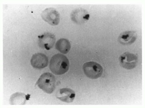
FIGURE 43.5 Heinz bodies in a newborn who developed hemolytic anemia after exposure to naphthalene in mothballs.


Glucose-6-Phosphate Dehydrogenase Deficiency and Neonatal Jaundice

Newborn erythrocytes have a diminished capacity to deal with oxidative stress as a result of lower levels of glutathione peroxidase and
catalase and a relative deficiency of vitamin E. Therefore, newborn infants with G6PD deficiency are at greater risk of developing hemolytic anemia than are adults. G6PD deficiency is associated with an increased incidence of neonatal hyperbilirubinemia, especially if they have class I and class II deficiency. Hyperbilirubinemia in G6PD-deficient newborn males has been reported in Eastern and Western countries (120,121,122,123,124,125,126). It has been reported that male infants of African descent with G6PD deficiency have a significantly higher incidence of hyperbilirubinemia than do controls (127). Although the hyperbilirubinemia is associated with G6PD deficiency, there is a tendency for the jaundice to occur more frequently in particular families and communities, indicating that genetic and environmental factors must influence the incidence of the disease (128).


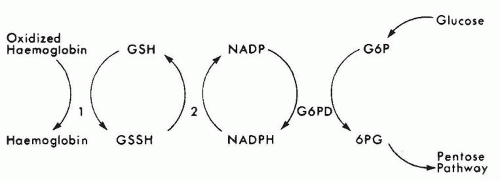
FIGURE 43.6 Protection against oxidative stress in erythrocytes. The erythrocyte is constantly exposed to oxygen; as a result, there is formation of hydrogen peroxide (H2O2), lipid peroxides in the membrane, and oxidized products of hemoglobin such as methemoglobin and Heinz bodies. To prevent the formation of, and to reduce, the levels of these oxidized products, the erythrocyte has a system by which a series of enzyme steps link the metabolism of glucose through the pentose pathway to the reduction of oxidized products (1, glutathione peroxidase; 2, glutathione reductase; G6PD, glucose-6-phosphate dehydrogenase; 6PG, 6-phosphogluconate; G6P, glucose-6-phosphate; GSH, reduced glutathione; GSSH, oxidized glutathione; NADP, nicotinamide adenine dinucleotide phosphate; NADPH, nicotinamide adenine dinucleotide phosphate, reduced).


TABLE 43.4 Drugs, Chemicals, and Other Factors That Cause Glucose-6-Phosphate Dehydrogenase Deficiency Hemolytic Disease


Antimalarials


Primaquine


Pamaquine


Pentaquine


Antipyretics and analgesics


Aspirina


Acetanilide


Acetophenetidin (phenacetin)a


Acetaminophena


Diabetic acidosis


Vitamin K analogs


Infections


Respiratory viruses


Infectious hepatitis


Infectious mononucleosis


Bacterial pneumonia


Nitrofurans


Nitrofurantoin (Furadantin)


Furazolidone (Furoxone)


Furaltadone (Altafur)


Nitrofurazone (Furacin)


Sulfonamides


Sulfanilamide


N2-acetylsulfanilamide


Sulfacetamide (Sulamyd)


Sulfamethoxazole (Gantanol)


Salicylazosulfapyridine (Azulfidine)


Sulfones


Thiazolesulfone


Others


Methylene blue


Toluidine blue


Naphthalene


Phenylhydrazine


Acetylphenylhydrazine


Fava beans


Nalidixic acid (Neggram)


Niridazole (Ambilhar)


Chloramphenicol


aOf doubtful significance.


In this group of patients, jaundice may be severe and may lead to kernicterus (122,129). In most cases, however, hemoglobin and reticulocyte counts are normal, although in some affected infants, the cord blood contains increased bilirubin and decreased hemoglobin levels, suggesting the presence of a mild hemolytic process in utero. There is no evidence of intravascular hemolysis in most of these patients. Slusher et al. (130) demonstrated elevated carboxyhemoglobin (a sensitive indicator of hemolysis) values in Nigerian children with G6PD deficiency and hyperbilirubinemia. Studies of Sephardic-Jewish neonates have yielded opposite results, with no elevation of carboxyhemoglobin values over nonjaundiced G6PD-deficient controls (131,132). The latter observation has been coupled with data suggesting deficient hepatic bilirubin conjugation in neonates with G6PD deficiency (133).

Clinical Manifestations The jaundice that occurs in these infants usually appears to be an accentuation of the physiologic jaundice of newborns with a late peak (around 5 to 6 days), although jaundice may appear in some during the first 24 hours of life. There is seldom evidence of a hemolytic process. Abnormal erythrocyte morphology has been documented during hemolytic episodes in adults, but this is seldom described in newborns. However, a more severe hemolytic anemia may appear, with evidence of abnormal erythrocyte morphology, Heinz bodies in the peripheral blood, and intravascular hemolysis. This may be the result of infection or exposure to drugs or chemicals (e.g., naphthalene in mothballs) (134). However, it is unusual to elicit the latter from the perinatal history.

Diagnosis The presence of unexplained hyperbilirubinemia in an infant of a high-risk population (racial intermarriage must be taken into account) may suggest G6PD deficiency, particularly if jaundice is noticed several days after birth. The enzyme defect can be detected by one of many screening tests, based on changes in either fluorescence or color resulting from the activity of NADPH, or by a direct assay of the G6PD enzyme activity based on spectrophotometric measurement of the reduction of NADP+ to NADPH (135). A false normal screen result can occur in infants with significant hemolysis, which destroys the older, more G6PD-deficient RBCs.

The finding of G6PD deficiency in a jaundiced infant does not in itself prove that the jaundice was caused by the enzyme defect. Other causes of jaundice must be excluded. In a study on Sephardic-Jewish neonates, neonates with both ABO incompatibility and G6PD deficiency showed no increased evidence of hemolysis when compared with neonates with only ABO incompatibility (135). G6PD deficiency is most severe and frequent in male infants because it is a recessive gender-linked disorder. However, females may also be affected because of the high mutant allele frequency and inheritance of two mutant alleles, one from an affected father and one from a carrier mother.

Treatment Treatment is the same as that for hyperbilirubinemia described in Chapter 32. Drugs and chemicals likely to produce hemolytic anemia (Table 43.4) should be avoided by these patients.

Other Glycolytic Enzyme Defects of the Erythrocyte Other abnormalities are far less common than G6PD deficiency and are unusual causes of a hemolytic process during the newborn period. Virtually all of the recognized defects have been associated with jaundice and anemia in neonates. Of this group, erythrocyte pyruvate kinase deficiency appears to be most commonly responsible for a severe hemolytic process during the first week of life. These disorders are usually characterized by the presence of a normal osmotic fragility of unincubated blood, few or no spherocytes in the peripheral blood smear. Unless the infant is a member of a high-risk group (e.g., the Amish population in the United States who typically carry the Arg479His mutation), it is practical to defer diagnosis of these infants until approximately 3 months of life, after it has been established that the hemolytic process observed in the neonatal period is chronic and that the more common reasons for it have been excluded.


Abnormalities of the Erythrocyte Membrane

Hereditary Spherocytosis In approximately 50% of patients with hereditary spherocytosis, a history of neonatal jaundice can be obtained. Hyperbilirubinemia may require exchange transfusions. Untreated hyperbilirubinemia has resulted in kernicterus in infants with hereditary spherocytosis.

Although most patients with hereditary spherocytosis are anemic, the degree of anemia, reticulocytosis, and hyperbilirubinemia is quite variable. The hemoglobin may fall rapidly during the first several weeks of life, reaching values of 5.0 to 7.0 g/dL by 1 month of age. Neither the hematologic values observed during the immediate newborn period, nor the values observed during the first several months of life are reliable indicators of the eventual severity of the disease. Hemoglobin levels of 4.0 to 7.0 g/dL during the first several months of life may subsequently stabilize in the range of 7.0 to 10.0 g/dL; therefore, repeated transfusions are rarely needed except during the course of infections or aplastic crises. Splenectomy, if indicated, should be deferred if possible, until at least 5 or 6 years of age so that the risk of postsplenectomy infections is minimized.

Hereditary spherocytosis can be diagnosed during the newborn period. Examination of the peripheral blood reveals characteristic microspherocytes, and the osmotic fragility of erythrocytes is increased (136). However, the diagnosis may be difficult since healthy neonates have some degree of spherocytes on the smear, and the osmotic fragility of the erythrocytes of normal newborn infants is lower than that of adults’ erythrocytes. If an infant is suspected of having spherocytosis, the osmotic fragility should be compared with normal newborn standards. The osmotic fragility test should be deferred, if possible, until the child can readily spare the necessary blood volume for the test. Family studies are extremely useful in confirming the diagnosis, although an affected parent is identified in only approximately 70% of cases. One group found that a mean corpuscular hemoglobin concentration of ≥36.0 g/dL (360 g/L) in a newborn is 82% sensitive and 98% specific for a diagnosis of hereditary spherocytosis in the newborn (137). Other screening tests for the disorder include acid glycerol lysis time test, cryohemolysis test, and eosin-5′-maleimide-binding test (136,138) (Fig. 43.7). In difficult diagnostic cases, Western blotting of erythrocyte membrane proteins can be used to identify the deficient protein.


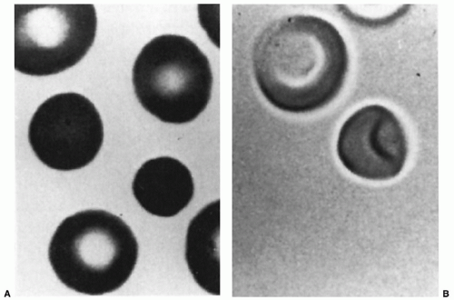
FIGURE 43.7 The erythrocytes of a patient with hereditary spherocytosis, as seen on a stained blood smear (A) and by three-dimensional viewing (B) of glutaraldehyde-fixed cells.

Hereditary Elliptocytosis Hereditary elliptocytosis may manifest in the newborn period as a hemolytic anemia. Only 12% to 15% of newborns with this morphologic abnormality have a shortened erythrocyte survival in later life, but many more appear to have a hemolytic anemia during the first several weeks or months of life. In the newborn period, hereditary elliptocytosis may manifest as hyperbilirubinemia and anemia associated with the presence of fragmented and deformed erythrocytes in the circulation. The erythrocytes of these infants are unusually susceptible to fragmentation after heating. This defect is related to destabilization of the RBC membrane proteins by the increased 2,3-DPG concentration in the newborn. This defect disappears within the first few months of life, and the erythrocytes assume an elliptic appearance, usually with no or minimal evidence of hemolytic disease. This temporary phenomenon resembles a more severe autosomal recessive variant of hereditary elliptocytosis—hereditary pyropoikilocytosis—both clinically and morphologically. The hemolytic anemia in hereditary pyropoikilocytosis, however, does not resolve and may necessitate a splenectomy in early childhood (139).

Most patients with hereditary elliptocytosis do not require treatment, although an exchange transfusion may be required for infants with hyperbilirubinemia. For patients with persistent hemolytic anemia, splenectomy has proved beneficial, but as in hereditary spherocytosis, it should be deferred, if possible, until the patient is about 5 or 6 years of age.

Disorders of Hemoglobin The predominant hemoglobin in the newborn infant is HbF (α2γ2); therefore, it is not surprising that abnormalities in β-chain production (e.g., sickle cell disease,
β-thalassemia) do not manifest during the first month of life. Patients with sickle cell disease are usually found to be anemic by 3 months of age, but cases of jaundice and systemic signs during the neonatal period have been reported (140).

The early identification of infants with severe sickle cell syndromes such as homozygous sickle cell disease and sickle β-thalassemia through universal newborn screening programs is strongly recommended. Morbidity and mortality from sepsis in infants in sickle cell disease can be substantially reduced by early identification of affected infants, enrollment in comprehensive care, and prophylactic treatment with penicillin (141,142). RBC transfusion before sampling as part of a newborn screening program can impair detection of sickle cell disease, and it is therefore recommended that repeat screening be performed in such infants 120 days following the last transfusion (143).

Abnormalities in γ-chain production have been described during the first month of life, although most of these are not clinically significant. Heinz body hemolytic anemia with an unstable γ-chain abnormality has been reported (144,145). Mutations in the α-globin gene can also cause unstable hemoglobin and presentation at the prenatal or neonatal period (146). However, similar to sickle cell anemia, unstable hemoglobin due to mutations in the β-globin gene does not cause anemia during the neonatal period due the low expression of the gene. Cases of microcytic anemia in newborns with reduced γ-chain synthesis have been described as part of a γ-β-thalassemia syndrome (147).

The α-thalassemia group of diseases represents abnormalities in the synthesis of the α-chains of hemoglobin and frequently present in the newborn period. Most are clinically insignificant, although some forms of α-thalassemia that manifest in the newborn period can be serious. Synthesis of these chains is determined by two pairs of α-gene loci (total of four gene copies). A deletion of one or more of these four α-genes results in one of the α-thalassemia disorders. The severity of the disease in the newborn and in the adult depends on the number of genes deleted. If one gene copy is lacking, the patient is hematologically normal except for a slight elevation of Bart hemoglobin (γ4) during the neonatal period. If two copies are absent (i.e., two missing from one chromosome or one missing from each of the two chromosomes), the patient has α-thalassemia trait, which manifests as microcytosis in the newborn (mean corpuscular volume < 95 µm3/cell) and elevation of Bart hemoglobin. If three genes are deleted, the patient has hemoglobin H (HbH; β4) disease, a lifelong hemolytic anemia that manifests in the newborn as jaundice and anemia. If all four genes are absent, the patient can form no α-chains and consequently lack HbA or HbF. The hemoglobin of these infants is predominantly Bart hemoglobin. As a result, the infant is usually born dead or severely hydropic, with death occurring several hours after birth. Treatment with regular transfusions starting at the prenatal period have changed the natural course of the disease, and children can be born alive and survive (148).

In patients with HbH disease, one parent is lacking one α-gene (i.e., a silent carrier) and the other is lacking two α-genes on one chromosome (i.e., α-thalassemia trait). In the patient with homozygous α-thalassemia, each parent is lacking two genes on one chromosome. The α-thalassemia trait that is found in 2% to 10% of individuals of African descent is in the trans form in which one abnormal gene is present on each of the two chromosomes (i.e., -α,-α) and that the cis form (- -,αα) does not occur in such individuals but does occur with various frequencies in populations in Southeast Asia and the Mediterranean region. This is the reason why homozygous α-thalassemia and HbH disease are very rare in individuals of African descent.

The incidence of α-thalassemia can be determined through measurement of levels of Bart hemoglobin in newborns. Silent carriers (i.e., -α,αα) have as much as 2% of Bart hemoglobin. Those with α-thalassemia trait (- -,αα or -α,-α) have 2% to 9% Bart hemoglobin. Those with HbH disease (-α,- -) have up to 20% of Bart hemoglobin as well as hemoglobin H.


Acquired Defects of the Erythrocyte

Infections Infections can induce a hemolytic anemia in the newborn infant who has no underlying inherited defect of erythrocyte metabolism. Such cases manifest with hyperbilirubinemia, which initially may be indirect and subsequently includes direct hyperbilirubinemia. Severe hemolytic anemia infrequently complicates sepsis. One exception is Clostridium welchii sepsis, in which anemia is caused by hemolysis and is associated with microspherocytosis.

Congenital syphilis, toxoplasmosis, cytomegalic inclusion disease, rubella, generalized coxsackie B infections, and Escherichia coli septicemia are examples of infections in which anemia and jaundice are common. Some of the nonhematologic manifestations of these diseases (e.g., rash, chorioretinitis, purpura, and hepatosplenomegaly) are useful in differentiating these disorders from alloimmunization or other primary erythrocyte abnormalities.

Drugs The erythrocytes of the newborn infant, particularly of premature babies, are sensitive to the toxic effects of oxidant drugs. In many respects, the cells of these infants mimic the metabolic abnormalities observed in cells from patients with G6PD deficiency. Severe Heinz body hemolytic anemia (Fig. 43.5), which occurs in infants with severe G6PD deficiency, is also seen in normal newborns exposed to oxidant drugs. The best and most frequent example of this is naphthalene-induced hemolytic anemia caused by exposure to mothballs. This disease is associated with a severe hemolytic anemia, hemoglobinuria, and the presence of fragmented erythrocytes and spherocytes in the circulation. If these are detected, a careful search for exposure to naphthalene or other oxidant drugs (Table 43.4) should be carried out. This increased susceptibility to oxidative damage may be related to the low levels of antioxidants, including glutathione peroxidase, catalase, and vitamin E, in the newborn infant. Idiopathic Heinz body hemolytic anemia probably reflects a similar mechanism, resulting in hyperbilirubinemia and anemia with Heinz bodies present, but the infant has normal G6PD levels, a normal hemoglobin electrophoresis, and a negative heat test for the presence of unstable hemoglobins (149). Evaluation of the family yields no evidence of an inherited disorder, and in the affected neonate, the disorder appears to be self-limited, disappearing within the first several months of life. Vitamin C supplementation may be a factor in the etiology of idiopathic Heinz body hemolytic anemia; however, a randomized controlled trial of vitamin C in small premature newborns was unable to demonstrate hemolysis in supplemented infants (150).


Anemia Caused by Impaired Erythrocyte Production


Diamond-Blackfan Syndrome

Inherited impaired erythrocyte production is a rare cause of anemia in the newborn. The most common cause is Diamond-Blackfan anemia (DBA), which is characterized morphologically by pure red cell aplasia or hypoplasia in the bone marrow. The incidence is about 10 cases per million live births (151). Before the discovery of most DBA genes and their application for clinical management, about half of the patients with DBA were found to be diagnosed by 3 months of age (10% with severe anemia at birth) and 92% within the first year (152). However, with the current genetic knowledge, increasing number of patients are diagnosed later on in childhood or adulthood (153). Mutations in at least 11 autosomal genes (all encode for ribosome proteins) that are transmitted in an autosomal dominant fashion and 1 X-chromosome gene (GATA1) have been identified. The most common mutated gene is RPS19, which is associated with 25% of DBA cases (154).

Neutrophil counts are mildly or moderately reduced in about 30% of the patients. Thrombocytosis is common, but thrombocytopenia is rare, particularly in the neonatal period. Physical anomalies are found in about 40% of patients. Anomalies apparent at birth include microcephaly, cleft palate, eye defects, web neck,
and abnormalities of the thumb including absent or triphalangeal thumbs (155). LBW is seen in about 10% of these patients.

The diagnosis may be established by demonstrating anemia, reticulocytopenia, and a marked decrease in the bone marrow erythroid-to-myeloid ratio in an otherwise healthy newborn. Erythroid-to-myeloid ratios range from 1:6 to more than 1:200. Clonogenic assays of marrow cells show decreased CFU-E and BFU-E progenitors (156). Red cell macrocytosis and high hemoglobin F levels that are typical features of the disease cannot be evaluated in neonates due to the normal high mean corpuscular volume and hemoglobin F levels in this age. The diagnosis is facilitated by demonstrated high level of erythrocyte adenosine deaminase (ADA) activity. Genetic testing frequently helps in establishing a diagnosis and is critical for family counseling.

Most patients require treatment for severe anemia. The initial management includes transfusion as necessary. At 6 to 12 months of age, a prednisone trial is recommended; the majority of the patients will show various degrees of response. Response, reflected by a reticulocytosis and a rise in the hemoglobin level, often occurs within 2 weeks. After the hemoglobin has reached its maximum, the medication is reduced to the lowest dose necessary to maintain a hemoglobin level in the acceptable range. Unfortunately, only about 30% to 40% of the patients remain responsive to prednisone and can be maintained on acceptably low doses. Most patients require a lifelong transfusion program. Spontaneous remissions occur in 15% of the patients and are unpredictable. Hematopoietic stem cell transplantation has been successful as a curative treatment for those who are transfusion dependent, with an overall survival of 72.7% ± 10.7% with a matched sibling donor (157). There is a slightly increased risk of myelodysplasia, leukemia, and solid tumors in individuals with DBA (158).


Inherited Sideroblastic Anemia

Inherited sideroblastic anemias are a heterogeneous group of disorders with defects in mitochondrial iron utilization resulting in accumulation of iron in the mitochondria of RBC precursors. In some, the clinical manifestations are confined to RBCs, while in others, multiple other systems are affected. The iron deposition can be diagnosed by Perl Prussian blue iron staining showing greater than 10% cells with circular or ringed staining around the nucleus. Among the inherited sideroblastic anemias are X-linked sideroblastic anemia (associated with the ALAS2 gene) (159,160), X-linked sideroblastic anemia with ataxia (associated with the ABC7 gene) (161,162), thiamine-responsive megaloblastic anemia (associated with the SLC19A2 gene) (163,164), and Pearson marrow-pancreatic syndrome (associated with the heteroplasmic mitochondrial DNA deletions). The various disorders are listed in Table 43.5. The associated genes encode for proteins that not only might be multifunctional but also promote transport of iron across the mitochondrial membrane and its utilization.


TABLE 43.5 Human Neutrophil Alloantigens (HNA) and Frequencies


Antigen Groups


Carrier Glycoproteins


Antigens


Frequencies


Amerindians


Asians


Africans


Whites


HNA-1


FcyRIIIb (CD16b)


HNA-1a


83-91


88-91


46-66


57-62


HNA-1b


36-80


51-54


78-84


88-89


HNA-1c


0-1


<1


23-31


5


HNA-2


NB1 glycoprotein (CD177)


HNA-2


Unknown


89-99


98


87-97


HNA-3


GP 70-95 (CLT2 gene)


HNA-3a


Unknown


Unknown


Unknown


89-96


HNA-4


α-M chain of β2-integrin (CD11b)


HNA-4a


>99


Unknown


Unknown


99


HNA-5


αL chain of β2-integrin (CD11a)


HNA-5a


79-97


81


88


86-92


Modified from Bux J. Human neutrophil alloantigens. Vox Sang 2008;94:277, with permission.


Although some features may suggest sideroblastic anemia (e.g., unexplained microcytic anemia), diagnosis is usually established only after the bone marrow is evaluated. Defining the specific sideroblastic anemia syndrome is facilitated by the presence or absence of nonhematologic manifestations and can be confirmed by molecular testing. Treatment depends on the specific syndrome. Patients with X-linked sideroblastic anemia respond to pyridoxine, and patients with thiamine-responsive megaloblastic anemia respond to pharmacologic doses of thiamine. In the other types of inherited sideroblastic anemia, RBC transfusions are the mainstay of treatment. Hematopoietic stem cell transplantation is curative (165,166). In Pearson syndrome, the cytopenia improves with age. It is broadly accepted that hematopoietic stem cell transplantation may not be required in this disease; however, improvement in hematologic and nonhematologic manifestations has been associated with hematopoietic stem cell transplantation in one patient (167).


Congenital Dyserythropoietic Anemia

Congenital dyserythropoietic anemias (CDA) are inherited disorders with ineffective erythropoiesis and striking morphologic dyserythropoiesis. Several types of CDA exist that differ in marrow morphology, serologic findings, and inheritance patterns. CDA types I and II are autosomal recessive. CDA types III and IV are autosomal dominant. The various types and genes are summarized in Table 43.6 and recently reviewed by Iolascon et al. (168).

The diagnosis may be suspected when chronic anemia, splenomegaly, hyperbilirubinemia, high LDH, and low reticulocytes are found. However, the diagnosis is usually established only after a morphologic examination of the bone marrow is done, which is sometimes further assisted by electron microscope. Most patients with CDA are diagnosed in late childhood or adolescence; however, some patients present in the neonatal period with variable splenomegaly, jaundice, and normocytic or macrocytic anemia. Cases of hydrops fetalis have also been described (169,170).

Most patients have mild anemia and do not require chronic therapy. In cases with severe anemia, a chronic RBC transfusion program, splenectomy, or hematopoietic stem cell transplantation should be considered. Later in life, patients can develop iron overload necessitating iron chelation as a result of ineffective erythropoiesis and multiple transfusions.


Parvovirus Infection

Parvovirus B19 is a single-stranded DNA virus, which can bind directly to the P antigen on RBCs. The expression of P antigen on erythroid and placental tissues may mediate transplacental fetal erythroid infection; however, there is accumulating evidence for the existence of a putative cellular coreceptor for efficient entry of parvovirus B19 into human cells (171). The virus may cause apoptosis of erythroid cells that is possibly induced by its nonstructural (NS1) protein.


Fetal parvovirus B19 infection may cause anemia, abortion, or stillbirth or be asymptomatic. Hydrops fetalis with lack of congenital malformations is the typical clinical presentation (172). Approximately 18% of cases of nonimmune hydrops fetalis are caused by parvovirus infection (173). Hydrops fetalis usually manifests during the second trimester of pregnancy and reflects a profound reduction of erythrocyte production in the fetal liver and marrow. This may result in severe anemia, high-output cardiac failure, and death. Myocarditis may also occur (174).

Bone marrow aspirates show a paucity of RBC precursors, with occasional giant pronormoblasts with large eosinophilic nuclear inclusion bodies, cytoplasmic vacuolization, and, occasionally, “dog-ear” projections (175). Diagnosis of parvovirus infection in older immunocompetent children can be made by positive IgM serology. However, this test is unreliable in infants, and in these cases, the diagnosis should be based on detection of viral DNA in peripheral blood or bone marrow samples by dot-blot hybridization or PCR (176). During pregnancy, maternal serum IgM is falsely negative in about 6% of the cases, and PCR is falsely negative in about 4%. Therefore, diagnosis of parvovirus infection in the prenatal period is ideally based on concurrent analysis of B19 IgM and DNA analysis (177). Viral studies of amniotic fluid or fetal blood may also be helpful in making the diagnosis before birth (178).


TABLE 43.6 Summary of the Inherited Bone Marrow Failure Syndromes and Genes


Disorder


Gene


Gene Locus


Inheritance


Reference


Fanconi anemia


FANCA


16q24.3


AR


(663)


FANCB


Xp22.31


XLR


(664)


FANCC


9q22.3


AR


(665)


FANCD1/BRCA2


13q12.3


AR


(666)


FANCD2


3p25.3


AR


(667)


FANCE


6p21.3


AR


(668)


FANCF


11p15


AR


(669)


FANCG/XRCC9


9p13


AR


(670)


FANCI


15q25-q26


AR


(671)


FANCJ/BRIP1


17q22


AR


(671)


FANCL/PHF9


2p16.1


AR


(672)


FANCM


14q21.3


AR


(673)


FANCN/PALB2


16p12


AR


(674)


FANCP/SLX4


16p13.3


AR


(675)


FANCO/RAD51C


17q22


AR


(676)


XRCC2


7q36.1


AR


(677)


ERCC1


19q13.32


AR


(678)


ERCC4


16p13.12


AR


(679)


Shwachman-Diamond syndrome


SBDS


7q11


AR


(680)


Dyskeratosis congenita


DKC1


Xq28


XLR


(681)


TINF2


14q12


AD


(682)


TERC


3q21-q28


AD


(683)


TERT


5p15.33


AD


(684)


NOP10


15q14-q15


AR


(685)


NHP2


5q35.3


AR


(686)


TCAB1


17p13


AR


(687)


RTEL1


20q13.3


AR


(688,689)


CTC1


17p13.1


AR


(690)


Congenital amegakaryocytic thrombocytopenia


MPL


1p34


AR


(410)


Reticular dysgenesis


AK2


1p34


AR


(274,691)


Pearson syndrome


mDNA


Mitochondrial DNA


Maternal


(446)


Lig4-associated aplastic anemia


LIG4


1q22-q34


(692)


Familial aplasia and myelodysplasia


SRP72


4q11


AD


(693)


Familial myelodysplastic syndrome (MonoMac syndrome, Emberger syndrome)


GATA2


3q21.3


AD


(694,695)


Rothmund-Thomson syndrome


RECQL4


8q24.3


AR


(696)


Diamond-Blackfan anemia


RPL5


1p22.1


AD


(697)


RPL11


1p36.1-p35


AD


(697)


RPL35A


3q29


AD


(698)


RPS7


2p25


AD


(699)


RPS17


15q AD (699)


RPS19


19q13.2


AD


(700)


RPS24


10q22


AD


(701)


RPS26


12q13


AD


(699)


RPS10


6p21.31


AD


(702)


RPL15


3p24.2


AD


(703)


RPL26


17p13


NNN


(704)


GATA1


Xp11.23


XL


(705)


Inherited sideroblastic anemia


ALAS2


Xp11.21


XL


(160)


ABC7


Xq13.1-q13.3


XL


(162)


SLC19A2


1q23.3


AR


(164)


GLRX5


14q32.13


AR


(706)


PUS1


2p16.1


AR


(707)


SLC25A38


3p22.1


AR


(708)


YARS2


12p11.21


AR


(709)


Congenital dyserythropoietic anemia type I


CDAN1


15q15


AR


(710)


C15ORF41


15q14


AR


(711)


Congenital dyserythropoietic anemia type II


SEC23B


20p11.2


AR


(712,713)


Congenital dyserythropoietic anemia type III


KIF23


15q23


AD


(714)


Congenital dyserythropoietic anemia—unclassified


KLF1


19p13.12-p13.13


AR


(715)


Kostmann/severe congenital neutropenia


ELA2


19p13.3


AD


(716)


HAX1


1q21.3


AR


(252)


GFI1


1p22


AD


(717)


WASP


Xp11.23-p11.22


XLR


(718)


G6PC3


17q21


AR


(253,719)


VPS45


1q21.2


AR


(720)


Cyclic neutropenia


ELA2


19p13.3


AD


(721)


WHIM syndrome


CXCR4


2q21


AD


(271)


Glycogen storage diseases Ib


G6PT (SLC37A4)


11q23


AR


(722)


Barth syndrome


TAZ


Xq28


XL


(723)


Poikiloderma with neutropenia


USB1


16q13


AR


(724)


Dominant intermediate Charcot-Marie-Tooth disease


DNM2


19p12-13.2


AD


(725)


Thrombocytopenia-absent radii syndrome


RBM8A


1q21.1


AR


(726)


Familial autosomal dominant nonsyndromic thrombocytopenia


MASTL


10p11-12


AD


(404)


ACBD5


10p12.1


AD


(405)


ANKRD26


10q22.1


AD


(727)


CYCS


7p15.3


AD


(728)


Thrombocytopenia with dyserythropoiesis


GATA1


Xp11.23


XL


(411)


Thrombocytopenia with associated myeloid malignancies


CBFA2/RUNX1


21q22.1-22.2


AD


(409)


X-linked thrombocytopenia


WASP


Xp11.23


XL


(718)


Thrombocytopenia with radioulnar synostosis


HOXA11


7p15-p14.2


AD


(412)


Mediterranean platelet disorder


GP1BA


17pter-p12


AD


(729)


Gray platelet syndrome (macrothrombocytopenia)


NBEAL2


3p21.31


AR


(402)


Epstein/Fechtner/Sebastian/May-Hegglin/Alport syndrome (macrothrombocytopenia)


MYH9


22q11-q13


AD


(730)


Familial macrothrombocytopenia


FLNA


Xq28


XL


(731)


TUBB1


20q13.32


AD


(702)


ITGA2(ITGA2B)


5q11.2


AD


(732)


ITGB3


17q21.32


AD


(733)


ABCG5


2p21


AR


(734)


ABCG8


2p21


AR


(734)


Modified from Dror InTech 2011.


Cordocentesis allows fetal blood sampling for hemoglobin measurement and PCR testing for parvovirus. During the procedure, the anemia can be corrected by intravenous RBC transfusion, which can lower the mortality rate from approximately 50% to 18% (179). Postnatal monitoring of the hemoglobin level and judicious transfusions results in resolution of the condition in the majority of cases with a good long-term outcome. Although rare, failure of red cell production has sometimes continued after birth (180).


Vitamin Deficiencies

Specific vitamin deficiencies may cause anemia in newborn infants because of decreased erythrocyte production, increased erythrocyte destruction, or a combination of these two mechanisms.

Nutritional anemia secondary to iron deficiency is uncommon in neonates (181). Studies by Seip and Halvorsen (182) indicate that stainable iron disappears from bone marrow aspirates by 12 weeks of age in premature infants and by 20 to 24 weeks in term infants, and it is only after this period that iron deficiency manifests in infants who do not receive supplemental iron. To prevent the development of iron deficiency, premature infants should receive supplemental iron from no later than 2 months of age.

Although most premature infants have low serum folate levels by 1 to 3 months of age, they rarely manifest evidence of a megaloblastic anemia. Cases of megaloblastic anemia resulting from folate deficiency typically involve infants receiving goat’s milk or phenytoin therapy and infants with chronic diarrhea or infection. Folic acid deficiency is a rare disorder of later infancy. It is noteworthy that starting folic acid around the time of conception has been shown to reduce the risk of neural tube defects (anencephaly, spina bifida, encephalocele). For example, a large randomized controlled trial of folic acid supplementation around the time of conception showed a significant reduction of 72% in the incidence of neural tube defects compared to a mixture of seven other vitamin (A, D, B1, B2, B6, C, and nicotinamide) or no supplements (183).

Anemia due to vitamin B12 deficiency is very rare in neonates but can appear in infants of mothers with severe B12 deficiency due to vegan diet without supplementation (184) or pernicious anemia (185). Megaloblastic anemia can also appear in cases with inborn defects in B12 absorption or metabolism. Defects in B12 absorption include hereditary intrinsic factor deficiency due to HIF gene mutations or Imerslünd-Grasbeck disease due to mutations in one of the two subunits of the intrinsic factor receptor: cubilin (CUBN gene) or amnionless (AMN gene). Defects in B12 metabolism include mutations in the cobalamin (cblC, cblD, cblF, and cblJ). Similar to folic acid, it has been shown that low levels of B12 can cause neural tube defects (186).

A syndrome that was attributed to vitamin E deficiency anemia in newborn infants was first described by Hassan et al. (187) and typically occurred in premature infants (birth weight < 1,500 g) at 6 weeks of age. Characteristic features included anemia, reticulocytosis, thrombocytosis, and shortened erythrocyte survival (188). Damage to the erythrocyte membrane by lipid peroxides, formed naturally during peroxidation of polyunsaturated fatty acids (PUFA) in the erythrocyte membrane, was thought to be the mechanism of the anemia. Vitamin E, a biologic antioxidant, inactivates lipid peroxides and protects against erythrocyte damage. Preterm infants have low body fat tissue and consequently reduced fat-soluble vitamin stores (189). The anemia may be exaggerated by increasing the PUFA content of the diet, particularly if infants are also given supplemental iron, a catalyst in the auto-oxidation of PUFA to free radicals, and lipid peroxides (190). After the association with the PUFA content of the diet, iron supplementation, and the vitamin E requirement of premature infants was recognized, the PUFA content of infant formulas was reduced. Other groups suggested administration of vitamin E while providing PUFA-enriched formula (191). Vitamin E deficiency as described above has become rare in premature infants, and there is no evidence that routine vitamin E supplementation is of benefit in preventing the anemia of prematurity (192,193).


Evaluation of Anemia in Neonates

Anemia is characterized by an abnormally low erythrocyte mass; in clinical practice, the hemoglobin concentration is assumed to reflect the circulating erythrocyte mass, and an abnormally low hemoglobin concentration defines the anemic state. After diagnosis, causes of anemia are traditionally considered under the pathophysiologic categories of decreased erythrocyte production, increased destruction (i.e., hemolysis), blood loss, and splenomegaly. In newborn infants, this classic approach to anemia is complicated by a hemoglobin concentration that undergoes constant physiologic change during the first few weeks of life. The site of blood sampling, quantity of blood sampled for laboratory monitoring, and the effect of rapid growth can significantly influence the hemoglobin values observed in newborn infants. Failure to consider these factors may lead to errors in diagnosis and result in unnecessary investigation and therapy.


Accuracy of Capillary Hemoglobin Levels

Blanchette and Zipursky (76) compared capillary hemoglobin values obtained by duplicate puncture of the right and left heels of 35 healthy full-term infants. The standard deviation of the difference in hemoglobin concentration of the duplicate samples was 0.8 g/dL; in an infant with a hemoglobin concentration of 17.0 g/dL, 95% of hemoglobin values obtained fall between 15.4 and 18.6 g/dL. It is evident that a difference as large as 1.5 g/dL of hemoglobin in consecutive laboratory reports may reflect the error inherent in the technique of capillary blood sampling in the newborn infant.


Effect of the Sampling Site on Hemoglobin Levels

In newborn infants, hemoglobin levels measured in capillary blood samples may be significantly higher than values obtained from simultaneously collected venous blood samples. Oettinger and Mills (194) found an average difference of 3.6 g/dL between simultaneous capillary and venous hemoglobin determinations in 24 infants studied on the first day of life. Other investigators have reported similar differences (Fig. 43.8) (76,195,196). These differences have been found in term and premature infants, and they persist through the first 6 weeks to 3 months of life (76,197). The difference in capillary and venous hemoglobin levels is most marked in the more premature infants (197). Linderkamp et al. (196) suggested that warming of the heel reverses the poor circulation and stasis in peripheral vessels that is largely responsible for capillary and venous differences. If the heel is prewarmed before collection of a capillary sample, the difference in capillary and venous hemoglobin values decreases significantly (195).


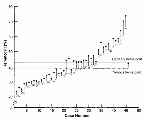
FIGURE 43.8 Simultaneous capillary (dark circles) and venous (open circles) hematocrit levels in 45 premature infants studied during the first 6 weeks of life. Each vertical line represents values for one infant, and the horizontal solid line represents mean capillary and venous hematocrit levels for the whole group. Data are not shown for five infants in whom capillary and venous hematocrit levels were identical.


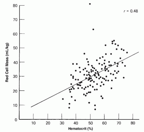
FIGURE 43.9 Simultaneous capillary hematocrit and circulating erythrocyte mass levels in 135 premature infants who had birth weights less than 1,500 g and were studied during the first week of life (r, correlation coefficient).


Correlation of Capillary Hematocrit Levels and Total Erythrocyte Mass

Erythrocyte mass is probably the best measurement of anemia. In adults, it correlates directly with hemoglobin values, which can be used as a valid means of determining anemia. In infants, the correlation between erythrocyte mass and hemoglobin values, although statistically significant, is poor (Figs. 43.9 and 43.10) (54,76). This is particularly true for ill infants, in whom a poor peripheral circulation may exaggerate capillary and venous hematocrit differences and for premature infants during periods of rapid body growth, when increases in the total circulating blood volume may influence hemoglobin levels through hemodilution (196).


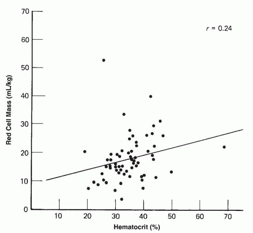
FIGURE 43.10 Simultaneous capillary hematocrit and circulating erythrocyte mass levels in 63 premature infants who had birth weights less than 1,500 g and were studied at 6 weeks of age (r, correlation coefficient).


Difficulties in Diagnosing Hemolytic Disease in Newborn Infants

Detection and diagnosis of hemolytic disease in newborn infants may be difficult because many of the tests used in older children and in adults are of little value during the first days of life. Hemolytic disease in adults and older children is diagnosed if there is evidence of a rapidly falling hemoglobin concentration, increased erythrocyte production in the absence of hemorrhage, abnormal erythrocyte morphology, and increased erythrocyte destruction within the bloodstream with the release of free hemoglobin or within the reticuloendothelial system with production of bilirubin. In the newborn infant, these signs of a hemolytic process are of limited value and require additional interpretation.

In adults and older children, an increased reticulocyte count with a stable or falling hemoglobin concentration is evidence of increased erythrocyte production and, in the absence of hemorrhage, is diagnostic of a hemolytic process. The reticulocyte count in normal newborns has a wide range, and the ability of the newborn to mount reticulocyte response is not consistent.

The shape of erythrocytes in newborns differs from that in adults. Variant erythroid morphologies are seen in blood smears of healthy newborn infants, particularly premature infants, up to certain frequencies as shown in Figure 43.11 and Table 43.7.

When erythrocytes are destroyed in the reticuloendothelial system, bilirubin is produced, with elevation of indirect bilirubin in the blood. Unusually rapid appearance of jaundice, particularly in the first 24 hours, suggests hemolytic disease. However, there are many other causes of hyperbilirubinemia in the newborn (see Chapter 32). Therefore, all newborns with abnormally high indirect bilirubin levels must be studied for evidence of hemolytic disease.

The catabolism of erythrocytes results in the equimolar production of bilirubin and carboxyhemoglobin (198). The concentration of blood carboxyhemoglobin or the rate of carbon monoxide excretion (199) also correlates with hemolysis (200).

In adults and children, intravascular hemolysis is evidenced by increased levels of hemoglobin in the plasma (i.e., hemoglobinemia), a fall in serum haptoglobin, and the appearance of hemoglobinuria and methemalbuminemia. In the normal newborn, haptoglobin levels may be zero, and plasma hemoglobin levels are above those found in adults. Gross elevations in plasma hemoglobin and hemoglobinuria are evidence of intravascular hemolysis, but the value of these tests in detecting mild hemolysis in newborn infants is limited.


Workup of a Newborn Infant with Anemia

At no other time does such a variety of disorders result in anemia as in the first week of life (201). The need for rapid treatment often adds to the diagnostic confusion. It is because of the multiple causes and the need for prompt therapy that the fundamentals of diagnosis should be appreciated and practiced without delay. Attempts at diagnosis begin with a history if the cause is not immediately apparent. In the family history, attention should be paid to anemia in other members of the family or to unexplained episodes of anemia, jaundice, cholelithiasis, or splenectomy. A positive family history is frequently obtained in cases of infants with hereditary spherocytosis, and a history of affected siblings may be encountered in patients with enzymatic defects of the erythrocyte.

In the maternal history, information should be obtained concerning both her and the biologic father’s ethnic origins including any consanguinity, and her medication history near term. Information about drugs known to initiate hemolysis in cases of G6PD deficiency should be sought and especially any history of recent exposure to mothballs containing naphthalene.

The obstetric history should provide information about vaginal bleeding during pregnancy, placenta previa, abruptio placentae, vasa previa, and CS (if the placenta was anterior and incised, was
the interval between its incision and delivery time >30 seconds as this may have resulted in significant fetal blood loss). Additional questions should be answered. Was the birth traumatic? Did the cord rupture? Was it a multiple birth?


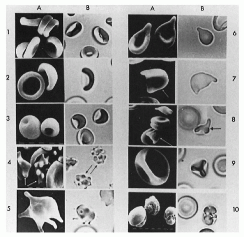
FIGURE 43.11 The three-dimensional appearance of erythrocytes as seen by scanning electron microscopy (A) and by light microscopy (B) of glutaraldehyde-fixed cells (1, discocytes; 2, bowls; 3, spherocytes; 4, echinocytes; 5, acanthocytes; 6, dacrocytes; 7, keratocytes; 8, schizocytes; 9, knizocytes; 10, immature erythrocytes).

The age at which anemia is first noticed is also of diagnostic value. Marked anemia at birth is usually the result of hemorrhage or severe alloimmunization. Anemia manifesting during the first 2 days of life is frequently caused by external or internal hemorrhages or severe alloimmune hemolytic disorder, while anemia appearing after the first 48 hours of life is most commonly hemolytic and is usually associated with jaundice.


TABLE 43.7 Erythrocyte Differential Counts in Adults and Neonates


Median (5%-95%)a


Erythrocytes


Adults


Full-Term Infantsb


Premature Infantsc


Number studied


53


31


52


Disks


78 (42,43,44,45,46,47,48,49,50,51,52,53,54,55,56,57,58,59,60,61,62,63,64,65,66,67,68,69,70,71,72,73,74,75,76,77,78,79,80,81,82,83,84,85,86,87,88,89,90,91,92,93,94)


43 (18,19,20,21,22,23,24,25,26,27,28,29,30,31,32,33,34,35,36,37,38,39,40,41,42,43,44,45,46,47,48,49,50,51,52,53,54,55,56,57,58,59,60,61,62)


39.5 (18,19,20,21,22,23,24,25,26,27,28,29,30,31,32,33,34,35,36,37,38,39,40,41,42,43,44,45,46,47,48,49,50,51,52,53,54,55,56,57)


Bowls


18 (4,5,6,7,8,9,10,11,12,13,14,15,16,17,18,19,20,21,22,23,24,25,26,27,28,29,30,31,32,33,34,35,36,37,38,39,40,41,42,43,44,45,46,47,48,49,50)


40 (14,15,16,17,18,19,20,21,22,23,24,25,26,27,28,29,30,31,32,33,34,35,36,37,38,39,40,41,42,43,44,45,46,47,48,49,50,51,52,53,54,55,56,57,58)


29 (13,14,15,16,17,18,19,20,21,22,23,24,25,26,27,28,29,30,31,32,33,34,35,36,37,38,39,40,41,42,43,44,45,46,47,48,49,50,51,52,53)


Ratio of disks to bowls


2 (0-4)


2 (0-5)


3 (0-10)


Spherocytes


0 (0-1)


0 (0-1)


0 (0-1)


Echinocytes


0 (0-3)


1 (0-4)


5.5 (1-43)


Acanthocytes


0 (0-1)


1 (0-2)


0 (0-2)


Dacrocytes


0 (0-1)


1 (0-3)


1 (0-5)


Keratocytes


0 (0-1)


2 (0-5)


3 (0-7)


Schizocytes


0 (0-1)


0 (0-2)


2 (0-5)


Knizocytes


1 (0-4)


3 (0-8)


1 (0-6)


Others


1 (0-4)


3 (0-7)


4 (0-11)


aAll values are expressed as a median plus the 5% to 95% range, because the distribution of most values was nongaussian.


bOf the sample, 29 were ABO compatible, 1 was AB with an A mother, and 1 was AB with a B mother.


cIncludes ABO-compatible and ABO-incompatible infants.


One approach to the differential diagnosis of anemia in the newborn period is presented in Figure 43.12. The physician should first decide whether the low hemoglobin level may be explained by blood loss from sampling. Cumulative losses, particularly in ill premature infants, may be extremely large, and correct interpretation of rapid changes in hemoglobin level can be made only if careful attention is paid to exact volumes of blood sampled and transfused. If the cause of anemia remains unknown, several laboratory tests may aid in diagnosis: reticulocyte count, a DAT of the infant’s blood, examination of a peripheral blood smear, and examination of the maternal blood smear for fetal erythrocytes. Ultrasound examination of the head or abdomen is useful to detect occult blood loss. From these studies and the history, a diagnosis often can be made, or at least the list of diagnostic possibilities can be greatly shortened.

Bone marrow aspiration is rarely needed in the neonatal period for the workup of a baby with anemia. However, if anemia persists without evidence for hemolysis or blood loss, a bone marrow should be considered to rule out conditions such as DBA.


Polycythemia

A venous hemoglobin exceeding 22.0 g/dL or a venous hematocrit more than 65% during the first week of life should be regarded as polycythemia. Although neonatal polycythemia may be the result of fetal disorders such as twin-to-twin transfusion, placental insufficiency, and certain metabolic disorders (Table 43.8), most cases occur in otherwise normal infants. Most of these infants have been full term, appropriate for gestational age, and without
asphyxia at birth. Polycythemia occurs in 1.5% to 4% of newborn infants (202).


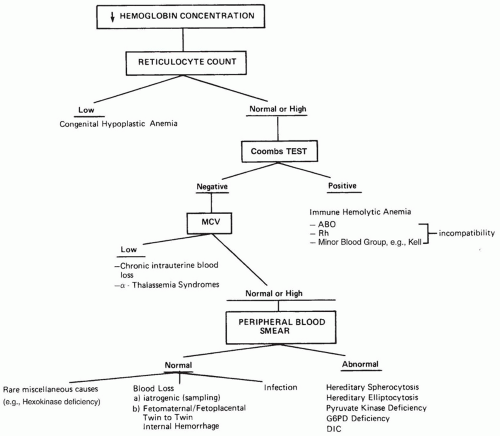
FIGURE 43.12 Diagnostic approach to anemia in the newborn infant.

The symptoms observed in the polycythemic infant appear to be primarily a consequence of hypervolemia and an increase in blood viscosity. After the central venous hematocrit reaches 60% to 65%, the increase in blood viscosity becomes much greater as a result of the exponential relationship between hematocrit and viscosity (203). Plasma and erythrocyte factors also affect the viscosity of neonatal blood (204,205,206).

Respiratory distress, thrombocytopenia, cyanosis, congestive heart failure, convulsions, priapism, jaundice, renal vein thrombosis, hypoglycemia, and hypocalcemia appear to be more common in infants with polycythemia (202). Many infants with polycythemia are asymptomatic.

In addition to supportive care, partial exchange transfusion (PET) has been used for the treatment of polycythemia. PET increases cerebral oxygenation and fractional tissue oxygen extraction in newborn with polycythemia, suggesting increased blood flow (207). However, the precise indications are still to be determined. While a central venous hematocrit of over 65% with symptoms is a widely accepted indication for PET, the threshold in an otherwise healthy-looking neonate is controversial. Some groups propose an exchange in neonates for central venous hematocrit over 70% (207); others have shown that a more restrictive approach of performing PET only for those who have hematocrit greater than or equal to 76% or symptoms may be safe (208). Reduction of the venous hematocrit to less than 60% may improve symptoms (209,210), but the long-term neurologic outcomes are still unclear. To perform a PET, the volume of blood to be exchanged (mls) is typically calculated from the following information: the weight of the infant, the actual hematocrit, the desired hematocrit (usually approximately 55%), and the blood volume (the blood volume of a term infant is 80 to 90 mL/kg and a preterm infant is 90 to 100 mL/kg). The formula for the volume (mls) of blood to be exchanged is:

[(Actual – Desired Hct) / Actual Hct] × Blood volume

Thus, a 3-kg term infant with a central Hct of 80% and a desired Hct of 55% would require a PET of approximately 84 mL. The blood
is typically drawn from the patient in 10 mL aliquots and replaced with normal saline.


TABLE 43.8 Neonatal Polycythemia


Possible causes by placental hypertransfusion


Twin-to-twin transfusion


Maternofetal transfusion


Delayed cord clamping


Intentional


Unassisted home delivery


Possible associations


Placental insufficiency


Small for gestational age infants


Postmaturity birth


Toxemia of pregnancy


Placental previa


Endocrine and metabolic disorders


Congenital adrenal hyperplasia


Neonatal thyrotoxicosis


Maternal diabetes


Miscellaneous


Trisomies 13, 19, and 21


Hyperplastic visceromegaly (i.e., Beckwith syndrome)


Erythroderma ichthyosiforme congenital


▪ LEUKOCYTE DISORDERS


Neutrophil Disorders

A diverse group of leukocyte disorders are encountered in newborn infants. Different blood cells are involved (e.g., neutrophils, lymphocytes, eosinophils), and the disorders may be quantitative or qualitative in nature. This section focuses on abnormalities that are frequent (e.g., neutrophil changes associated with bacterial infections) or are unique to this age group (e.g., neonatal alloimmune neutropenia, inherited neutropenia, and congenital leukemia).


Normal Leukocyte Count in the Neonatal Period

The mature neutrophil has a nucleus that is distinctly segmented into two or more lobes connected by a thin filament. Cells with no lobulation and those in which the width of the narrowest segment of the nucleus is greater than one-third the width of the broadest segment are referred to as nonsegmented neutrophils or bands. During the first 2 weeks of life of full-term and premature infants, a band (young)-to-segmented neutrophil ratio greater than 0.3 should be considered abnormal (211). Examination of a peripheral blood smear during the first few days of life characteristically reveals an excess of neutrophils. Particularly, in premature infants, some immature forms (e.g., promyelocytes, myelocytes) may be seen. Sometime between the 4th and 7th days of life, the lymphocyte becomes the predominant cell and remains so until the 4th year of life.

Counts of segmented and band neutrophils of full-term and VLBW infants have been reported by a number of investigators (211,212,213,214,215,216,217,218). The lower limit of normal for neutrophil counts in VLBW infants is significantly below those for term infants. Reference values of neutrophils in term and preterm babies are in Figures 43.13, 43.14, 43.15 (215,217).

Although the definition of neutropenia is essentially a statistical consideration based on data obtained from studies of healthy term and premature newborn infants, there is consensus among experts that an absolute neutrophil count (ANC, band plus mature neutrophils) below 1 × 10⁹/L increases the risk of infections in fullterm and premature newborn infants (219) and that ANC below 0.5 × 10⁹/L is considered severe neutropenia (220). Neutropenia that is milder than the above requires a follow-up and possibly a search for a cause. Although most often in neonates neutropenia is transient and the main concern is an increased risk of infection or
an undiagnosed infection, sometimes neutropenia is a sign or a serious underlying disorder that needs urgent diagnosis and treatment.


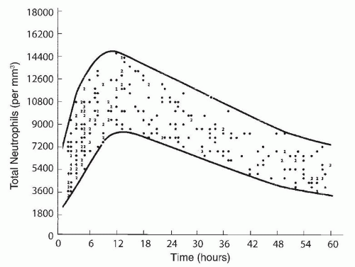
FIGURE 43.13 The total neutrophil count reference range in the first 60 hours of life. Subjects were 434 newborn infants (birth weight 2,685 ± 683 g; range 29-44 weeks of gestational age). Solid circles represent single values; numbers represent the number of values at the same point. Heavy lines represent the envelope bounding these data. Reproduced with permission from Manroe BL, Weinberg AG, Rosenfeld CR, et al. The neonatal blood count in health and disease. I. Reference values for neutrophilic cells. J Pediatr 1979;95:89.


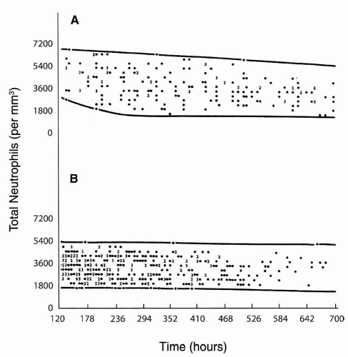
FIGURE 43.14 The reference range for the total neutrophil count for (A) infants 60 to 120 hours of life and (B) 120 hours to 28 days of life. Subjects were 434 newborn infants (birth weight 2,685 ± 683 g; range 29-44 weeks of gestational age). Solid circles represent single values; numbers represent the number of values at the same point. Heavy lines represent the envelope bounding these data. Reproduced with permission from Manroe BL, Weinberg AG, Rosenfeld CR, et al. The neonatal blood count in health and disease. I. Reference values for neutrophilic cells. J Pediatr 1979;95:89.


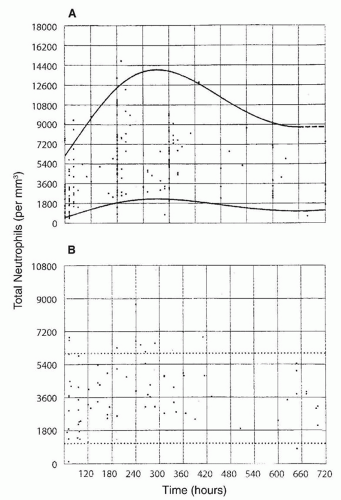
FIGURE 43.15 Reference ranges for total neutrophil values in VLBW infants from (A) birth to 60 hours of life and (B) 61 hours to 28 days of life. Subjects were 193 newborn infants: 50 at 1,000 g and 143 at 1,001-1,500 g. Bold lines (A) and dotted lines (B) represent the envelopes bounding these data, respectively. Reproduced with permission from Mouzinho A, Rosenfeld CR, Sanchez PJ, et al. Revised reference ranges for circulating neutrophils in very-low-birth-weight neonates. Pediatrics 1994;94:76.

Physiologic neutrophilia is common in neonates in the first week of life. According to Thilaganathan et al. (221) total leukocyte counts in umbilical cord blood ranged between 7.25 and 48 × 109/L with a mean of 13.8 × 10⁹/L. After birth, neutrophil counts increase to levels up to 23,000 23 × 109/L at 16 hours postlabour and then gradually decrease to less than 9.5 × 10⁹/L at 5 days of age. The mechanism for the physiologic neutrophilia seems to be a surge in cytokine secretion (222,223); granulocyte colony-stimulating factor (G-CSF) levels increase on day 1 after birth and then gradually decrease (223). Interestingly, the major cause of physiologic neutrophilia in newborns was found to be related to an increase production of G-CSF by the placenta (trophoblasts and decidual stromal cells).


Neutropenia

Neutropenia occurs in 6% to 8% of babies admitted to the NICU. Causes of neutropenia include decreased production, increased destruction, margination in the microvascular endothelium, sequestration in the spleen, or a combination of mechanisms (Table 43.9). Most episodes occur during the first week of life and are related to low gestational age, intrauterine growth restriction, infections, pregnancy-induced hypertension (PIH), severe neonatal asphyxia, drug therapy, or other perinatal events (224). Late-onset neutropenia occurs at a postnatal age of more than 3 weeks and has been reported in premature babies with anemia and marked reticulocytosis (225). The mechanism is unknown but may be related to induced expression of transcription factors that promote erythropoiesis while suppressing granulopoiesis. This physiologic response is typically transient, not severe, and is not associated with an increased risk of infection.


TABLE 43.9 Causes of Neonatal Neutropenia


Decreased Production of Neutrophils (See also Table 43.13)


Infants of hypertensive women


Donors of twin-twin transfusion


Rhesus hemolytic disease


Kostmann/severe congenital neutropenia


Cyclic neutropenia


Shwachman-Diamond syndrome


Barth syndrome


Glycogen storage disease type 1b


Organic aciduria (propionic aciduira, methylmalonic aciduria, fumarase deficiency)


Cartilage-hair hypoplasia


Reticular dysgenesis


Chédiak-Higashi syndrome


Excessive Neutrophil Margination


Endotoxemia (e.g., NEC)


Drug-induced neutropenia


Idiopathic neutropenia of prematurity


Increased Destruction of Neutrophils


Neonatal alloimmune neutropenia


Neonatal autoimmune neutropenia


Drug-induced neutropenia


Neutropenia associated with immunodeficiency syndromes


Decreased Production and Increased Destruction of Neutrophils


Infections


Congenital, usually viral


Acquired, usually bacterial


Drug induced neutropenia


Hypersplenism


The various causes of neutropenia in neonates are summarized in Table 43.9. The commonest causes include neutropenia associated with infection, neutropenia in premature infants, neutropenia in infants of hypertensive mothers, allo-autoimmune neutropenia, and twin-to-twin transfusion (219). Other causes for destruction or underproduction of neutrophils are less common and include inherited bone marrow failure syndromes.


Neutropenia due to Bacterial Infection

Neutropenia frequently occurs in the setting of neonatal sepsis. It can be the cause of sepsis, but more commonly, it is the consequence. It is noteworthy that neutrophil function, particularly chemotaxis and phagocytosis, is reduced in newborn infants and may contribute to susceptibility to infection (226). In infants with systemic bacterial infection, the total neutrophil count is usually decreased but can be increased or normal. In a small study of 24 newborn infants with sepsis and documented positive cultures, neutropenia was observed in 5, neutrophilia in 3, and normal neutrophil counts in the remaining 16 (212). In studies where neonates with both confirmed and suspected bacterial disease were
included (215,224), neutropenia was observed in about three-quarters of the subjects. In another study in 65% of 63 neonates with neutropenia and sepsis, the neutropenia was present on the day of the clinical onset of sepsis (224), in 13% of the cases neutropenia developed within 3 days of the onset of sepsis, and in 22% neutropenia was present before the clinical onset of sepsis. Seventy-seven percent of the neutropenic episodes occurred during the first week of life; in 75% of affected neonates, the duration of neutropenia ranged from 0 to 8 days, with 75% having neutropenia for less than 24 hours.

In addition to neutropenia, increased numbers of immature neutrophils and an elevated band-to-segmented neutrophil ratio are seen in neonates with sepsis (212,213). In a study of premature infants with proven bacterial infection, 73% of the infants had elevated band counts and a reversed band-to-segmented neutrophil ratio (213). The maximal normal immature-to-total ratio is 0.16 in the first 24 hours after birth that gradually decreases to 0.12 after the 5th day of life (215).

During infection, the neutrophils of newborn infants have increased numbers of Döhle bodies (i.e., aggregates of rough endoplasmic reticulum), vacuoles, and toxic granules (213).

In terms of prediction of sepsis, several factors should to be taken into account. First, two negative blood counts performed 8 to 12 hours apart and a negative blood culture at 24 hours improve the ability to rule out sepsis in the first day of life to 100% (227). Second, immature neutrophil to total neutrophil of greater than 0.2 is suggestive of sepsis (228). Third, high or low leukocytes or neutrophils counts; the sensitivity of a low leukocyte count is only 29%, but the specificity is as high as 91% (229). Fourth, morphologic changes in neutrophils as discussed above have positive predictive value (Table 43.10) (230,231).


Decreased Production of Neutrophils

Idiopathic Neutropenia of Prematurity Neutropenia in NICUs is most commonly seen in premature infants (typically <30 weeks of gestational age), particularly those of VLBW. It presents at 4 to 10 weeks after birth. The blood smear does not show immature neutrophils. The underlying mechanisms appear to be a combination of reduced total body neutrophil mass, together with reduced numbers of committed neutrophil precursors in the bone marrow at birth and an inability to mount a granulopoietic response (232). Intravenous or subcutaneous administration of the granulocytic growth factors usually produces significant increases in the level of circulating neutrophils (G-CSF at daily doses of 5 to 10 µg/kg) (222,233) or both neutrophils and monocytes (granulocyte-macrophage colony-stimulating factor [GM-CSF] at daily doses of 5 to 10 µg/kg) (234). Toxicity is minimal, particularly when G-CSF is administered. Most investigators found an improved outcome when G-CSF (235,236) or GM-CSF (237) was administered, but some did not (238). Granulocyte transfusions have been used in neonates with sepsis, but the role of this modality still needs to be established (239). These studies largely included patients with varying degrees of neutropenia, which some authors postulate may have lessened the therapeutic effect seen among babies who may have benefited the most from this therapy.


TABLE 43.10 Hematologic Scoring System in Neonates with Suspected Sepsis


Abnormality


Score


Immature:total neutrophil ratioa


1


Total neutrophil counta,b


↑ or ↓


1


I:M ratio


≥0.3


1


Immature PMN count


1


Total WBC countc


↑ or ↓


1


Degenerative changes in PMNsd


≥3+e


1


Platelet count


<150,000/mm3


1


aNormal values as defined by Manroe et al. (312).


bIf no mature neutrophils are seen on the blood film, score 2 rather than 1 for total PMN count.


c≤5,000/mm3 or ≥25,000, 30,000 and 21,000/mm3 at birth, 12-24 h and day 2 onward, respectively.


dQuantitated on 0 to 4+ scale according to classification by Zipursky et al. (310).


eFor vacuolization, toxic granulation, or Döhle bodies.


1, immature; M, mature; PMN, polymorphonuclear leukocytes; WBC, white blood cell.


There have been investigators who studied the ability of prophylactic administration of cytokines to prevent infections and reduce mortality. However, no consistent reduction in infection rates and mortality was demonstrated when G-CSF was given prophylactically in preterm infants without neutropenia or with mild neutropenia. Improvement was shown by some (233) but not by others (240) The study by Kuhn et al. (240) is a large, multicenter, randomized, double-blind placebo-controlled trial that included 200 subjects. Similarly, no consistent reduction in infections and rates and mortality was demonstrated when GM-CSF was given prophylactically in preterm infants without neutropenia or with mild neutropenia. Improvement was shown in some studies (241) but not in others (234,242). The publication by Carr et al. (234) included results from a large, single-blind, multicenter, randomized controlled trial that included 280 subjects as well as a meta-analysis.

From the above studies, it is reasonable to draw the following conclusions: (a) the treatment of preterm infants with sepsis and severe neutropenia should include the usage of granulocyte growth factors such as G-CSF, (b) prophylactic administration of granulocyte growth factor to prevent sepsis in preterm babies without neutropenia or with mild neutropenia is not recommended, and (c) prophylactic administration of granulocyte growth factors to reduce infections and mortality in preterm infants with severe idiopathic neutropenia and no clinical infection still needs to be studied.

Infants of Hypertensive Women Neutropenia occurs in 50% of infants born to mothers with PIH. The mechanism is reduced production as a result of an inhibitor interfering with normal granulopoiesis (243,244). Initially, the neutropenia can be very low (e.g., 0.5 ×10⁹/L), but it resolves spontaneously in 3 to 5 days, and the risk of infections is unclear (245,246).

Kostmann/Severe Congenital Neutropenia Kostmann/severe congenital neutropenia (K/SCN) is an inherited bone marrow failure syndrome that affects only the granulocytes. Neutropenia is severe, typically less than 0.2 × 10⁹/L from the first day of life (247). Bone marrow smears typically reveal a maturation arrest at the promyelocyte -myelocyte level but normal overall cellularity. Inheritance depends on the mutant gene (Table 43.6). Monoallelic mutations in ELANE (248) lead to abnormally activated neutrophil elastase with exclusive membrane localization (249), misfolded protein (250), and increased apoptosis of myeloid precursors (250,251). Biallelic mutations in HAX1 are associated with autophagy and activation of the mitochondrial apoptosis pathway (252). Thirty percent of the patients have neurologic manifestations such as developmental delay and seizures. Mutations in the G6PC3 gene lead to neutropenia in addition to atrial septal defects, mild-moderate immunodeficiency, and prominent vasculature (253). Other causes of severe congenital neutropenia are listed in Table 43.6. Patients with K/SCNs suffer from severe bacterial infections from infancy. About 50% of the patients with mutations in the ELANE and HAX1 genes suffer from infections in the first month of life, and the disorder is usually fatal if not diagnosed and treated early. The risk of leukemia increases with age (254); however, the appearance of leukemia in early childhood has been reported. Early diagnosis is critical for proper treatment, prevention of early deaths, genetic counseling, and prompt initiation of a cancer surveillance program. Diagnosis is facilitated by clinical features, complete blood counts,
bone marrow testing, and genetic testing. Treatment with G-CSF increases neutrophil counts and prevents infection in 90% of cases (255,256). Patients who do not respond to G-CSF may benefit from the addition of low-dose prednisone to the G-CSF regimen or hematopoietic stem cell transplantation (257,258,259). Transformation to myelodysplastic syndrome and acute myeloid leukemia is a major complication in K/SCN regardless of treatment with G-CSF and is an indication for hematopoietic stem cell transplantation.

Cyclic Neutropenia Cyclic neutropenia is an inherited disorder characterized by a regular, repetitive decrease in peripheral blood neutrophils at approximately 21-day intervals (260,261,262). Typical cycling pattern may be apparent in early infancy and can start in the neonatal period. The disorder is caused by ELANE gene mutations at the active site of neutrophil elastase causing defective membrane localization of the enzyme (249) and a cycling increase in apoptosis of myeloid precursors (251). Patients may develop severe infections and mouth sores during the neutrophil nadir leading to chronic gingivitis. Diagnosis requires the demonstration of regular neutrophil cycles and is supported by genetic testing. Daily treatment with G-CSF improves symptoms in most patients. Although the mutations are in the same gene that causes K/SCN, the disorder is not associated with a high risk of leukemic transformation.

Glycogen Storage Disease Type Ib Patients with this autosomal recessive disorder have classical metabolic manifestations as seen in glycogen storage disease type Ia. These include hepatomegaly, hypoglycemia, and lactic acidosis. In contrast to glycogen storage disease type Ia, patients with the disorder have neutropenia and impaired neutrophil chemotaxis and respiratory burst. Granulocytes are reduced and dysfunctional possibly because of accelerated apoptosis (263,264). The increased cell death might be related to inability to meet intracellular glucose requirements (264) and translocation and activation of the Bax proapoptotic protein (263). The genetic defect resides in the gene that encodes the glucose-6-phosphate translocase. Neutropenia may be severe and cause serious infection and inflammatory bowel disease. Most patients require G-CSF, which successfully increases the neutrophil counts, improves function, and prevents infection (265).

Barth Syndrome Barth syndrome is an X-linked recessive disorder with dilated cardiomyopathy and left ventricular noncompaction, skeletal myopathy, 3-methylglutaconic aciduria, and neutropenia (266,267). The disorder is associated with the TAZ gene mutation (268). The gene encodes 10 different proteins called “taffazins” that are involved in remodeling of cardiolipin, an essential component of the mitochondrial inner membrane that is necessary for proper function of the respiratory chain. Neutropenia varies from mild to very severe. The mechanism for the neutropenia seems to involve increased dissipation of mitochondrial membrane potential and apoptosis of (269). Marrow specimens are of normal cellularity but may show maturation arrest at the myeloid stage. Most patients do not require continuous therapy for their neutropenia. However, in cases with severe bacterial infections, G-CSF can be given with a very good response (267).

WHIM Syndrome Patients with this autosomal dominant syndrome (Warts, Hypogammaglobulinemia, Infections, and Myelokathexis) may present in the first months of life (270). The neutropenia results from a defective release of marrow cells into the peripheral blood due to mutations in the chemokine receptor gene CXCR4 (271). CXCR4 interference by either a direct antagonist (272) or G-CSF has therapeutic value.

Reticular Dysgenesis Reticular dysgenesis is one of the rarest and most extreme forms of neutropenia associated with severe combined immunodeficiency (SCID). It is characterized by congenital agranulocytosis, lymphopenia, and lymphoid and thymic hypoplasia (273). Most patients have mutations in the gene encoding the mitochondrial adenylate kinase 2 (274). Neutrophil counts usually do not improve with administration of G-CSF, and the patients often die within the first weeks of life unless they receive hematopoietic stem cell transplantation (275). Myelodysplastic syndrome has been reported in patients who had mixed chimerism posttransplant (276).

Cartilage-Hair Hypoplasia Severe neutropenia and macrocytic or normocytic anemia occur in patients with cartilage-hair hypoplasia (CHH). The cytopenia is due to either an autoimmune mechanism or bone marrow failure. The disorder is characterized by fine hair, short-limbed dwarfism, metaphyseal dysplasia (not often evident in the first year of life), and T-cell abnormalities. This autosomal recessive condition is caused by mutations in the RMRP gene (277), which plays a role in rRNA processing and ribosome biogenesis. Although common in the Finish and Amish, the disorder has been reported in other populations (278). In those patients with severe immunodeficiency, hematopoietic stem cell transplantation can correct the hematologic and immune dysfunction.

Chédiak-Higashi Syndrome Chédiak-Higashi syndrome is an autosomal recessive disorder caused by mutations in the lysosomal trafficking regulator gene, LYST (279). It is characterized by variable degrees of oculocutaneous albinism, easy bruising, and bleeding due to platelet dysfunction (280). Patients suffer from recurrent infections as a result of neutropenia, impaired chemotaxis and bactericidal activity, and abnormal natural killer (NK) cell function. Large cytoplasmic granules in circulating granulocytes are a clue to the diagnosis (281).

Griscelli Syndrome Patients with Griscelli syndrome have some features similar to those with the Chédiak-Higashi syndrome including partial albinism, frequent episodes of fever and pyogenic infections, neutropenia, and thrombocytopenia, but they lack abnormal cytoplasmic granules (282,283). T-lymphocyte proliferation and macrophage activation syndrome is a complication. The disorder is caused by mutations in the myosin VA (Griscelli syndrome type 1), RAB27A (Griscelli syndrome type 2), or melanophilin (Griscelli syndrome type 3) genes, which play a role in vesicle transport and membrane trafficking processes. Hematopoietic stem cell transplantation is curative (284).

Hemophagocytic Lymphohistiocytosis Hemophagocytic lymphohistiocytosis (HLH) is characterized by pancytopenia, fever, hepatosplenomegaly, neurologic findings, liver and coagulation abnormalities, and elevated triglyceride and ferritin levels (285,286). An important presentation during the neonatal period is progressive liver failure, which mimics neonatal hemochromatosis (287). Most or all neonatal cases are probably inherited. Germline mutations in genes that protect immune cells from apoptosis have been identified in patients with familial and sporadic HLH. The most commonly mutated genes are PRF1, UNC13D, STX11, and STXBP2. Early diagnosis is critical to institute immunosuppressive therapy with or without hematopoietic stem cell transplantation.


Increased Destruction of Neutrophils

Neonatal Alloimmune Neutropenia Alloimmune neutropenia occurs when a mother becomes sensitized to an antigen of paternal origin that is expressed on the neutrophils of her infant and forms specific immunoglobulin G (IgG) antibody directed against this fetal antigen. Transplacental passage of IgG antibody into the fetal circulation results in accelerated destruction of neutrophils in the reticuloendothelial system with consequent neutropenia. The condition is self-limiting, and neutropenia persists for only a few weeks or months. The severity of neutropenia is influenced by the titer and subclass of the maternal IgG neutrophil antibody, the phagocytic activity of the infant’s reticuloendothelial system, and the capacity of the infant’s marrow to compensate for the shortened survival of antibody-sensitized neutrophils. The frequency of clinical
alloimmune neutropenia was estimated as 1 in 500 newborn infants (288) to less than 0.1% (289). Using modern testing to prospectively analyze 247 cord blood samples of full-term babies and evaluation of neutropenia, the incidence was found to be 0.81% (290).

The study of neonatal alloimmune neutropenia has contributed much to the current knowledge of neutrophil-specific antigens (Table 43.5) (291). The most common antibodies found in patients are against neutrophil-specific antigens, particularly the HNA-1a, HNA-1b, and HNA-1c antigens. However, rare cases of alloimmunization due to anti-HLA antibodies and anti-Fc gamma receptor IIIb were also reported.

Unfortunately, recent and large clinical studies are unavailable, but the reported clinical course of infants with alloimmune neutropenia is of interest. Neutropenia is usually severe. Symptomatic infants may present with delayed separation of the umbilical cord, skin infections, pneumonia, or otitis media within the first 2 weeks of life (289,292). The duration of neutropenia ranges from 2 to 28 weeks, with a mean of about 7 weeks. Mild infections are common. Severe or overwhelming infections were reported in up to 5% of the cases (293), and most were caused by Staphylococcus aureus. Deaths due to disseminated bacterial infections were reported. While most infections in newborn infants with neonatal alloimmune neutropenia are mild, affected infants with severe neutropenia are at risk for serious bacterial infections, and therapeutic intervention should be considered. Intravenous antibiotic therapy should be initiated for infants with suspected or proven infection. The preferred strategy to prevent infections and treat neonates with severe alloimmune neutropenia is administration of rhG-CSF. The initial recommended dose is 5 µg/kg/d given by intravenous or subcutaneous injection for 3 days with titration of additional doses to keep the blood neutrophil count greater than 1,000/µL (292). The response to rhG-CSF is usually rapid and evident within 24 to 48 hours; generally, 2- to 3-week treatment is sufficient. It is important that infants be monitored for recurrence of neutropenia once rhG-CSF therapy is stopped (294). For infants who fail to respond to initial therapy with rhG-CSF, a trial of IVIG (1 g/kg/d for 2 to 5 days) alone or in combination with rhG-CSF should be considered. The use of exchange transfusion or transfusion with compatible antigen-negative neutrophils should be reserved for those rare infants who have failed an adequate trial of IVIG and rhG-CSF (10 µg/kg/d or higher doses), who are clinically extremely ill, and who are not responding to broad-spectrum intravenous antibiotic therapy. There is little evidence that corticosteroids are of value in this condition.

As the condition may recur in subsequent pregnancies, we would recommend testing subsequent offspring’s neutrophil counts immediately after birth and at 1 week of age. However, asymptomatic babies may not need treatment.

Rare cases of alloimmune neutropenia have been reported in newborn infants following transfusion of blood components or IVIG (295).

Neonatal Autoimmune Neutropenia Transient neutropenia in the neonatal period may reflect transfer of IgG neutrophil autoantibodies from the mother to the fetus during pregnancy (296,297). In these cases, the maternal serum contains the pathologic neutrophil antibodies, and the mother may be neutropenic and may have a history of an autoimmune disorder, such as systemic lupus erythematosus (297,298). Most children are asymptomatic, and the neutropenia resolves spontaneously by the 3rd to 4th months of life. However, severe neutropenia and life-threatening infections have been described, and prophylactic G-CSF after birth or third trimester should be considered (299,300).

Autoimmune Neutropenia of Infancy Autoimmune neutropenia of infancy typically presents in children between the ages of 3 and 30 months (301,302). The mechanism involves production of autoreactive antibodies usually against antigens of the NA1 or NA2 antigens on the Fcγ receptor IIIb (303). Rarely, it is seen in the neonatal period (304). In such cases, an underlying immunodeficiency should be ruled out. Unless associated with immunodeficiency or generalized autoimmune disorder, the condition is self-limiting, and only 5% to 10% of the children require treatment with G-CSF or IVIG (305).

Immunodeficiency Syndromes Associated with Autoimmune Neutropenia Immunodeficiency can be associated with underproductive neutropenia (e.g., reticular dysgenesis and CHH) or destructive autoimmune neutropenia (e.g., hyper-IgM syndrome) or both (306). Autoimmune neutropenia associated with immunodeficiency syndromes can present in the first few weeks of life. Therefore, it is imperative to assess total lymphocyte counts and immunoglobulin levels in neonates with neutropenia, particularly as some of these disorders require urgent initiation of a search for a suitable donor for hematopoietic stem cell transplantation and prophylaxis against Pneumocystis carinii pneumonia. Unusual infections (e.g., Pneumocystis carinii pneumonia or systemic candidiasis), immune abnormalities, extrahematologic manifestations, and family history may be clues to primary immunodeficiency states.

Autoimmune Lymphoproliferative Syndrome An important immunodeficiency syndrome that is associated with autoimmune process causing neutropenia, hemolytic anemia, and thrombocytopenia concurrently or sequentially is autoimmune lymphoproliferative syndrome (ALPS). Neutropenia is a frequent finding in patients with this disorder (307). In this disorder, neutropenia is often associated with other autoimmune disorders. Defects in genes associated with lymphocyte apoptosis have been identified in the disorder. Lymphadenopathy and splenomegaly are common. Treatment of severe cytopenia is based on immunosuppressive therapy with drugs such as mycophenolate, sirolimus, and rituximab.

Other primary immunodeficiencies that can be associated with early-onset neutropenia include conditions such as agammaglobulinemia, hyper-IgM syndrome, and Wiskott-Aldrich syndrome and are described in the Lymphopenia section below.


Evaluation of the Infant with Neutropenia

The unexpected finding of severe or prolonged neutropenia in a newborn infant should prompt evaluation. Infections, particularly bacterial, should always be considered. A peripheral blood smear should be carefully examined for Döhle bodies, vacuolization, and toxic granulation, and the band-to-segmented neutrophil ratio should be determined. In infants with neutropenia, an increased band-to-segmented neutrophil ratio, and morphology suggestive of bacterial infection, empirical broad-spectrum antibiotic therapy should be started and continued until the results of cultures are known, and infection is not the etiology. If there is no clinical or laboratory evidence of infection, other causes of neutropenia must be considered.

As noted, preeclampsia and/or hypertension in the mother is a common cause of neutropenia and should be considered in the differential diagnosis. A maternal history, including drug exposure, should be obtained. Maternal history of autoimmune disorders such as systemic lupus erythematosus, particularly if it is accompanied by neutropenia, suggests vertical transfer of autoimmune antibody. The physician should obtain a careful family history of neutropenia, severe or unusual infections, and early neonatal deaths. Early neonatal deaths may be caused by overwhelming infection secondary to inherited neutropenia before any diagnosis is made. Physical examination of affected infants suggests or excludes hypersplenism and congenital viral infections as the likely cause of neutropenia. In well infants with no apparent cause for the neutropenic state, neonatal alloimmune neutropenia should be considered. In such cases, a search should be made for neutrophil antibodies in a sample of maternal and/or baby’s serum by methods such as granulocyte
agglutination test, granulocyte immunofluorescence test, monoclonal antibody immobilization of granulocyte antigens, or a multiplex assay for antibody detection using microbead coupled to purified antigens (292,293,308). These antibody detection assays are best performed in neutrophil reference laboratories and should be complemented by genotyping of the neutrophil-specific antigen of the biologic mother, affected neonate, and/or biologic father acting as a surrogate. It is important to stress that treatment of the infant should not be delayed while confirmatory serologic studies are in progress. Finally, a bone marrow aspirate should be considered if neutropenia is severe (<0.5 × 10⁹/L) and persists for longer than 1 week. Bone marrow biopsy is helpful but may not be feasible in young infants.


Neutrophilia and Neonatal Leukemoid Reactions

The incidence of leukemoid reactions with leukocyte counts more than 50 × 109/L among neonates in the NICU varies between 1.3% and 15% (309,310,311,312,313). It is most commonly seen in the first week of life. The most common causes include administration of betamethasone antenatally and infections. Congenital infections such as CMV disease, toxoplasmosis, and syphilis may manifest as hepatosplenomegaly with a pronounced leukemoid response in the peripheral blood. Severe bacterial infections also may be associated with a leukemoid blood picture. It is noteworthy that concurrent pathology may not be found in a proportion of the cases; for example, 15% of 60 patients in one series (310). The mechanism involves accelerated neutrophil production. Serum cytokine measurement did not show a consistent increase in G-CSF or GM-CSF; thus, marrow paracrine secretion or other mechanisms are possible. The leukemoid reaction resolves within several days to weeks, without evidence of sequelae due to the neutrophilia even in extreme leukocytosis (314). The main clinical significance is the possibility of an underlying pathology. Rastogi et al. (310) noted that the neonates who had been able to mount a leukemoid response had a better chance of survival than those who did not. An association has been reported between leukemoid reaction with leukocyte counts more than 50 × 109/L and the development of bronchopulmonary dysplasia and chronic lung disease (312).


Lymphopenia

Lymphocytes account for approximately 30% of the circulating white blood cells in newborn infants. Lymphopenia should always be considered when the total lymphocyte count is below the lower 5th percentile for age and particularly if the absolute lymphocyte count is less than 1.5 × 10⁹/L (315). Lymphopenia may occur in a number of immunodeficiency diseases, during infections or as part of an autoimmune process. Disorders in which lymphopenia is often diagnosed with neutropenia are detailed in the section “Neutropenia” associated with immunodeficiency, and those in which isolated lymphopenia is the hallmark of the disease are reviewed below. Of importance in the investigation of these disorders is the assessment of lymphocyte subset numbers adjusted to age (316). Usually in the newborn, 35% to 64% of lymphocytes express CD4 and are designated as “helper” T lymphocytes, and 12% to 18% of lymphocytes express CD8, which is a marker for “suppressor/cytotoxic” T lymphocytes. CD19 (a B-cell marker) is found on 6% to 32% of lymphocytes, and CD16/56 that designate NK cells is detected on 4% to 18% of cells. Typical laboratory features that may help to clarify the etiology of lymphopenia in newborn infants and the molecular defects are summarized in Table 43.11.


Severe Combined Immunodeficiency

SCID is a rare genetically heterogeneous group of serious disorders that carry a grave prognosis unless recognized early in life and treated promptly (317). Diagnostic criteria for SCID and related disorders have been developed and studied (318). The most frequent cause of SCID is mutations in the gene that encodes the gamma (also known as common) chain of interleukin (IL)-2 receptor on the X chromosome. Affected males often present in the first months of life with life-threatening infections and lymphopenia. T- and NK cell numbers and functions are significantly decreased. B-cell numbers and immunoglobulin levels can be reduced, normal, or elevated. The thymus is often undetected by chest radiograph or ultrasound, and lymphatic tissue such as lymph nodes or tonsils are absent. Early diagnosis in the neonatal period improves survival (319). Supportive care including isolation, prophylaxis to prevent Pneumocystis carinii pneumonia, nutrition, and early treatment of infections is critical before definitive treatment is available. If newborn infants with SCID require blood transfusions, they should be given only irradiated blood products to prevent graft versus host disease. The main curative treatment is hematopoietic stem cell transplantation. Successful replacing of the abnormal protein by viral-mediated gene therapy can also provide cure (320).

Mutations in other genes that are important for lymphocyte development and function can cause similar SCID phenotype. Mutations in Jak-3, which is a signaling molecule downstream of the IL-2 receptor, cause a similar phenotype both in males and in females (321). Defects in another lymphocyte receptor, the alpha chain of the IL-7 receptor, can also result in SCID with low T-cell numbers, variable B-cell numbers, but normal NK activity (322). Mutations in several components of the CD3 complex expressed on T cells result in abnormal T-cell development with normal B- and NK cell functions (323). Similarly, mutations in the transmembrane protein tyrosine phosphatase (CD45) result in diminished T-cell number and function and normal B-cell number; however, patients also have decreased serum immunoglobulin levels (324). Mutations in another T-cell signaling molecule, ZAP-70, affect primarily CD8 development (325).

Mutations in the gene encoding ADA that leads to low enzyme activity account for 15% of patients with SCID. Infants with ADA deficiency have a more profound lymphopenia than do children with other types of SCID, because the accumulation of ADA substrates or their metabolites is toxic to T, B, and NK cells. Patients with ADA deficiency may have extraimmunologic manifestations such as chondro-osseous dysplasia and a pulmonary disease (326). Enzyme replacement therapy with polyethylene glycol-modified bovine ADA or hematopoietic stem cell transplantation or gene therapy (327) provides clinical and immunologic improvement.


Omenn Syndrome

Omenn syndrome is an immunodeficiency syndrome that presents with erythroderma, hepatosplenomegaly, lymphadenopathy, and eosinophilia. It is often caused by mutations in the recombinaseactivating genes (RAG1 and RAG2). The majority of the mutations are missense mutations that allow limited T-cell development. Other mutations include nonsense, frameshift, or splicing mutations that result in severely reduced B- and T-cell numbers. Abnormal function of another gene involved in recombination, Artemis, was identified as leading to an early arrest of both B- and T-cell maturation (328). An infant with the phenotype picture of Omenn syndrome and IL7RA gene mutation has been described (329).

Agammaglobulinemia Patients with agammaglobulinemia have low or absent IgG production; however, the passage of IgG from the mother to the fetus may result in detectable IgG in the first months of life. In contrast, detection of IgM, which does not cross the placenta, is a reliable indicator of B-cell function even at an early age. Since B cells normally constitute only 5% to 20% of total lymphocytes, lymphopenia is usually not evident. Autoimmune cytopenia is common. Approximately one-fourth of patients with agammaglobulinemia develop neutropenia in the first year of life during periods of infection (330,331). In most cases, the disorder is caused by inactivating mutations in the Bruton tyrosine kinase (BTK) gene (332). The treatment consists of monthly IVIG replacement.


TABLE 43.11 Lymphopenia and Immunodeficiency Syndromes in the Neonatal Period


Disorder


Typical Immune Abnormalities


Associated Features


Inheritance


Genetic Defect


SCID, gamma, common chain deficiency type


SCID (T-, NK-, B+)


None


X-linked recessive


IL-2R gamma


SCID, Jak-3 deficiency type


SCID (T-, NK-, B+)


None


Autosomal recessive


JAK-3


SCID, IL-7 R alpha deficiency type


SCID (T-, NK+, B+/-)


None


Autosomal recessive


IL-7R alpha


SCID, CD3 TCR deficiency type


SCID (T-, NK+, B+)


None


Autosomal recessive


CD3 epsilon/gamma/delta


SCID, CD45 deficiency type


SCID (T-, NK+, B+)


None


Autosomal recessive


CD45


SCID, ZAP-70 deficiency type


SCID (CD8-, NK+, B+)


None


Autosomal recessive


ZAP-70


SCID, Omenn type


SCID (T+/-, NK+, B-)


Erythroderma, splenomegaly, lymphadenopathy


Autosomal recessive


RAG1, RAG2, Artemis


SCID, ADA deficiency type


SCID (T-, NK-, B-)


Bone dysplasia, pulmonary disease


Autosomal recessive


ADA


SCID, PNP deficiency type


SCID (T-, NK-, B-)


Autosomal recessive


PNP


Reticular dysgenesis


SCID


None


Autosomal recessive


AK2


Agammaglobulinemia


Low/absent IgG


None


X-linked; autosomal recessive


BTK


Low/absent IgG


None


Autosomal recessive


IgHM


Common variable immunodeficiency


Low IgG


None


Autosomal dominant/recessive or sporadic


ICOS, TNFRSF13B, CD19, BAFFR, unknown


IgA deficiency


Low IgA


None


Autosomal recessive/dominant


IGAD1, TNFRSF13B, unknown


Hyper IgM syndrome


High IgM. Low/normal IgG


Liver disease, ectodermal dysplasia


X-linked; autosomal recessive


CD40L, AID, CD40, NEMO


WHIM syndrome


Low IgG


Warts, myelokathexis


Autosomal dominant


CXCR4


Wiskott-Aldrich syndrome


Variable T/B cells


Eczema, thrombocytopenia, small platelets


X-linked recessive


WAS


DiGeorge syndrome


Variable (T+/-, NK+, B+)


Hypocalcemia, cardiac abnormalities


Autosomal dominant, autosomal recessive


22q11.2 chromosome deletion


Cartilage-hair hypoplasia


Variable T/B cell counts


Dwarfism, fine hair


Autosomal recessive


RMRP


Autoimmune lymphoproliferative syndrome


Variable T/B-cell count


Hepatosplenomegaly, lymphadenopathy


Autosomal dominant


CD95, CD95L, CASP8, CASP10


Chédiak-Higashi syndrome


NK cells


Bleeding, albinism, cytoplasmic granules


Autosomal recessive


LYST


Griscelli disease


T cell


Albinism; HLH


Autosomal recessive


RAB27a


Ataxia-telangiectasia


Variable T/B cells


Ataxia, increased AFP, telangiectasia


Autosomal recessive


ATM


Nijmegen syndrome


Variable T/B cells


Microcephaly


Autosomal recessive


NBS1


IPEX syndrome


XR


FoxP3, unknown


SCID, severe combined immunodeficiency syndrome; AT, ataxia-telangiectasia; ADA, adenosine deaminase deficiency; AFP, alpha-fetoprotein; NK, natural killer cells.


Hyper-IgM Syndrome

The underlying defect is in the immunoglobulin class switch recombination, which prevents generation of an appropriate antibody repertoire. IgM levels are high, and IgG and IgA levels are low or near normal. Several subtypes exist. Type 1 hyper-IgM syndrome is caused by mutations in the gene that encodes for CD40 ligand on chromosome X (333). Affected males suffer from recurrent bacterial and opportunistic infections (e.g., Pneumocystis carinii pneumonia and watery diarrhea as a result of cryptosporidium infection) from a young age. Severe liver disease is also a feature of the disorder. Type 2 is autosomal recessive and is caused by mutations in the activation-induced cytosine deaminase gene (334). The patients present with enlarged tonsils and lymph nodes, sinopulmonary infections but without opportunistic infections. Patients with type 3 have mutations in the gene that encodes for the B-cell receptor CD40. Their clinical condition is similar to type 1 (335). Hyper-IgM syndrome type 4 affects males and is characterized by hypogammaglobulinemia and hypohidrotic ectodermal dysplasia (336). It is caused by mutations of the NEMO gene. Heterozygous mutations in the same gene in females cause incontinentia pigmenti. Autoimmune cytopenia often occurs. Neutropenia is often observed in patients with the hyper-IgM syndrome.


Wiskott-Aldrich Syndrome

Up to one-quarter of males with Wiskott-Aldrich syndrome manifest neutropenia (337). Characteristic features of the disorder include eczema and thrombocytopenia with small platelets on a peripheral blood smear. The identification of the gene responsible for the Wiskott-Aldrich syndrome protein (WASP) facilitates the diagnosis. Cytopenia is probably caused by a mixed mechanism of peripheral destruction and hypoproduction. Thrombocytopenia is the most common and life-threatening cytopenia in the disorder, but autoimmune hemolytic anemia and neutropenia are also common. Cytopenia can be treated temporarily with IVIG or rituximab, but ultimately, hematopoietic stem cell transplantation is required. Splenectomy is not recommended due to the risk of overwhelming sepsis. Interestingly, activating mutations in WASP cause excess actin polymerization and severe congenital neutropenia, which is a completely different disorder from Wiskott-Aldrich syndrome (338).


DiGeorge Syndrome

DiGeorge syndrome, also referred to as velocardiofacial syndrome, is caused by microdeletion in the 22q11.2 region (339,340). It is accompanied by reduced T-cell numbers in more than 50% of affected patients. In the minority of subjects, complete absence of T cells was reported, and bone marrow or thymus transplantation was attempted (341,342). The commonly associated hypocalcemia, cardiac defects, and typical facial dysmorphism assist in the diagnosis. Single lineage or multilineage autoimmune cytopenia are common.


Ataxia-telangiectasia

Ataxia-telangiectasia (AT) often presents in the 3rd to 4th year of life with cerebellar ataxia followed by the appearance of cutaneous telangiectasia, and variable, humoral, and cellular immunodeficiencies (343). A minority of patients may present in the first year of life with lymphopenia and increased susceptibility to infection (344). Elevated serum α-fetoprotein is characteristic and assists in the diagnosis that should be made as early as possible to minimize exposure to radiation. AT is caused by mutations in the ATM gene. Patients with Nijmegen breakage syndrome caused by defects in the NBS1 gene also have an increased sensitivity to ionizing radiation. Similar to patients with AT, they too may present very early in life with lymphopenia. However, those with Nijmegen breakage syndrome often have microcephaly and normal serum α-fetoprotein levels, which allow a distinction between two diseases (345).


Eosinophilia

The mean value of neutrophils in neonates and preterms are higher than older children (Fig. 43.16) (346). However, most investigators defined an absolute eosinophil count of more than 0.7 × 10⁹/L as abnormally high. Using this definition, eosinophilia is very common in neonates and appeared in 22% of all neonates in one study (347). Importantly, the frequency is particularly high in preterms, and in two studies, 45% to 69% of premature babies were reported to have at least one detected episode of eosinophilia (348,349). Association was found between the development of eosinophilia and a number of infections, NEC, packed RBC transfusion, and a family history of atopic eczema. The causative nature of these associations is uncertain because these features are common in ill, premature infants. Prolonged processing of antigens at the cellular level is required for the development of eosinophilia, and the investigators suggested that eosinophilia in the premature infant may be a physiologic process needed to handle foreign antigens. The fact that eosinophilia is more frequent in premature infants than in term infants may reflect immaturity of barrier mechanisms in the gastrointestinal tract, respiratory tract, or both.


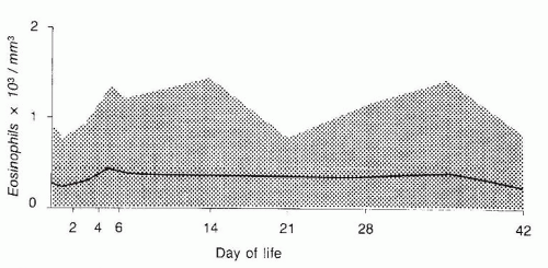
FIGURE 43.16 Eosinophil counts in 142 healthy, premature infants. Data at the first point, day 0, are cord blood values. Subsequent points represent data from capillary blood samples on 1, 5, 7, 14, 28, 35, and 42 days of life. The heavy line represents the mean of each point. The shaded area includes 95% of the infants studied, excluding the top and bottom 2.5% of the group.


▪ PLATELET DISORDERS

A platelet count less than 150 × 109/L is abnormal in term and premature infants (350). Based on very large population-based databases from preterm babies and determination of 5% to 95% margins for normal platelet counts, investigators from the Primary Children’s Medical Center, Salt Lake City, Utah, challenged this traditional definition of thrombocytopenia and proposed a threshold of 100 × 109/L (351). However, it is still controversial whether the reference platelet count data in preterm babies should be derived from CBCs of healthy individuals or CBCs of all preterm babies, where a large proportion of them have pathologic decrease in the platelet counts.

Thrombocytopenia may result from decreased production, increased destruction, sequestration, or some combination of these mechanisms (350). Examination of a peripheral blood smear to assess platelet morphology may yield important information concerning the mechanism of thrombocytopenia; however, examination of megakaryocytes by bone marrow sampling is challenging in newborn infants, and an adequate specimen is often not obtained. Sola et al. (352) have published a technique for bone marrow biopsies in neonates that yields small but high-quality specimens thus allowing accurate assessment of cellularity and megakaryocyte numbers.

There are many causes of thrombocytopenia in the newborn. The most common of these disorders are highlighted in Table 43.12 and have been reviewed elsewhere (350,353,354). The most common disorders with neonatal thrombocytopenia or those that are critically important to diagnose in this age group are reviewed or mentioned in detail below.


Neonatal Immune Thrombocytopenia

Immune thrombocytopenia occurs when antibody-sensitized platelets are prematurely destroyed in the reticuloendothelial system, particularly the spleen. Characteristic laboratory features include isolated thrombocytopenia and an increased number of immature megakaryocytes in a bone marrow aspirate.

A variety of conditions are associated with the transplacental passage of maternal antiplatelet antibodies into the fetus, resulting in immunologic destruction of platelets and fetal thrombocytopenia. The antibody may be formed against an antigen on the platelets of the infant (isoimmune or alloimmune thrombocytopenia, in which case the mother’s platelet count is normal) or an antigen
present on the platelets of the mother (autoimmune thrombocytopenia, in which case both the mother and the child may have thrombocytopenia), as occurs in maternal immune thrombocytopenic purpura (ITP) or thrombocytopenia associated with a collagen vascular disorder such as systemic lupus erythematosus.


TABLE 43.12 Causes of Neonatal Thrombocytopenia


Decreased production of platelets


Inherited thrombocytopenias (see further details in Table 43.13)


Thrombocytopenia-absent radius syndrome


Amegakaryocytic thrombocytopenia


Nonsyndromic familial thrombocytopenia


MYH9-related macrothrombocytopenia


Wiskott-Aldrich syndrome


Drug-induced thrombocytopenia


Congenital leukemia


Both decreased production and increased destruction of platelets


Infections


Congenital (CMV, rubella, toxoplasmosis, herpes, others)


Acquired after birth (usually bacterial, most often gram-negative rods or streptococcus group B)


Increased destruction of platelets


Alloimmune


Maternal autoimmune


Neonatal autoimmune


Disseminated intravascular coagulopathy Kasabach-Merritt syndrome


Neonatal Alloimmune Thrombocytopenia

Neonatal alloimmune thrombocytopenia (NAIT) is a rare but potentially severe bleeding disorder with a mechanism analogous to that causing hemolytic disease of the newborn. In NAIT, the infant possesses a platelet antigen of paternal origin that is lacking in the mother. Typically, the infant’s platelets cross the placenta into the maternal circulation during pregnancy or at the time of delivery and cause immunization of the mother, with the formation of antibodies against the foreign platelet antigen. Less frequently, the cause of immunization is exposure of an antigen-negative mother to antigen-positive platelets during transfusion. During pregnancy, transplacental passage of the maternal IgG antibodies leads to sensitization of fetal platelets. Sensitized platelets are rapidly destroyed in the fetal reticuloendothelial system, particularly the spleen, and the result may be thrombocytopenia in utero and in the infant at the time of delivery. These antibodies often develop during a first pregnancy.

Human platelet antigens (HPAs) are expressed on platelet membrane glycoproteins such as GPIIb/IIIa that facilitate hemostasis. Thirty-three human platelet cell surface antigens have been identified, 20 of which reside on the GPIIb/IIIa complex (355). The HPA system of nomenclature has assigned numbers to the genetic polymorphisms that encode these proteins, with the most common allele denoted “a” and the less common allele denoted “b” (356,357,358,359). For example, the most common genotype of HPA-1 is HPA-1a1a. Therefore, NAIT may occur in the setting of a father who is HPA-1a1a, a mother who is HPA-1b1b, and a baby who is HAP-1a1b. HPA-1a (formerly denoted PlA1) is the platelet-specific antigen involved in approximately three-quarters of the cases of NAIT. The second most common NAIT-associated antigen in 15% of the cases is HPA-5a (Table 43.13) (357,359). Other platelet-specific antigens (HPA-5b, HPA-15a, HPA-15b, HPA-3a, HPA-2a, HPA-2b, HPA-4a) are involved less frequently (357,358,360). Human leukocyte antigen (HLA) alloantibodies often develop as a result of pregnancy and have been proposed to be the cause of NAIT in some cases (357,358,361). However, this requires further investigation.


TABLE 43.13 Platelet-Specific Alloantigens


Biallelic HPA


Allele frequencya


Antigens


Caucasian (%)


African (%)


Asian (%)


Glycoprotein/Amino Acid Change


Encoding Gene/Nucleotide Change


Immune Platelet Disorder Reports


HPA-1a


72 a/a


90


100


GPIIIa/L33P


ITGB3/T196C


NAIT, PTP, MPR


HPA-1b


26 a/b


10


0


2 b/b


HPA-2a


85 a/a


71


95


GPIbα/T145M


GPIBA/C524T


NAIT, PTP, MPR


HPA-2b


14 a/b


29


5


1 b/b


HPA-3a


37 a/a


68


59.5


GPIIb/I843S


ITGA2B/T2621G


NAIT, PTP, MPR


HPA-3b


48 a/b


32


40.5


15 b/b


HPA-4a


>99.9 a/a


100


99.5


GPIIIa/R143Q


ITGB3/G526A


NAIT, PTP, MPR


HPA-4b


<0.1 a/b


0


0.5


<0.1 a/b


HPA-5a


88 a/a


82


98.6


GPIa/E505K


ITGA2/G1648A


NAIT, PTP, MPR


HPA-5b


20 a/b


1 8


0.4


1 b/b


HPA-15a


35 a/a


65


53


CD109/Y703S


CD109/A2108C


NAIT, PTP, MPR


HPA-15b


42 a/b


35


47


23 b/b


aPhenotypic frequencies are Caucasian (North America);, African (Benin), and Asian (China). HPA frequencies in other races and ethnic groups can be found at: http://www.ebi.ac.uk/ipd/hpa/freqs_1.html. NAIT, neonatal alloimmune thrombocytopenia; PTP, posttransfusion purpura; MPR, multi-platelet transfusion refractoriness. Adapted from Curtis BR, McFarland JG. Human platelet antigens—2013. Vox Sang 2014;106:93. doi:10.1111/vox.12085, with permission.


The incidence of NAIT, based on data from prospective studies, is estimated as 1 in 1,000 to 2,000 live-born infants (362,363). The typical infant with NAIT is term and generally well appearing. Cutaneous manifestations of severe thrombocytopenia including petechiae (seen in 90% of cases) and hematoma (66% of cases) are often the only abnormalities found on physical examination (359,364). A complete blood count shows severe isolated thrombocytopenia with a normal hemoglobin and leukocyte count. NAIT should be considered as a diagnosis in an otherwise healthy newborn infant with severe isolated thrombocytopenia with or without bleeding and a normal maternal platelet count (357). Infants affected by NAIT are at risk of serious hemorrhage, particularly into the central nervous system (CNS) (365). NAIT is the most common cause of severe thrombocytopenia (<50 × 109/L) with intracranial hemorrhage (ICH) in term infants (366,367). The incidence of ICH in NAIT is about 20%, and over half of the bleeds occur in utero (364,368).

Early diagnosis and treatment of infants with NAIT are critical (369). In a study from Norway screening for NAIT in 100,448 pregnant women, early intervention resulted in only 5% severe NAIT-related complications compared to about 20% in pooled historical controls (363). Intervention in immunized women included delivery by CS 2 to 4 weeks prior to term, and preparation of platelets from HPA-1a-negative donors to be transfused immediately after birth if petechiae were present and/or if platelet count was less than 35 × 109/L. In this population, 2.1% of women were found to be HPA-1a negative, and anti-HPA-1a was detected in 10.6% of these cases. Fifty-seven of the 161 HPA-1a-positive children had severe thrombocytopenia, and severe bleeding complications occurred in 3. A systematic review of this study and nine other
studies (with a total of 3,028 pregnancies) noted that screening for HPA-1a alloimmunization detects approximately 2 cases of NAIT in 1,000 pregnancies and concluded that screening along with antenatal treatment may indeed reduce the morbidity and mortality associated with NAIT (370). Two additional important points emerge from these prospective screening studies. First, not all pregnancies involving an HPA-1a (PlA1)-alloimmunized mother result in a thrombocytopenic fetus/neonate. Second, HPA-1a (PlA1) alloantibodies may first be detected in the postpartum period as was the case in 39 pregnancies in the large Norwegian study, with the potential to affect future pregnancies (362,363). A recombinant HPA-1a antibody, which competes for binding to the HPA-1a epitope but carries a modified constant region that does not bind to Fcγ receptors, was developed (371). The antibody was studied in healthy HPA-1a1b volunteer subjects and showed improved survival of the subjects’ platelets after ex vivo incubation with the recombinant antibody and reinfusion, as compared to incubation with a destructive IgG1 antibody. The same survival benefit was seen after the platelets were incubated with a mixture of the recombinant antibody and a destructive antibody. The study provides the groundwork for clinical trials studying the ability of the antibody to prevent alloimmunization in HPA-1a-negative mothers.

Infants affected by NAIT with clinical bleeding or platelet counts less than 50 × 109/L should receive antigen-negative, compatible platelets harvested from the mother or a phenotyped (typically HPA-1a negative and HPA-5b negative) blood donor if available. If maternal platelets are used, supernatant plasma with pathologic antibody should be removed by centrifugation or washing and the compatible maternal platelets infused after irradiation. In clinical practice, infants with bleeding and/or severe thrombocytopenia are often initially transfused with a unit of random donor platelets. Kiefel et al. retrospectively analyzed 27 cases of infants with NAIT who received a transfusion of antigen-positive platelets from a random donor. They found an increment in platelet count by at least 30 × 109/L and recommended this approach while waiting for compatible platelets (372). In another study, random donor platelets resulted in platelet increment of about half of that achieved with antigen-matched donor (373). Platelet survival is also different when random platelets are transfused. In the study published by Allen et al., the time to drop in platelet counts to less than 50 × 109/L in the patients who were transfused with random platelets was 3 days compared to 6 days in those cases where compatible platelets were transfused (373). The data demonstrate superiority of compatible donor, but this study and others (374) suggest that random donor platelets can be used when compatible platelets are not available in a timely fashion. Administration of IVIG in addition to platelet transfusions may improve platelet increment and survival with random donor platelet transfusions in NAIT (374,375). IVIG is given at a dose of 1 g/kg over 6 to 8 hours on 2 consecutive days; methylprednisolone (1 mg every 8 hours) may also be used concurrently with platelet transfusions (376).

Ninety-eight percent of individuals of European descent carry the HPA-1a (PlA1) antigen. In other ethnic groups, other platelet-specific alloantigens should be considered. The rarity of the HPA-1a (PlA1 negative) phenotype in individuals of African and Asian descent explains why HPA-1a-associated NAIT is extremely rare in these populations. Among Japanese patients, the majority of cases of NAIT are caused by antibodies to HPA-4b (377).

In patients of Caucasian descent, transfusion with HPA-1a- or HPA-5b-negative platelets is effective in over 95% of cases of NAIT, which is considered the initial treatment of choice for this condition (378). Regional blood centers serving large NICUs should be encouraged to maintain an ongoing list of potential donors who are readily available for donation and have platelet phenotypes that are most commonly required for neonates with NAIT in this population.

The risk of NAIT to recur in subsequent pregnancies is high and depends on the platelet genotype of the pregnant woman’s partner. If the partner is homozygous (HPA-1a/1a), the rate of recurrence is essentially 100%, whereas the risk is 50% of the partner in heterozygous (HPA-1a/1b). This has led to the recommendation that in at-risk pregnancies, especially when a previous sibling was affected, fetal platelet antigen typing should be performed on DNA of the fetal amniocytes obtained following amniocentesis at 15 to 18 weeks of gestation (362). If the fetus is determined to be HPA-1a negative, no further intervention is needed. If the fetus is HPA-1a positive, a cordocentesis may be planned at 20 to 24 weeks of gestation to determine the fetal platelet count and to guide antenatal management; however, this invasive practice is falling out of favor, and prenatal treatment is often now started empirically (379,380). Recent efforts to avoid invasive testing have included the study of predictive parameters of severe fetal thrombocytopenia from NAIT such as the maternal anti-HPA-1a antibody concentration (381,382) as well as the development of noninvasive fetal genotyping of HPA-1a (383).

A subsequent child affected by NAIT typically has thrombocytopenia that is at least as severe as in the first affected sibling (384). In North America, the most common antenatal regime is weekly administration of IVIG to the mother with or without the addition of corticosteroids, although the optimal prenatal management of NAIT is not yet clear (385). Treatment in terms of the timing, dosing, and inclusion of steroids may be stratified based on the history of the affected sibling and may be initiated as early as 12 weeks of gestation if there is a history of ICH in a previously affected sibling (386,387).

The mode of the delivery of babies that are at risk of developing NAIT can also be managed by a noninvasive approach based on risk stratification and fetal blood sampling. A cesarean delivery is often recommended, particularly if the pregnancy is deemed high or very high risk. Alternatively, if a trial of vaginal labor is desired, fetal blood sampling may be performed with the administration of platelets via intrauterine fetal transfusion for platelets less than 50 × 109/L followed by induction of vaginal labor and delivery, ideally without instrumentation (386,388). The optimal timing of delivery is not clear; however, a risk-based approach is often used whereby higher-risk cases will be delivered earlier weighing the risks of ICH against prematurity (388).

At the time of delivery, a cord blood platelet count should be obtained, and thrombocytopenia should be verified in a peripheral blood sample obtained shortly after delivery. If the infant is severely thrombocytopenic, platelets should be infused immediately and a head ultrasound should be performed as soon as possible (389). These pregnancies are high risk and should be managed by a team of perinatologists, neonatologists, and hematologists.


Neonatal Autoimmune Thrombocytopenia

Clinical and laboratory features of neonatal autoimmune thrombocytopenia parallel those of the alloimmune state. In both disorders, the observation of ecchymoses, a petechial rash, or both in an otherwise well infant may be the first clue to the disorder. Measurement of a maternal platelet count and examination of a peripheral blood smear obtained from the mother can help to differentiate autoimmune from alloimmune neonatal thrombocytopenia. In NAIT, the maternal peripheral blood smear and platelet count are normal, whereas in the autoimmune condition (e.g., mothers with autoimmune ITP), the platelet count is reduced, and the existing platelets are often large (i.e., megathrombocytes). Occasionally, the finding of unexpected thrombocytopenia in an infant may lead to the diagnosis of previously unrecognized ITP in the mother. Autoimmune thrombocytopenia may occur in infants of mothers with ITP who have normal platelet counts after splenectomy. Occasionally, in mothers with ITP, increased bone marrow activity can compensate for accelerated destruction of antibody-sensitized platelets. These women may have normal platelet counts, but their infants are at risk of developing thrombocytopenia.

The risk of thrombocytopenia in the infant born to a mother with ITP has been reported as 5%, 10%, and 25% for platelet counts of less than 20, 50, and 150 × 109/L, respectively (390,391). Importantly, the risk of ICH is very low in this setting (0% to 1.5%) and does not
seem to increase with vaginal delivery (392). Maternal history of ITP, platelet count, and treatment are not necessarily predictive of the newborn’s platelet counts and risk of hemorrhage (393). However, a recent analysis of 127 pregnancies of women with ITP showed that having either a previously affected offspring, or ITP that was refractory to splenectomy are risk factors for significant neonatal autoimmune thrombocytopenia (394). Having a previously affected offspring and a low maternal platelet nadir were found to be significant risk factors in a similar study of 67 neonates (395).

Management of infants with autoimmune neonatal thrombocytopenia differs from that of those with the alloimmune form of the disease. Compatible platelets cannot be found because platelet autoantibodies react with all donor platelets. In infants with significant thrombocytopenia (i.e., platelet counts <50 × 109/L) or clinical bleeding, administration of IVIG (typically 1 g/kg daily for 1 or 2 days) results in a marked increment in platelet count within 24 to 48 hours in 75% of patients (395,396,397). Steroids have also been used in this setting; however, they are not considered stand-alone treatment in the thrombocytopenic neonate born to a mother with ITP (398).

Current practice dictates that CS should be reserved for obstetric indications only, and instrumentation is avoided when possible (392,399,400). The platelet count of affected infants should be monitored on a daily basis in the first few day of life as the platelet nadir tends to occur between days 2 and 5. In contrast to NAIT, ICH is more likely to occur between 24 and 28 hours after birth (392,401).


Decreased Production of Platelets

Decreased production of platelets in the bone marrow in the neonatal period can be as a result of various causes including perinatal infections (prenatal, natal, or postnatal), maternal drug use (e.g., azathioprine), inherited marrow failure syndromes, and metabolic defects (e.g., branched amino acid metabolism defects).

Inherited Bone Marrow Failure with Predominantly Thrombocytopenia Several inherited bone marrow failure syndromes are associated with thrombocytopenia. In some of them (e.g., thrombocytopenia -absent radii syndrome), only defects in thrombopoiesis are manifested. In others (e.g., congenital amegakaryocytic thrombocytopenia or Fanconi anemia), the first manifestation is usually thrombocytopenia, but later on, the complete stem cell phenotype is affected with the appearance of multilineage cytopenia.

The genes associated with some of the reported syndromes are known (Table 43.6). Patients with thrombocytopenia-absent radii syndrome have multiple physical malformations. The syndrome is caused by biallelic mutations in RBM8A (402,403). All patients described to date have both thrombocytopenia and absent radii. Thrombocytopenia is commonly severe at birth and gradually improves in the first year or two. There is a small increase in the risk of myelodysplastic syndrome and acute myeloid leukemia in this disorder. Nonsyndromic autosomal dominant familial thrombocytopenia is associated with mutations in the MASTL gene (404), ACBD5 (405) or ANKRD26 (406). Thrombocytopenia is usually mild to moderate and treatment is rarely required. Patients with macrothrombocytopenia related to MYH9 (May-Hegglin/Sebastian/Fechtner/Epstein/Alport syndromes) (407) usually have mild-tomoderated thrombocytopenia. However, occasional patients may need transfusions. Platelet dysfunction is associated with this disorder. Patients with gray platelet syndrome have macrothrombocytopenia, gray platelets, and thrombasthenia. However, clinical bleeding is rare. Patients with mutations in the WAS gene may have the full phenotypic spectrum of Wiskott-Aldrich syndrome or isolated X-linked thrombocytopenia (XLT) (408). In these disorders with WAS gene mutations, the platelets are typically small.

Mutations in CBFA2 cause familial thrombocytopenia with a predisposition to acute myeloid leukemia (409). Patients with C-MPL mutations have congenital amegakaryocytic thrombocytopenia (410) that may present with isolated thrombocytopenia but most commonly progress to aplastic anemia. Development of myelodysplastic syndrome and AML may also occur. Mutations in GATA1 are associated with thrombocytopenia with dyserythropoiesis (411). Patients with HOXA11 mutations have thrombocytopenia associated with radioulnar synostosis (412). Noonan syndrome is caused by mutations in one of the genes in the RAS pathway. Patients may have prolonged and severe thrombocytopenia. The thrombocytopenia may be isolated or associated with broader hematologic disorder that mimics juvenile myelomonocytic leukemia (JMML) (see below). In one child, a 6-week trial of eltrombopag (an agonist of the thrombopoietin receptor) failed to increase the platelet count (413).

Clues for the specific diagnosis of the inherited marrow failure syndrome can be provided by a family history suggestive of an inherited pattern (e.g., autosomal recessive in amegakaryocytic thrombocytopenia and autosomal dominant in familial thrombocytopenia with a predisposition to acute myelogenous leukemia); age at diagnosis (e.g., birth in thrombocytopenia-absent radii syndrome and adulthood in thrombocytopenia with a predisposition to acute myelogenous leukemia); associated nonhematologic manifestations (e.g., limb abnormalities in thrombocytopenia-absent radius and radioulnar synostosis syndromes and renal anomalies in Alport syndrome) (414).

A battery of tests, including bone marrow aspiration and biopsy, are usually required to establish a diagnosis, and early referral to a hematologist is advisable. Management varies from supportive care during surgical procedures and avoidance of antiplatelet agents to regular platelet transfusions or hematopoietic stem cell transplantation. The efficacy of thrombopoietic growth factors in this group of patients has to be determined.


Thrombocytopenia in Premature Newborn Infants

The commonest example of thrombocytopenia in neonates is that seen in premature LBW infants admitted to NICUs. In this patient population, the frequency of thrombocytopenia, defined by a platelet count of less than 150 × 109/L, is of the order of 25% (415,416). In the prospective study of 807 consecutive newborn infants reported by Castle et al. (415), the incidence of thrombocytopenia was 22%; in 38% of thrombocytopenic infants, the platelet count was 50 to 100 × 109/L, and in 20%, the platelet count was less than 50 × 109/L.

A practical classification of thrombocytopenia in neonates has been proposed by Roberts et al. (353). In this classification, a distinction is made between early-onset and late-onset thrombocytopenia. Early-onset thrombocytopenia is present at birth or occurs within 72 hours of life. Only a minority of these cases will have immunologic disorders (e.g., neonatal auto-/alloimmune thrombocytopenia) or coagulopathy (e.g., DIC) as a cause for thrombocytopenia. The vast majority of cases are preterm infants born following pregnancies complicated by placental insufficiency or fetal hypoxia, for example, maternal PIH, fetal intrauterine growth restriction, and maternal diabetes (417,418). Typically, these infants have modest thrombocytopenia (platelet counts 100 to 150 × 109/L). Following birth, their platelet count falls, reaching a nadir by postnatal days 4 to 5 before recovering to greater than 150 × 109/L by 7 to 10 days of age (415). Although earlier studies suggested that both decreased platelet production (419) and increased platelet destruction (415) contribute to the thrombocytopenia in neonates admitted to NICUs, later studies suggested that early-onset thrombocytopenia in LBW infants is primarily the result of a transient impairment of megakaryocytopoiesis (420). In contrast, late-onset neonatal thrombocytopenia defined by a platelet count of less than 150 × 109/L occurring after the first 72 hours of life most commonly results from sepsis or NEC. This late-onset thrombocytopenia has a significant natural history (421). Thrombocytopenia usually progresses rapidly with a platelet nadir that is reached within 24 to 48 hours and is frequently severe with the platelet nadir often falling below 50 × 109/L. Affected infants often require support with platelet transfusion. Platelet recovery is slow, occurring over 5 to 7 days as sepsis or NEC is controlled. Neonates with this type of thrombocytopenia initially require careful monitoring of their platelet count (at least every 12 hours) to
track their platelet nadir and to time appropriate intervention with platelet transfusions. In neonates with NEC, a rapid fall in platelet count to a level well below 100 × 109/L may be a useful marker of intestinal gangrene (422). Finally, in neonates with thrombocytopenia and no apparent cause, thrombosis should be considered.


Evaluation of the Neonate with Thrombocytopenia

An approach to the multiple diagnostic possibilities is outlined in Figure 43.17. It is important to study the mother, as it is to study the infant and to examine the placenta (for multiple hemangiomas). Points requiring specific inquiry include a maternal history of previous bleeding in the form of purpura, bruising, or nose bleeds that might suggest a diagnosis of ITP at some time in the past or more rarely familial thrombocytopenia; ingestion of drugs that may cause thrombocytopenia in the mother and infant (e.g., quinidine, quinine). Maternal exposure to infections or development of skin rash during pregnancy should prompt investigation toward infectious causes including serologic evidence of congenital infections (e.g., syphilis, CMV, herpesvirus, toxoplasmosis). An accurate maternal platelet count should be performed as soon as possible after delivery so that immune neonatal thrombocytopenia caused by maternal ITP or familial thrombocytopenia can be differentiated from that caused by platelet alloimmunization, in which case the mother’s platelet count is normal. A history of previous siblings affected with transient bleeding and/or thrombocytopenia in infancy raises the possibilities of alloimmune or autoimmune thrombocytopenia. Chronic thrombocytopenia in relatives may indicate an inherited thrombocytopenia.

Physical findings of importance in the differential diagnosis of the affected newborn include hepatosplenomegaly and congenital anomalies. Hepatosplenomegaly is often accompanied by jaundice and suggests an infectious process as the most likely cause of thrombocytopenia. In some cases, congenital leukemia may have to be considered. Among the congenital anomalies associated with neonatal thrombocytopenia, the commonest group recognizable at birth is that occurring in the rubella syndrome (congenital heart defects, cataracts, and microcephaly). Physical malformation without clear association with infections indicates an investigation for inherited thrombocytopenias. Deformity and shortening of the forearms due to bilateral absence of the radii are associated with thrombocytopenia -absent radii. A single large hemangioma or multiple smaller hemangiomas point to possible platelet trapping and should prompt a search for bruits produced by internal hemangiomas.


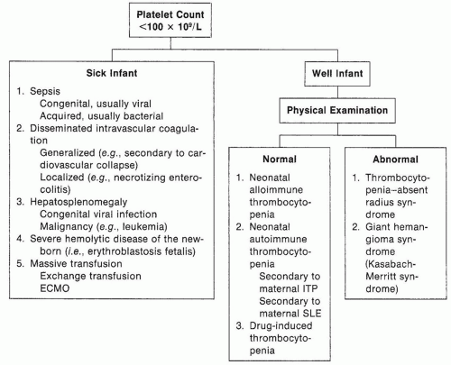
FIGURE 43.17 Approach to the diagnosis of the thrombocytopenic newborn (ECMO, extracorporeal membrane oxygenation; ITP, immune thrombocytopenic purpura; SLE, systemic lupus erythematosus).

A complete blood count on the infant should include a hemoglobin determination, leukocyte count, platelet count, platelet size, and a blood smear. Associated anemia may result from blood loss, concurrent hemolysis (e.g., infection associated), marrow failure, or marrow infiltration caused by congenital leukemia. Leukocytosis may accompany infection or blood loss; however, prominent leukocytosis that exceeds 40 to 50 × 109/L or persistent leukocytosis requires investigation for an underlying blood dyscrasia. Careful examination of the smear is critically important. Large platelets should raise the possibility of inherited macrothrombocytopenia. Small platelets may indicate XLT or Wiskott-Aldrich syndrome. Bone marrow examination should be considered if thrombocytopenia is persistent and a specific cause cannot be identified. Serologic tests for platelet antibodies and platelet antigen typing are generally only available in reference laboratories. If alloimmune thrombocytopenia is suspected by the finding of an otherwise normal newborn with thrombocytopenia and a healthy mother with a normal platelet count, blood should be drawn from the parents soon after delivery for serologic testing. Characteristically, the maternal serum contains an antibody reactive against paternal platelets. Platelet antigen typing should be performed on both parents, if available; in cases of NAIT, the mother will type negative and the father positive for the pathologic platelet-specific antigen. In this situation, the infant’s platelet type is assumed to be identical to that of the father because it is usually not possible to obtain sufficient blood from severely thrombocytopenic newborn infants for extensive serologic testing. Results of platelet studies may not be available for some time, and therapy should not be delayed pending their results.


Disorders with Pancytopenia

Reduced levels of multiple blood cell lineages can be because of underproduction of cells in the bone marrow, peripheral destruction,
and sequestration in specific organs such as the spleen. In this section of the chapter, the focus is on disorders that have not been covered by other sections.


Nonimmune Peripheral Blood Cell Destruction

Peripheral destruction of blood cells can be caused by different etiologies. A major cause is perinatal infections due to microorganism such as streptococcal group B, CMV, rubella, herpesviruses, and toxoplasmosis. Any neonate with pancytopenia should be investigated for the possibility of infections even if the cause is clearly an inherited disorder that causes low blood counts. Disseminated intravascular coagulopathy is commonly caused by infections but can also be caused by variety of other disorders that affect the coagulation system (see section “Disorders of Coagulation Factors”). Large vascular anomalies such as those occur in congenital hemangiomas may lead to intravascular clot formation and local consumption and destruction of RBCs and platelets (see section “Disorders of Coagulation Factors”). Low fibrinogen and high fibrin degradation products are typical findings.


Immune-Mediated Pancytopenia

Multilineage cytopenia can be due to transplacental transfer of antibodies from a mother who suffers from an autoimmune disease such as systemic lupus erythematosus. Transplacental transfer of alloantibodies against several blood lineages is another cause of immune-mediated multilineage cytopenia. Severe cytopenia due to both maternal transfer of autoantibodies or alloantibodies can be treated with transfusions of compatible RBCs and platelets products, IVIG, and G-CSF according to the cell line that is severely affected. Patients with immunodeficiency such as ALPS may develop autoimmune pancytopenia early in life. These conditions are discussed in the section on Lymphopenia.


Hypersplenism

Sequestration of blood cells in a very large and hyperactive spleen may lead to significant cytopenia. Typically, blood cell counts are mildly to moderately reduced. Severe reduction in blood cells indicates an investigation for other or concomitant causes. Potential causes in the neonatal period are infections and metabolic and hemolytic disorders. Treatment should be mainly directed at the underlying cause.


Shwachman-Diamond Syndrome

Shwachman-Diamond syndrome (SDS) is a multisystem autosomal recessive disease characterized by varying degrees of bone marrow failure and cytopenia, most commonly neutropenia (423). Exocrine pancreatic dysfunction and metaphyseal dysplasia are other major features of the disorder. Various B-, T-, and NK cell abnormalities are common (424). Patients may present in the neonatal period with failure to thrive, a small chest with thoracic dystrophy, or infection as a result of neutropenia. The bone marrow may show hypocellularity with maturation arrest of myeloid elements, but it may also appear normal or even hypercellular in the first several years of life (425) The bone marrow failure may results in severe cytopenia as a result of increased apoptosis (426) through the Fas pathway (427,428) and abnormal marrow stromal function. The syndrome is associated with biallelic mutations in the SBDS gene (429) that plays a role in maturation of the large ribosome unit (430). Patients with SDS are susceptible to recurrent viral, bacterial, and fungal infections. Overwhelming sepsis is a well-recognized fatal complication of the disorder particularly early in life. Transformation to myelodysplastic syndrome and acute myeloid leukemia are major causes of morbidity and mortality (425); hence, early diagnosis is important for initiating cancer surveillance and providing family counseling. Transfusions, cytokine therapy, or androgens may be required to treat the cytopenia; however, the only curative treatment for the hematologic complications is allogeneic hematopoietic stem cell transplantation.


Dyskeratosis Congenita

Typically, patients have cytopenia, macrocytosis, high fetal hemoglobin, mucous membrane leukoplakia, dystrophic nails, reticulated skin pigmentation, and increased lacrimation as a result of atresia of the lacrimal ducts, which appear in early or late childhood (431). Severe cases commonly present in the neonatal period with thrombocytopenia, immunodeficiency, and varying CNS malformations. In the X-linked subtype, severe immunodeficiency may occur (432,433). Multiple genes have been found to be associated with the diseases: DKC1 (X-linked recessive); TINF2, hTR, and TERT (autosomal dominant); and NHP2, HOP10, CTC1, and WRAP53 (autosomal recessive). The gene products are involved in telomere maintenance. Treatment of the hematologic complications includes transfusions, cytokines, and androgens (434), but only allogeneic hematopoietic stem cell transplantation is curative (435). Due to the characteristic genomic instability in the disorder, the management should include minimal and judicious usage of radiation imaging to reduce the risk of cancer.


Congenital Amegakaryocytic Thrombocytopenia

Children with this disorder typically present with thrombocytopenia at birth, which gradually progresses to pancytopenia. Initially the bone marrow shows a paucity of megakaryocytes, but later on varying degrees of decreased cellularity develops (436). Nonhematologic manifestations can occur but are not frequent. This disorder is associated with mutations in the gene (C-mpl) encoding for the thrombopoietin receptor (437). The only curative treatment is allogeneic hematopoietic stem cell transplantation (438).


Fanconi Anemia

Fanconi anemia is an inherited marrow failure syndrome with a defect in DNA repair and accelerated apoptosis. Thus far, 17 different genes have been found to be mutated in patients with the disease (reviewed in (439)). Some children present with only hematologic abnormalities, although others have multiple congenital anomalies including thumbs, kidneys, cardiac, and skin defects. Patients may have characteristic facies. Leukemia and solid tumors are common and can appear at birth (440) or shortly after. Although early diagnosis is critically important for proper care, initiation of cancer surveillance, and family counseling, it is often delayed since only about 4% of the children have cytopenia in the neonatal period (441). Diagnosis should be suspected in any patient with characteristic congenital malformation. Currently, diagnosis is usually made at the age of 4 to 8 years, when children develop cytopenia, macrocytosis, and a high fetal hemoglobin. The chromosomal fragility testing of peripheral blood cells is positive in almost all patients. Treatment of the cytopenia includes transfusion, cytokines, and androgens, but only allogeneic hematopoietic stem cell transplantation is curative (442,443). The Fanconi anemia genes are critical for DNA repair, replication fork progression, cytokinesis, and cell survival. Therefore, the management should include minimal and judicious usage of radiation imaging to reduce the risk of cancer.


Cytopenia Related to Metabolic Disorders

Cytopenia can be seen in inherited defects in the metabolism of branched amino acids such as propionic aciduria and methylmalonic aciduria. The cytopenia is probably related to a toxic effect of specific metabolites whose serum levels are increased causing underproduction (444,445). Deterioration in the cytopenia can occur with episodes of metabolic crisis. Pearson disease is caused by deletional mutations in mitochondrial DNA (446) and is associated with variable degrees of exocrine pancreatic dysfunction, pancytopenia, and metabolic acidosis (447). Marrow aspirates typically show ring sideroblasts and vacuoles in myeloid and erythroid precursors. However, the absence of these findings in the bone
marrow does not exclude the diagnosis, and genetic testing should be pursued if the clinical presentation is suggestive of the disorder. The cytopenia tends to improve with age (448,449). Using induced pluripotent cells from patients with Pearson syndrome, defects in cell growth and mitochondrial function have been demonstrated during hematopoietic cell differentiation (450).


Evaluation of the Child with Pancytopenia

Any neonate with pancytopenia should be investigated for the possibility of infections even if the cause seems clearly to be associated with a noninfectious disorder. Maternal history of drug usage as well as history of cytopenia in the mother or other family members should always be taken. Physical examination showing large spleen size may suggest hypersplenism. Anemia, thrombocytopenia, low fibrinogen, and high D-dimers with or without skin hemangiomas indicate investigation for disseminated intravascular coagulopathy and large internal vascular anomalies. Short stature, small head circumference, and physical malformations may suggest inherited bone marrow failure disorder. Large liver and spleen may suggest leukemia. Large liver size, low glucose levels, and acidosis may suggest metabolic disorders.

Evidence for peripheral blood cell destruction comes from high unconjugated bilirubin and LDH. High reticulocyte count is supportive, and high haptoglobin levels are suggestive of no peripheral destruction, but normal reticulocytes and low haptoglobin are not helpful clues. Free hemoglobin in the plasma and antiglobulin test are useful in this setting. Evidence for immune-mediated peripheral blood destruction can come from detection of antibodies against RBCs, platelets, and neutrophils and from genetic testing of mismatched antigens between the mother and baby. Evaluation of the immune system by testing the immunoglobulin as well as the T, B, and NK cell lymphocytes can be helpful for both autoimmune cytopenia as well as inherited bone marrow failure syndromes.

Imaging such as ultrasonographic examination of the abdomen, skeletal survey, echocardiography, and magnetic resonance imaging (MRI) of the brain may be indicated to rule out malformations that are typical for inherited bone marrow failure disorders. However, if an inherited bone marrow failure syndrome with genomic instability is considered, radiation should be avoided whenever possible to minimize an additive risk cancer. Screening for common inherited bone marrow failure syndromes with pancytopenia include pancreatic enzyme, vitamin A/D-1,25 or D-25 and E for SDS, and telomere length for dyskeratosis congenita. Chromosome fragility testing should be done in case of physical malformation suggestive of Fanconi anemia even if cytopenia is not evident. Analysis of specific candidate genes or utilization of more comprehensive next-generation sequencing panels should be considered. Peripheral blood karyotype, comparative genomic hybridization, or single nucleotide polymorphism arrays may be necessary to rule out large deletions of genes.

Deficiency of vitamin B12 and folic acid is rare in the neonatal period but can be tested if suspected by evaluating blood smears, bilirubin and LDH levels, and the vitamins’ levels in RBCs.

If leukemia is suspected, uric acid and LDH should be done in addition to electrolytes, phosphorus, renal function, and liver function. Bone marrow aspirate is a useful to direct investigations toward leukemia, bone marrow failure, and other bone marrow pathologies. The test should be performed when a specific bone marrow disorder is highly suspected or should be ruled out. The time of the test should be determined based on the urgency of establishing a diagnosis and initiating treatment.

Biopsy of the skin, muscle, or liver may be necessary to identify metabolic disorders and sometimes inherited bone marrow failure syndromes that sometimes are associated with specific tissue phenotype or a mixed population of cells and growth advantage of normal cells.


Leukemia and Myeloproliferative Syndromes


Congenital Leukemia

Congenital leukemia is rare, occurring in up to five cases per million births. It is defined as leukemia that is diagnosed before the age of 4 weeks (451,452). Infants show a larger proportion (50% to 80%) of acute myeloid leukemia, commonly either monoblastic or myelomonocytic leukemia. The rest are pre-B acute lymphoblastic (20% to 50%) and mixed lineage (about 5%) leukemias. The clinical presentation is defined by a high white blood cell count, hepatosplenomegaly, and in many cases, CNS involvement. Skin involvement is common in myeloid and in lymphoid leukemia.

In both, ALL and AML, there is high frequency of the chromosomal translocation t(4;11) that is associated with internal duplication or deletion involving the mixed lineage leukemia (MLL) gene at chromosome band 11q23 (452,453,454). Intensive chemotherapy combined with hematopoietic stem cell transplantation is the mainstay of treatment. However, the prognosis of congenital leukemia remains unfavorable. In contrast to older children, ALL confers worse prognosis than AML, and long-term survival is achieved in less than a quarter of the patients with ALL and in less of half of the patients with AML (452,453).

The study of newborns has significantly advanced our understanding of leukemia. The prenatal origin of leukemia became evident from studies showing that ALL may arise in monozygotic twins from a prenatally shared population of leukemic blasts after a variable latency period (455,456).


Transient Myeloproliferative Disorder in Infants with Down Syndrome

Children with Down syndrome (constitutional trisomy 21) have a 10- to 20-fold increased overall risk of developing acute lymphoblastic leukemia and acute myeloid leukemia (457,458). Unique to individuals with Down syndrome, a transient myeloproliferative syndrome (TMD) is observed in at least 10% of newborns (459). TMD can develop also in patients with mosaicism for trisomy 21. Newborns with Down syndrome and TMD are typically well and show circulating blasts in their peripheral blood that are indistinguishable from those of acute megakaryoblastic leukemia (acute myeloblastic leukemia type 7). The number of blasts may exceed 100 × 103/L or may be barely detectable. Some infants have hepatosplenomegaly or skin infiltrates. Remarkably, the blasts of TMD in Down syndrome disappear spontaneously in the majority of cases in the first 3 to 4 months of life. After this spontaneous resolution of the TMD, however, approximately 20% of these children go on to develop acute megakaryoblastic leukemia later in life. Therapeutic intervention is reserved for the approximately 15% to 20% of cases of TMD who develop life-threatening complications such as hyperleukocytosis, pericardial or pleural infusions, and acute liver failure (460). Progressive liver failure as a result of blast infiltration or fibrosis is frequently fatal (461) despite therapeutic reduction of the circulating and infiltrating blasts.

Inactivating truncation mutations within the gene encoding the hematopoietic transcription factor GATA1 were shown to be specific for the blast cells with trisomy 21 at the TMD phase (462,463,464) and also later on if the children develop acute megakaryoblastic leukemia (465). In a portion of the patients with TMD and in all cases of acute myeloblastic leukemia, additional mutations/deletions in additional genes that had been proven as transformation drivers in leukemia in the general population (e.g., EZH2, APC, FLT3, and JAK1) have been found (466).

Management of newborns with Down syndrome should include a complete blood count with a careful review of the blood smear. Immunophenotyping and cytogenetic evaluation of any blast population is recommended. Although observation is sufficient for the majority of newborns with Down syndrome and TMD, neonatologists need to be aware of the potential complications that were mentioned above and indicate chemotherapeutic intervention.


Juvenile Myelomonocytic Leukemia and Juvenile Myelomonocytic Leukemia-Like Syndromes

JMML previously referred to as juvenile chronic myelogenous leukemia (JCML) is a rare myeloproliferative disorder in the neonatal period and early infancy (467,468). Suggestive clinical features include hepatosplenomegaly, lymphadenopathy, pallor, fever, and skin rash. The peripheral blood shows leukocytosis with immature myeloid cells and monocytosis. Fetal hemoglobin may be elevated but is not a useful indicator for this diagnosis in the neonatal period. Clonal chromosomal abnormalities, for example, monosomy 7, support the diagnosis. The Philadelphia chromosome and BCR-ABL fusion transcript, which are typical of adult chronic myelogenous leukemia, are absent in JMML. In vitro bone marrow cultures show growth factor-independent growth (467) and hypersensitivity to GM-CSF (469). JMML is associated with various inherited disorders such as neurofibromatosis (468) and Noonan syndrome (470). Somatic mutations of NF1 (471) and PTPN11 (470), of other genes in the RAS pathway, and of SETBP1 and JAK3 (472) have also been found. Urgent and comprehensive clinical evaluation and genetic investigation are critical for the management of the patients, since certain inherited types of JMML, such as those associated with germline mutations in PTPN11 (473,474) and CBL (475), may resolve spontaneously, while other patients who have other inherited or idiopathic JMML may be rapidly progressive and cannot be cured without hematopoietic stem cell transplantation. JMML cases that are not associated with massive hepatosplenomegaly and significant morbidity and tend to regress spontaneously over time are sometimes referred to as JMML-like syndromes. For those who need aggressive hematopoietic stem cell transplantation, pretransplant chemotherapy or splenectomy does not seem to improve overall survival. However, if patients are symptomatic, mild chemotherapeutic agents such as 6-mercaptopurine and 13-cis-retinoic acid or more aggressive chemotherapy such as high-dose cytarabine and fludarabine in nonresponders can be considered until transplant can be implemented.


▪ DISORDERS OF COAGULATION FACTORS


Developmental Hemostasis

Hemostasis is a physiologic process that aims at stopping blood flow if an injury occurs, while maintaining blood in fluid state under normal circumstances (476). The hemostatic system has three different components: platelets, coagulation factors, and fibrinolytic system. These are referred to as primary, secondary, and tertiary hemostasis, respectively.


TABLE 43.14 APTT Reference Values for Neonates and Children Using Four Different Reagents in Two Landmark Studies


APTT Results (s)


Age


Day 1


Day 3


1 mo-1 y


1-5 y


6-10 y


11-16 y


Adults


PTT-A


38.7a (34.3-44.8)


36.3a (29.5-42.2)


39.3a (35.1-46.3)


37.7a (33.6-43.8)


37.3a (31.8-43.7)


39.5a (33.9-46.1)


33.2 (28.6-38.2)


Monagle et al.


N = 21 (10F/11M)


N = 25 (13F/12M)


N = 35 (3F/30M)


N = 56 (26F/30M)


N = 71 (27F/44M)


N = 54 (12F/42M)


N = 42


CK Prest


Not available


Not available


34.4a (31.1-36.6)


32.3a (29.8-35.0)


32.9a (30.8-34.8)


34.1a (29.4-40.4)


29.1 (25.7-31.5)


Monagle et al.


N = 20 (3F/17M)


N = 22 (11F/11M)


N = 22 (12F/10M)


N = 39 (8F/31M)


N = 40


Actin FSL


Not available


Not available


37.4a (33.4-41.4)


36.7a (31.8-42.8)


35.4a (30.1-40.4)


38.1a (32.2-42.2)


30.8 (27.1-34.3)


Monagle et al.


N = 20 (3F/17M)


N = 20 (10F/10M)


N = 21 (12/9M)


N = 39 (9F/30M)


N = 40


Platelin L


Not available


Not available


36.5a (33.6-40.4)


37.3a (32.5-43.8)


35a (31.0-39.3)


39.4a (32.6-49.2)


31.3 (27.2-35.4)


Monagle et al.


N = 20 (3F/17M)


N = 21 (11F/10M)


N = 22 (12F/10M)


N = 35 (7F/28M)


N = 38


Andrew et al.


42.9b (31.3-54.5)


42.6b (25.4-59.8)


35.5 (28.1-42.9)


30 (24,25,26,27,28,29,30,31,32,33,34,35,36)


31 (26,27,28,29,30,31,32,33,34,35,36)


32 (26,27,28,29,30,31,32,33,34,35,36,37)


33 (27,28,29,30,31,32,33,34,35,36,37,38,39,40)


Note: Andrew et al. results shown for day 3 are actually day 5 results. M, males; F, female. For each reagent, the first row shows the mean and boundaries including 95% of the population. The second row shows the number of individual samples and the ratio of males to females for each group.


aDenotes values that are significantly different from adult values (p < 0.05).


bDenotes values that are significantly different from adult values for Andrew et al. data.


Table is from Monagle P, et al. Developmental haemostasis. Impact for clinical haemostasis laboratories. Thromb Haemost 2006;95:362, with permission.


For many decades, it has been recognized that the hemostatic system is different in neonates and children compared to adults (477,478). The concept of progressive maturation in the hemostatic system from intrauterine life to the adulthood has been termed developmental hemostasis (479).

Maternal proteins involved in hemostasis do not cross the placenta (480) but are synthesized during intrauterine life by the fetus. Functional levels of most of these hemostatic proteins are low in the fetus and progressively increase throughout the neonatal period approximating adult values at around the age of 6 months. This increment is also seen in healthy premature infants, who show a compensatory accelerated maturation of the hemostatic system (481).

Briefly, vitamin K-dependent factors (II, VII, IX, and X), and factors of the contact pathway (XI, XII, prekallikrein, and high molecular weight kininogen), are decreased in the term newborn to around half adult values. Conversely, the levels of coagulation factors V, VIII, and XIII, and von Willebrand factor (vWF) near or exceed adult values at birth. Fibrinogen levels are increased in neonates due to the presence of a fetal variant with increased sialic acid and phosphorus content, but fibrinogen activity is decreased (482).

Both thrombin generation and thrombin inhibition are slower in neonates compared to adults (483). Indeed, the levels of two of the natural inhibitors of thrombin, antithrombin and heparin cofactor II, are low at birth. However, the level of a third natural thrombin inhibitor, alpha-2-macroglobulin, is higher in neonates and children compared to adults. The higher levels in young patients would seemingly compensate for the low antithrombin levels (484). Lastly, the levels of the natural anticoagulants, protein C and S, are approximately 30% of the adult values. Nonetheless, protein S activity is relatively high due to the low levels of its carrier: C4b-binding protein (485).

The levels of the fibrinolytic system proteins are also lower in neonates compared to adults; alpha 2-antiplasmin levels are slightly decreased, whereas neonatal plasminogen levels are half the adult values. Moreover, plasminogen also has a different glycosylation pattern, which may result in a less efficient conversion to plasmin (486).

The immaturity of the hemostatic system, characterized by a low functional level of many of the coagulant and anticoagulant proteins,
is reflected in coagulation tests, and the physician should be aware of the expected physiologic differences when interpreting laboratory results (487). Importantly, despite the immaturity, healthy neonates do not tend to develop bleeding or thrombotic complications (488).


TABLE 43.15 TCT, PT, INR, and Fibrinogen Reference Values for Neonates and Children in Two Landmark Studies


Coagulation Tests


Age


Day 1


Day 3


1 mo-1 y


1-5 y


6-10 y


11-16 y


Adults


TCT (s)


Not available


Not available


17.1a (16.3-17.6)


17.5a (16.5-18.2)


17.1 (16.1-18.5)


16.9 (16.2-17.6)


16.6 (16.2-17.2)


Monagle et al.


N = 20 (10F/10M)


N = 21 (11F/10M)


N = 21 (11F/10M)


N = 22 (11F/11M)


N = 20


TCT Andrew et al.


23.5 (19.0-28.3)


23.1 (18.0-29.2)


24.3 (19.4-29.2)


Not available


Not available


Not available


Not available


PT (s)


15.6a (14.4-16.4)


14.9a (13.5-16.4)


13.1 (11.5-15.3)


13.3a (12.1-14.5)


13.4a (11.7-15.1)


13.8a (12.7-16.1)


13.0 (11.5-14.5)


Monagle et al.


N = 21 (10F/11M)


N = 25 (13F/12M)


N = 35 (8F/27M)


N = 43 (23F/20M)


N = 53 (22F/31M)


N = 23 (7F/16M)


N = 51


PT Andrew et al.


13 (11.6-14.43)


12.4 (10.5-13.86)


12.3 (10.7-13.9)


11 (10.6-11.4)


11.1 (10.1-12.1)


11.2 (10.2-12.0)


12.0 (11.0-14.0)


INR


1.26a (1.15-1.35)


1.20a (1.05-1.35)


1.00 (0.86-1.22)


1.03a (0.92-1.14)


1.04a (0.87-1.20)


1.08a (0.97-1.30)


1.00 (0.80-1.20)


Monagle et al.


N = 21 (10F/11M)


N = 25 (13F/12M)


N = 35 (8F/27M)


N = 43 (23F/20M)


N = 53 (22F/31M)


N = 23 (7F/16M)


N = 51 (43F/8M)


INR Andrew et al.


1b (0.53-1.62)


0.91b (0.53-1.48)


0.88b (0.61-1.17)


1 (0.96-1.04)


1.01 (0.91-1.11)


1.02 (0.93-1.10)


1.10 (1.0-1.3)


Fibrinogen (g/L)


2.80 (1.92-3.74)


3.30 (2.83-4.01)


2.42a (0.82-3.83)


2.82a (1.62-4.01)


3.04 (1.99-4.09)


3.15 (2.12-4.33)


3.1 (1.9-4.3)


N = 22 (10F/12M)


N = 21 (10F/11M)


N = 34 (7F/27M)


N = 43 (23F/20M)


N = 52 (22F/30M)


N = 21 (7F/14M)


N = 55 (47F/8M)


Fibrinogen


2.83 (2.25-3.41)


3.12 (2.37-3.87)


2.51 (1.5-3.87)


2.76 (1.70-4.05)


2.75 (1.57-4.0)


3 (1.54-4.48)


2.78 (1.56-4.0)


Andrew et al.


Note: Andrew et al. results shown for day 3 are actually day 5 results. M, males; F, females.


For each assay, the first row shows the mean and boundaries including 95% of the population. The second row shows the number of individual samples and the ratio of males to females for each group.


aDenotes values that are significantly different from adult values (p < 0.05).


bDenotes values that are significantly different from adult values for Andrew et al. data.


Current reference ranges of screening laboratory assays and coagulant and anticoagulant proteins for neonates are shown in Tables 43.14, 43.15, 43.16. Reference values in premature babies are shown in Table 43.17. These reference values serve as a guideline only, as ideally, laboratories should develop their local reference ranges since the results of the laboratory tests depend not only on the age of the patients but also on the reagents and the analyzer used to perform the tests (489).


TABLE 43.16 Coagulation Factor Reference Values for Neonates and Children from Two Landmark Studies


Coagulation Factors (%)


Age


Day 1


Day 3


1 mo-1 y


1-5 y


6-10 y


11-16 y


Adults


II


54a (41,42,43,44,45,46,47,48,49,50,51,52,53,54,55,56,57,58,59,60,61,62,63,64,65,66,67,68,69)


62a (50,51,52,53,54,55,56,57,58,59,60,61,62,63,64,65,66,67,68,69,70,71,72,73)


90a (62,63,64,65,66,67,68,69,70,71,72,73,74,75,76,77,78,79,80,81,82,83,84,85,86,87,88,89,90,91,92,93,94,95,96,97,98,99,100,101,102,103)


89a (70,71,72,73,74,75,76,77,78,79,80,81,82,83,84,85,86,87,88,89,90,91,92,93,94,95,96,97,98,99,100,101,102,103,104,105,106,107,108,109)


89a (67,68,69,70,71,72,73,74,75,76,77,78,79,80,81,82,83,84,85,86,87,88,89,90,91,92,93,94,95,96,97,98,99,100,101,102,103,104,105,106,107,108,109,110)


90a (61,62,63,64,65,66,67,68,69,70,71,72,73,74,75,76,77,78,79,80,81,82,83,84,85,86,87,88,89,90,91,92,93,94,95,96,97,98,99,100,101,102,103,104,105,106,107)


110 (78,79,80,81,82,83,84,85,86,87,88,89,90,91,92,93,94,95,96,97,98,99,100,101,102,103,104,105,106,107,108,109,110,111,112,113,114,115,116,117,118,119,120,121,122,123,124,125,126,127,128,129,130,131,132,133,134,135,136,137,138)


Monagle et al.


N = 23 (13F/10M)


N = 22 (11F/11M)


N = 22 (7F/15M)


N = 67 (26F/41M)


N = 64 (23F/41M)


N = 23 (6F/17M)


N = 44


II


48b (37,38,39,40,41,42,43,44,45,46,47,48,49,50,51,52,53,54,55,56,57,58,59)


63b (48,49,50,51,52,53,54,55,56,57,58,59,60,61,62,63,64,65,66,67,68,69,70,71,72,73,74,75,76,77,78)


88b (60,61,62,63,64,65,66,67,68,69,70,71,72,73,74,75,76,77,78,79,80,81,82,83,84,85,86,87,88,89,90,91,92,93,94,95,96,97,98,99,100,101,102,103,104,105,106,107,108,109,110,111,112,113,114,115,116)


94b (71,72,73,74,75,76,77,78,79,80,81,82,83,84,85,86,87,88,89,90,91,92,93,94,95,96,97,98,99,100,101,102,103,104,105,106,107,108,109,110,111,112,113,114,115,116)


88 (67,68,69,70,71,72,73,74,75,76,77,78,79,80,81,82,83,84,85,86,87,88,89,90,91,92,93,94,95,96,97,98,99,100,101,102,103,104,105,106,107)


83b (61,62,63,64,65,66,67,68,69,70,71,72,73,74,75,76,77,78,79,80,81,82,83,84,85,86,87,88,89,90,91,92,93,94,95,96,97,98,99,100,101,102,103,104)


108 (70,71,72,73,74,75,76,77,78,79,80,81,82,83,84,85,86,87,88,89,90,91,92,93,94,95,96,97,98,99,100,101,102,103,104,105,106,107,108,109,110,111,112,113,114,115,116,117,118,119,120,121,122,123,124,125,126,127,128,129,130,131,132,133,134,135,136,137,138,139,140,141,142,143,144,145,146)


Andrew et al.


V


81a (64,65,66,67,68,69,70,71,72,73,74,75,76,77,78,79,80,81,82,83,84,85,86,87,88,89,90,91,92,93,94,95,96,97,98,99,100,101,102,103)


122 (92,93,94,95,96,97,98,99,100,101,102,103,104,105,106,107,108,109,110,111,112,113,114,115,116,117,118,119,120,121,122,123,124,125,126,127,128,129,130,131,132,133,134,135,136,137,138,139,140,141,142,143,144,145,146,147,148,149,150,151,152,153,154)


113 (94,95,96,97,98,99,100,101,102,103,104,105,106,107,108,109,110,111,112,113,114,115,116,117,118,119,120,121,122,123,124,125,126,127,128,129,130,131,132,133,134,135,136,137,138,139,140,141)


97a (67,68,69,70,71,72,73,74,75,76,77,78,79,80,81,82,83,84,85,86,87,88,89,90,91,92,93,94,95,96,97,98,99,100,101,102,103,104,105,106,107,108,109,110,111,112,113,114,115,116,117,118,119,120,121,122,123,124,125,126,127)


99a (56,57,58,59,60,61,62,63,64,65,66,67,68,69,70,71,72,73,74,75,76,77,78,79,80,81,82,83,84,85,86,87,88,89,90,91,92,93,94,95,96,97,98,99,100,101,102,103,104,105,106,107,108,109,110,111,112,113,114,115,116,117,118,119,120,121,122,123,124,125,126,127,128,129,130,131,132,133,134,135,136,137,138,139,140,141)


89a (67,68,69,70,71,72,73,74,75,76,77,78,79,80,81,82,83,84,85,86,87,88,89,90,91,92,93,94,95,96,97,98,99,100,101,102,103,104,105,106,107,108,109,110,111,112,113,114,115,116,117,118,119,120,121,122,123,124,125,126,127,128,129,130,131,132,133,134,135,136,137,138,139,140,141)


118 (78,79,80,81,82,83,84,85,86,87,88,89,90,91,92,93,94,95,96,97,98,99,100,101,102,103,104,105,106,107,108,109,110,111,112,113,114,115,116,117,118,119,120,121,122,123,124,125,126,127,128,129,130,131,132,133,134,135,136,137,138,139,140,141,142,143,144,145,146,147,148,149,150,151,152)


Monagle et al.


N = 22 (13F/9M)


N = 22 (11F/11M)


N = 20 (6F/14M)


N = 75 (26F/41M)


N = 64 (23F/41M)


N = 20 (5F/15M)


N = 44


V


72b (54,55,56,57,58,59,60,61,62,63,64,65,66,67,68,69,70,71,72,73,74,75,76,77,78,79,80,81,82,83,84,85,86,87,88,89,90)


95b (70,71,72,73,74,75,76,77,78,79,80,81,82,83,84,85,86,87,88,89,90,91,92,93,94,95,96,97,98,99,100,101,102,103,104,105,106,107,108,109,110,111,112,113,114,115,116,117,118,119,120)


91b (55,56,57,58,59,60,61,62,63,64,65,66,67,68,69,70,71,72,73,74,75,76,77,78,79,80,81,82,83,84,85,86,87,88,89,90,91,92,93,94,95,96,97,98,99,100,101,102,103,104,105,106,107,108,109,110,111,112,113,114,115,116,117,118,119,120,121,122,123,124,125,126,127)


103 (79,80,81,82,83,84,85,86,87,88,89,90,91,92,93,94,95,96,97,98,99,100,101,102,103,104,105,106,107,108,109,110,111,112,113,114,115,116,117,118,119,120,121,122,123,124,125,126,127)


90b (63,64,65,66,67,68,69,70,71,72,73,74,75,76,77,78,79,80,81,82,83,84,85,86,87,88,89,90,91,92,93,94,95,96,97,98,99,100,101,102,103,104,105,106,107,108,109,110,111,112,113,114,115,116)


77b (55,56,57,58,59,60,61,62,63,64,65,66,67,68,69,70,71,72,73,74,75,76,77,78,79,80,81,82,83,84,85,86,87,88,89,90,91,92,93,94,95,96,97,98,99)


106 (62,63,64,65,66,67,68,69,70,71,72,73,74,75,76,77,78,79,80,81,82,83,84,85,86,87,88,89,90,91,92,93,94,95,96,97,98,99,100,101,102,103,104,105,106,107,108,109,110,111,112,113,114,115,116,117,118,119,120,121,122,123,124,125,126,127,128,129,130,131,132,133,134,135,136,137,138,139,140,141,142,143,144,145,146,147,148,149,150)


Andrew et al.


VII


70a (52,53,54,55,56,57,58,59,60,61,62,63,64,65,66,67,68,69,70,71,72,73,74,75,76,77,78,79,80,81,82,83,84,85,86,87,88)


86a (67,68,69,70,71,72,73,74,75,76,77,78,79,80,81,82,83,84,85,86,87,88,89,90,91,92,93,94,95,96,97,98,99,100,101,102,103,104,105,106,107)


128 (83,84,85,86,87,88,89,90,91,92,93,94,95,96,97,98,99,100,101,102,103,104,105,106,107,108,109,110,111,112,113,114,115,116,117,118,119,120,121,122,123,124,125,126,127,128,129,130,131,132,133,134,135,136,137,138,139,140,141,142,143,144,145,146,147,148,149,150,151,152,153,154,155,156,157,158,159,160)


111a (72,73,74,75,76,77,78,79,80,81,82,83,84,85,86,87,88,89,90,91,92,93,94,95,96,97,98,99,100,101,102,103,104,105,106,107,108,109,110,111,112,113,114,115,116,117,118,119,120,121,122,123,124,125,126,127,128,129,130,131,132,133,134,135,136,137,138,139,140,141,142,143,144,145,146,147,148,149,150)


113a (70,71,72,73,74,75,76,77,78,79,80,81,82,83,84,85,86,87,88,89,90,91,92,93,94,95,96,97,98,99,100,101,102,103,104,105,106,107,108,109,110,111,112,113,114,115,116,117,118,119,120,121,122,123,124,125,126,127,128,129,130,131,132,133,134,135,136,137,138,139,140,141,142,143,144,145,146,147,148,149,150,151,152,153,154,155,156)


118 (69,70,71,72,73,74,75,76,77,78,79,80,81,82,83,84,85,86,87,88,89,90,91,92,93,94,95,96,97,98,99,100,101,102,103,104,105,106,107,108,109,110,111,112,113,114,115,116,117,118,119,120,121,122,123,124,125,126,127,128,129,130,131,132,133,134,135,136,137,138,139,140,141,142,143,144,145,146,147,148,149,150,151,152,153,154,155,156,157,158,159,160,161,162,163,164,165,166,167,168,169,170,171,172,173,174,175,176,177,178,179,180,181,182,183,184,185,186,187,188,189,190,191,192,193,194,195,196,197,198,199,200)


129 (61,62,63,64,65,66,67,68,69,70,71,72,73,74,75,76,77,78,79,80,81,82,83,84,85,86,87,88,89,90,91,92,93,94,95,96,97,98,99,100,101,102,103,104,105,106,107,108,109,110,111,112,113,114,115,116,117,118,119,120,121,122,123,124,125,126,127,128,129,130,131,132,133,134,135,136,137,138,139,140,141,142,143,144,145,146,147,148,149,150,151,152,153,154,155,156,157,158,159,160,161,162,163,164,165,166,167,168,169,170,171,172,173,174,175,176,177,178,179,180,181,182,183,184,185,186,187,188,189,190,191,192,193,194,195,196,197,198,199)


Monagle et al.


N = 22 (12F/10M)


N = 22 (11F/11M)


N = 20 (6F/14M)


N = 66 (25F/41M)


N = 64 (23F/41M)


N = 22 (6F/16M)


N = 44


VII


66b (47,48,49,50,51,52,53,54,55,56,57,58,59,60,61,62,63,64,65,66,67,68,69,70,71,72,73,74,75,76,77,78,79,80,81,82,83,84,85)


89b (62,63,64,65,66,67,68,69,70,71,72,73,74,75,76,77,78,79,80,81,82,83,84,85,86,87,88,89,90,91,92,93,94,95,96,97,98,99,100,101,102,103,104,105,106,107,108,109,110,111,112,113,114,115,116)


87b (47,48,49,50,51,52,53,54,55,56,57,58,59,60,61,62,63,64,65,66,67,68,69,70,71,72,73,74,75,76,77,78,79,80,81,82,83,84,85,86,87,88,89,90,91,92,93,94,95,96,97,98,99,100,101,102,103,104,105,106,107,108,109,110,111,112,113,114,115,116,117,118,119,120,121,122,123,124,125,126,127)


82b (55,56,57,58,59,60,61,62,63,64,65,66,67,68,69,70,71,72,73,74,75,76,77,78,79,80,81,82,83,84,85,86,87,88,89,90,91,92,93,94,95,96,97,98,99,100,101,102,103,104,105,106,107,108,109,110,111,112,113,114,115,116)


85 (52,53,54,55,56,57,58,59,60,61,62,63,64,65,66,67,68,69,70,71,72,73,74,75,76,77,78,79,80,81,82,83,84,85,86,87,88,89,90,91,92,93,94,95,96,97,98,99,100,101,102,103,104,105,106,107,108,109,110,111,112,113,114,115,116,117,118,119,120)


83b (58,59,60,61,62,63,64,65,66,67,68,69,70,71,72,73,74,75,76,77,78,79,80,81,82,83,84,85,86,87,88,89,90,91,92,93,94,95,96,97,98,99,100,101,102,103,104,105,106,107,108,109,110,111,112,113,114,115)


105 (67,68,69,70,71,72,73,74,75,76,77,78,79,80,81,82,83,84,85,86,87,88,89,90,91,92,93,94,95,96,97,98,99,100,101,102,103,104,105,106,107,108,109,110,111,112,113,114,115,116,117,118,119,120,121,122,123,124,125,126,127,128,129,130,131,132,133,134,135,136,137,138,139,140,141,142,143)


Andrew et al.


VIII


182 (105,106,107,108,109,110,111,112,113,114,115,116,117,118,119,120,121,122,123,124,125,126,127,128,129,130,131,132,133,134,135,136,137,138,139,140,141,142,143,144,145,146,147,148,149,150,151,152,153,154,155,156,157,158,159,160,161,162,163,164,165,166,167,168,169,170,171,172,173,174,175,176,177,178,179,180,181,182,183,184,185,186,187,188,189,190,191,192,193,194,195,196,197,198,199,200,201,202,203,204,205,206,207,208,209,210,211,212,213,214,215,216,217,218,219,220,221,222,223,224,225,226,227,228,229,230,231,232,233,234,235,236,237,238,239,240,241,242,243,244,245,246,247,248,249,250,251,252,253,254,255,256,257,258,259,260

Only gold members can continue reading. Log In or Register to continue

Stay updated, free articles. Join our Telegram channel

May 30, 2016 | Posted by in PEDIATRICS | Comments Off on Hematology

Full access? Get Clinical Tree

Get Clinical Tree app for offline access