Endocrine Disorders of the Newborn


Endocrine Disorders of the Newborn


Penny M. Feldman

Mary Min-chin Lee


From the moment of conception, physiologic endocrine processes are actively involved in embryonic and fetal growth and development. Disturbances in these complex hormonal processes can affect the fetus and newborn infant. Clinical disorders of endocrine function in the neonate, therefore, can reflect an altered physiologic state in the fetus, the mother, or the fetal-maternal unit. Moreover, perturbations of endocrine function at different stages of fetal development result in diverse clinical manifestations and developmental programming, which may predispose to increased risk for disease in adolescence or adulthood. Knowledge of the ontogeny of the endocrine glands and their physiologic function during fetal development facilitates understanding disorders of endocrine function in the newborn.


▪ DISORDERS OF SEX DEVELOPMENT


Normal Sexual Differentiation

The normal regulation of sexual differentiation is broadly illustrated in 
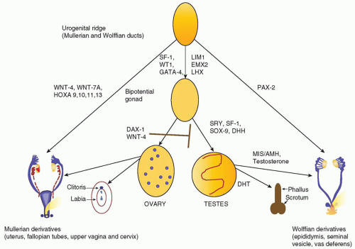
Figure 36.1. All embryos are initially undifferentiated, having a bipotential gonad and the anlagen of both male and female reproductive tracts and genitalia (1). Differentiation of the gonads as testes or ovaries dictates the subsequent development of the internal and external genitalia. The gonad forms when germ cells migrate from the dorsal endoderm of the yolk sac to populate the genital ridges. At the fifth to sixth week of gestation, these primitive bipotential gonads consist of both cortical (ovarian) and medullary (testicular) components. The genital ridge is composed of three cell types:


  • Primordial germ cells destined to become prespermatogonia in the male or oogonia in the female


  • Supporting epithelial somatic cells, destined to become Sertoli cells (male) or granulosa cells (female)


  • Mesenchymal somatic cells destined to become the steroid-producing Leydig cells (male) or theca cells (female)

Figure 36.1 illustrates the genes involved in the differentiation of the genital ridge into a female or male reproductive tract. WT1 and steroidogenic factor 1 (SF-1) play a role early on in the development of the genital ridge and are critical for gonadal development (2). Mutations in these two genes are clearly associated with gonadal dysgenesis:


  • Wilms tumor suppressor gene (WT1) mutations are associated with three related syndromes (the WAGR contiguous gene syndrome, Denys-Drash, and Frasier) that affect renal function and gonadal development (3).


  • Mutations in the transcription factor, SF-1, cause agenesis of the adrenal glands and gonads (4).

Development of the supporting cells as Sertoli or granulosa cells is critical for determining whether the germ cells differentiate as spermatogonia or oogonia. Sexually dimorphic differentiation of the gonads and reproductive system commences when the testis-determining gene is first expressed. In 1959, Ford and colleagues determined that the Y chromosome was necessary for male development (5); in 1966, the critical region for testis determination was localized to the short arm of the Y chromosome (6); and in 1990, the primary testis-determining gene was identified at Yp11.3 by positional cloning in patients with 46,XX testicular disorders of sex development (DSD) (7). This gene, termed SRY (sex-determining region of the Y chromosome), is a member of the SOX family of transcription factors that all contain a high mobility group (HMG) DNA-binding motif (7). Activation of SRY initiates differentiation of the bipotential gonad as a testis. Loss of function mutations in SRY or a delay in its onset of expression can cause 46,XY complete gonadal dysgenesis, while translocation of SRY to the X chromosome or an autosome causes 46,XX testicular DSD.

SOX9, a presumptive target for SRY, is a related HMG box gene that induces the supporting cells of the gonadal ridge to differentiate as Sertoli cells (8). SRY and SF-1 work in concert to activate SOX9 gene expression. Inactivating mutations of SOX9 cause campomelic dysplasia, a syndrome of skeletal anomalies and 46,XY gonadal dysgenesis (9). Mutations affecting genes located downstream of SOX9, SRY, SF-1, and desert hedgehog (Dhh) may also disrupt normal testicular determination (Fig. 36.1).

Sexually dimorphic differentiation of the wolffian (male anlagen) and mullerian (female anlagen) internal genital tracts depends on the hormonal milieu established by the somatic cells. If SRY is expressed, the primary sex cords develop into testes and the somatic cells differentiate as Sertoli and Leydig cells. SOX9 acts synergistically with WNT1, GATA4, and SF-1 to induce Sertoli cell expression of anti-mullerian hormone (AMH) also known as mullerian inhibiting substance (MIS), a 140-kDa glycoprotein in the TGF-&bgr; family. AMH causes degeneration of the mullerian ducts by inducing apoptotic cell death of the ductal epithelial cells. The Leydig cells secrete testosterone, which stimulates the wolffian ducts to differentiate into the vas deferens, seminal vesicle, and epididymis and virilizes the external genitalia.

Differentiation of the external genitalia requires the activation of testosterone by 5&agr;-reductase-2 to its more active metabolite, dihydrotestosterone (DHT). DHT stimulates fusion of the urethral folds and the labioscrotal swellings to form the corpus spongiosa and scrotum. DHT also stimulates growth of the genital tubercle and prostate. Sexually dimorphic differentiation of the internal ducts and the external genitalia is complete by 12 weeks of gestation. During the latter part of gestation, the testes descend into the scrotum and the phallus enlarges as testosterone production increases under the stimulus of pituitary gonadotropins.

At least two genes, WNT4, a locally secreted signaling glycoprotein, and DAX1, a nuclear hormone receptor in the dosagesensitive sex reversal (DSS) region of the X chromosome, Xp21, are critical for ovarian development. WNT4 prevents Leydig cell differentiation and testosterone production, possibly by suppressing SF-1 activity. DAX1 represses both SF-1 and SOX9 activity and AMH expression (2). A duplication of either of these genes interferes with normal testicular development to cause a dosagesensitive form of 46,XY gonadal dysgenesis (4,10). Furthermore, DAX1 mutations cause adrenal hypoplasia congenita (AHC) and hypogonadotropic hypogonadism, the former is life threatening, and early diagnosis in the first few days of life is critical for prompt institution of treatment with corticosteroid and mineralocorticoid replacement therapy (11).

In 46,XX embryos, the primary sex cords form follicles by 10 weeks of gestation and the primordial germ cells differentiate as oogonia. Both X chromosomes are needed for oocyte survival. In the absence of one X chromosome, as in Turner syndrome, the ovaries form initially, but degenerate before birth. Fetal ovaries do not secrete AMH; thus, the mullerian ducts differentiate to form the uterus, fallopian tubes, and upper vagina. The absence of testosterone and DHT production by the fetal ovary leads to degeneration of the wolffian ducts and female external genitalia.


FIGURE 36.1 Schematic of the sex differentiation pathway. The urogenital ridge and gonad are initially undifferentiated. In male embryos, the induction of SRY expression initiates testis determination. The testicular hormones, MIS/AMH, and androgens stimulate male phenotypic development of the internal and external genitalia. In female embryos, the absence of SRY in concert with the expression of DAX1 and WNT-4, which inhibit testicular determination, enables the gonad to develop as an ovary. In the absence of androgens and MIS/AMH, the internal and external genitalia differentiate as female. Note the various genes involved in gonadal differentiation and development of the genital tracts from the mullerian and wolffian derivatives.


▪ DISORDERS OF CHROMOSOMAL SEX

A number of sex chromosome aberrations have been reported (see Chapter 35): some are embryonic lethal (e.g., 45,Y), others cause minimal somatic or hormonal manifestations in the newborn (e.g., 47,XXY and 47,XYY), and a few perturb gonadal and genital development (45,X/46,XY,45,X). In contrast to the autosomes, extra genetic material from the X chromosome can be tolerated with minor untoward effects as a result of inactivation of the second and additional X chromosomes. Although ovarian formation and function are intact in patients with X polyploidy, early menopause may occur (12). In contrast, a Y chromosome is generally necessary for testicular development, although rare cases of 46,XX testicular DSD with normal testicular function and male genitalia are reported (13).

The classic sex chromosomal anomalies occur relatively frequently. In the New Haven Study, the frequency of the following karyotypes in newborns were 1/545 males with 47,XXY karyotype, 1/728 males with 47,XYY, 1/727 females with 47,XXX, and 1/2,181 female newborns with 45,X karyotype (14). The incidence of 45,X is higher than this, but leads to increased fetal demise and is found in 10% of spontaneous abortions (15). The diagnosis of Turner syndrome, however, is made with greater frequency than the other sex chromosomal aberrations because of the associated somatic abnormalities.


Turner Syndrome

The classic and most common chromosomal abnormality in conceptuses is total loss of one X chromosome. Over 50% of girls with Turner syndrome have a 45,X karyotype, 17% have mosaicism with an isochromosome 46,X,i(Xq), 8% are chimeras with 45,X/46,XX, and the remainder have other forms of mosaicism with loss of X material (16). The presence of a mosaic 46,XX cell line has little bearing on stature or somatic abnormalities, but does influence gonadal development. A retrospective multicenter Italian Study Group for Turner’s Syndrome found the highest rates of complete pubertal development among girls with mosaicism and a structurally normal second X chromosome or girls with two X chromosomes and structural abnormalities in only one of the X chromosomes (17).

The Turner phenotype in the newborn is secondary to lymphangiectasia and lymphedema of the dorsa of the hands and feet. The webbed neck is most often seen as redundant folds about the posterior neck. Many of the somatic defects described in this syndrome become more evident with increasing age (15). The most common are triangular facies with low-set ears, high-arched palate, low hairline, shield-like chest with wide spaced and hypoplastic areolae, and cubitus valgus. Skin manifestations include hemangiomas, cutis laxa, pigmented nevi, dysplastic nails, and tendency to keloid formation. Skeletal abnormalities include “beaking” of the medial tibial condyle, drumstick-shaped distal phalanges, vertebral anomalies, Madelung deformity, and short metacarpals (18). Dermatoglyphic abnormalities include single palmar crease, distal axial triradius, and an increased number of digital ulnar whorls. Declining growth can manifest in young children and is the most consistent characteristic in the older child.

After diagnosis, screening for associated disorders such as cardiac and renal defects is imperative. Bicuspid aortic valve (16%) and coarctation of the aorta (11%) are most common, but atrial septal defect, ventricular septal defect, and partial anomalous
pulmonary venous return occur as well. Families should be educated about potential associated problems, such as recurrent otitis media, chronic lymphocytic thyroiditis, celiac disease, sensorineural and conductive hearing loss, and idiopathic hypertension. The incidence of intellectual disability is slightly increased with specific X chromosome rearrangements. In most of these girls, cognition is normal with good verbal skills, but some have selected visual-spatial deficits and a nonverbal learning disorder (15).

A major concern for girls with Turner syndrome is extreme short stature with a mean adult height of 148 cm. Recombinant growth hormone (GH) increases final height and is approved for treatment of short stature in Turner syndrome. The combination of early use of GH (before 5 years of age) and low-dose estrogen replacement at an appropriate age is thought to give the best outcome in terms of height and psychosexual development (19).

Questions regarding fertility may arise, even in the newborn period, because primary gonadal failure occurs in more than 90% of individuals with Turner syndrome. Women with Turner syndrome, however, have successfully carried offspring to term using donor oocytes, at a rate similar to couples with other causes of infertility (20).

The presence of Y material in the karyotype raises concerns about testicular elements that are at risk for malignant transformation. In patients with mosaicism that includes Y material, gonadectomy is therefore recommended, both to eliminate the risk for gonadoblastoma and to avoid the virilizing effects of hormonally active residual testicular elements (21).


▪ DISORDERS OF GONADAL DEVELOPMENT

Disorders of gonadal development can occur in association with autosomal or sex chromosomal anomalies and/or loss of function mutations or deletions of SRY, SOX9, and the Dhh gene (22). Mutations have also been identified in other genes essential for gonadal formation such as WT1 and SF-1. The clinical phenotype of these single gene mutations varies from complete gonadal dysgenesis to lesser degrees of testicular damage. Teratogens such as radiation, viruses, and drugs have also been implicated in in utero gonadal damage. Differentiation and development of the internal ducts and external genitalia in these infants depend on the timing and extent of insult to the developing gonad. DSD can present as normal-appearing genitalia with a discordant karyotype (listed in Table 36.1).


Complete Gonadal Dysgenesis

Complete dysgenesis of the genital ridges results in normal female genitalia with no associated somatic findings; thus, the diagnosis may not be clinically evident at birth. Infants with 46,XY complete gonadal dysgenesis, however, may be identified because the genitalia are discordant with the prenatal karyotype. Affected girls tend to be tall with eunuchoid proportions and often present with primary amenorrhea and sexual infantilism. Perrault syndrome is an autosomal recessive form of 46,XX gonadal dysgenesis that is associated with sensory neural deafness and in some individuals with a progressive sensory and motor peripheral neuropathy (23). The majority of 46,XY gonadal dysgenesis is sporadic, but SRY mutations are present in 10% to 20%, and familial forms can be sex limited, autosomal recessive, X linked, or autosomal dominant in inheritance (24,25).


TABLE 36.1 Etiology of Male or Female Genital Phenotype Inconsistent with the Genotype


Disorder


Genotype


Phenotype


Etiology


Pure gonadal dysgenesis


XY


Female


Mutations in SRY, SOX9 WT1, SF-1


46,XX Males


XX


Male


SRY translocation


46,XY Females


XY


Female


SRY deletion


Congenital lipoid hyperplasia


XY


Female


CAH (StAR)


17,20-Lyase deficiency


XY


Female


CAH (17&bgr;-HSD)


17&agr;-Hydroxylase deficiency


XY


Female


CAH (p450c17)


Androgen resistance syndrome


XY


Female


Mutation in AR


Partial Gonadal Dysgenesis

Incomplete loss of function of genes essential for testicular differentiation or exposure to teratogens that damage the developing testis causes partial gonadal dysgenesis. If the testicular loss occurs later than 9 to 10 weeks of gestation, regression of the mullerian structures has already been initiated, but midline fusion and development of the external genitalia, which depends on ongoing testosterone stimulation, is perturbed. Thus, the external genitalia are severely undervirilized, but the gonads, uterus, and fallopian tubes are absent or rudimentary, and the wolffian structures are incompletely developed.


Ovotesticular DSD

In ovotesticular DSD, both ovarian and testicular elements are present. Findings may consist of an ovary on one side and a testis on the contralateral side, an ovary or a testis and a contralateral ovotestis, or two ovotestes (26). Most patients with ovotesticular DSD have genital ambiguity, although the extent of differentiation of the wolffian structures and external genitalia depends on the amount of functioning testicular tissue. In those reared as female, the testicular component of the gonad may secrete androgens at puberty to cause unwanted virilization; thus, gonadectomy should be performed early. Although some patients have sex chromosome abnormalities, 46,XX is the most common karyotype, followed by 46,XY. The pathogenesis of ovotesticular DSD is not well understood, but is not consistently linked to alterations in SRY expression. Ovotesticular DSD secondary to 46,XX/46,XY chimerism from in vitro fertilization has been reported as well (27).


▪ DISORDERS OF PHENOTYPIC SEX

Disorders of phenotypic sex result when the anatomic development of the external genitalia does not correspond to the chromosomal and gonadal sex. The external genitalia may be truly ambiguous— that is, the sex of the infant cannot be ascertained by physical examination. Alternatively, the phenotype may be normal male or female, but inappropriate for the genotype. These conditions may be secondary to teratogens or virilizing maternal hormones or to genetic defects affecting hormonal synthesis or action, problems in timing or regulation of hormonal secretion, or defects in receptor binding or signaling defects. A genotypic (46,XY) male with testes and inadequate virilization is classified as 46,XY DSD, whereas a virilized genotypic (46,XX) female with ovaries is considered to have 46,XX DSD (Table 36.1).


46,XX DSD

The female fetus can be virilized by fetal adrenal androgens due to congenital adrenal hyperplasia (CAH) or maternal androgens transferred across the placenta, such as progestational agents used to prevent spontaneous abortion or a rare androgen-producing maternal tumor (28).

Maternal androgen-producing tumors are almost always caused by an ovarian lesion—arrhenoblastomas, Krukenberg tumors, luteomas, or lipoid or stromal cell tumors, although adrenal adenomas are also reported (29). These tumors cause clitoromegaly, acne, deepening of the voice, decreased lactation, and hirsutism in the mothers and are associated with elevated serum androgens and
elevated excretion of urinary 17-ketosteroids (30). Fetal exposure to androgens prior to week 12 of gestation results in fusion of the urogenital sinus and genital folds. Exposure to androgens after week 12 of gestation or after birth causes milder manifestations of clitoral enlargement, labial hyperpigmentation, and posterior labial fusion.

In contrast to untreated infants with CAH, infants exposed to maternal androgens through the placenta do not have progressive virilization or continued acceleration of growth and skeletal maturation after birth. No medical intervention is needed as androgens are not elevated, but surgical correction might be warranted. These children will feminize normally at puberty and achieve normal fertility. The enzymatic defects causing virilization due to CAH (21-hydroxylase, 11-hydroxylase, and 3&bgr;-hydroxysteroid dehydrogenase defects) are more fully discussed in the section on adrenal disorders.


Aromatase Deficiency

Rare genetic defects in the fetal or placental aromatase gene impair aromatization of maternal and placental androgens to estrogens and cause in utero elevations of androgens (31). Both fetal and maternal virilization can occur.


Idiopathic 46,XX DSD

Idiopathic virilization may be caused by nonhormonal factors if exposure to androgens cannot be documented. This can occur in isolation or in conjunction with congenital anomalies of the gastrointestinal (GI) and urinary tracts that include imperforate anus, renal agenesis, urinary tract obstructions, urethrovaginal fistulas, and/or defective mullerian duct formation.


46,XY DSD

Incomplete masculinization of the male fetus may arise from a myriad of causes that disrupt either androgen action or the response of target tissues to androgens during sexual differentiation. The differential diagnosis of 46,XY DSD is extensive, including enzymatic defects of testosterone synthesis, unresponsiveness to testosterone action (androgen resistance syndromes), hypothalamic or pituitary dysfunction, and vascular or teratogenic insult to the testis. The full details of disorders of testosterone synthesis (17&agr;-hydroxylase/lyase, 17&bgr;-hydroxysteroid dehydrogenase, steroidogenic acute regulatory protein [StAR], and 3&bgr;-hydroxysteroid dehydrogenase) including diagnosis and treatment are outlined in the section on adrenal disorders.


Syndromes of Androgen Resistance

Androgen resistance is characterized by undervirilized genitalia with normal mullerian duct regression and normal testosterone synthesis (32). The term androgen resistance encompasses androgen receptor or postreceptor defects (androgen insensitivity syndrome [AIS]) and 5&agr;-reductase deficiency in which the conversion of testosterone to its more active metabolite, DHT, is affected. In both conditions, AMH is produced normally by the fetal testis and causes involution of the mullerian structures. In AIS, an X-linked condition, although testosterone is produced, the defect resides in the receptor or its signaling; thus, target tissue response is compromised. Consequently, all aspects of male development mediated by androgens, including development of the wolffian structures and external genitalia, are affected.

Patients with complete androgen insensitivity syndrome (CAIS) have female external genitalia with a blind vaginal pouch, abdominal or inguinal testes, and absent wolffian and mullerian structures. At puberty, peripheral conversion of the high testosterone concentrations to estradiol stimulates breast development and estrogenization of the vaginal mucosa. Most patients have little pubic hair and some have total absence of sexual hair. In all other respects, including height, habitus, voice, breast development, and gender identity, these individuals are feminine. The diagnosis is made in infancy or childhood as a result of female genitals that are discrepant with a 46,XY karyotype or when testicular tissue is found during hernia repair. Adolescent patients present with primary amenorrhea. Genetic mutations of the androgen receptor are identified in only two-thirds of individuals with suspected AIS. The gonads in CAIS have a 9% risk for malignant transformation and thus should be removed surgically (33). A wide spectrum of phenotypes is observed in individuals with incomplete forms of androgen insensitivity. Partial androgen insensitivity syndrome (PAIS) can range from a female phenotype with clitoromegaly and posterior labial fusion to a male phenotype with oligospermia. Sex assignment may be difficult in patients with PAIS. In some cases, assessment of responsiveness of the phallus to androgens is helpful.

In contrast, in 5&agr;-reductase deficiency, the testosterone produced is sufficient for differentiation of the wolffian structures, but is not converted to DHT, which is essential for midline fusion and phallic growth (34). Thus, patients with 5&agr;-reductase deficiency typically have a blind vaginal pouch, a small phallus with chordee, a hooded prepuce, and perineoscrotal hypospadias. At puberty, the marked rise in testosterone secretion and induction of 5&agr;-reductase and the androgen receptor expression in genital tissues stimulate growth of pubic hair, penile enlargement, and testicular descent. 5&agr;-reductase deficiency is suspected in 46,XY patients with perineoscrotal hypospadias and an elevated testosterone to DHT ratio of greater than 35 under basal conditions and greater than 74 after human chorionic gonadotropin (hCG) stimulation. The diagnosis is confirmed by genetic testing or finding reduced 5&agr;-reductase activity in genital skin fibroblasts.


▪ OTHER CONDITIONS INVOLVING GENITOURINARY DEVELOPMENT


Hypospadias and Cryptorchidism

Isolated hypospadias occurs in 0.8% of newborn infants, and isolated cryptorchidism is present in approximately 5% of full-term and up to 15% of premature infants. Generally, neither condition by itself is associated with an endocrine abnormality. The incidence of DSD, however, is greater if the hypospadias is severe (on the shaft or perineum) or if the testes are nonpalpable. If cryptorchidism and hypospadias are both present, 25% of infants have a DSD.


Micropenis

Isolated micropenis with otherwise normally formed genitalia generally is not considered as ambiguous genitalia. This condition is associated with insufficient testosterone secretion during the third trimester. The evaluation of micropenis is discussed under hypopituitarism, the most common treatable cause of this condition.


▪ EVALUATION

The evaluation and appropriate sex assignment of a newborn with ambiguous genitalia should be managed expediently by a team of experienced providers comprised of an endocrinologist, urologist, geneticist, psychiatrist or psychologist, the pediatrician, and clergy or other support personnel. Parents should be reassured that incomplete or excessive differentiation of the genitals occurred as part of a continuum in the developmental process and that the appropriate sex will be determined within several days. It is our general philosophy not to discuss pending studies in detail because there are occasions for gender assignment that are inconsistent with either chromosomal or gonadal sex. Presentation of all data available enables a more cohesive explanation. As in any diagnostic problem, the approach to the infant with ambiguous genitalia should begin with a thorough history, a careful physical examination, and appropriate laboratory and radiologic testing. Table 36.2 outlines the different causes of sexual ambiguity.


TABLE 36.2 Etiology of Ambiguous Genitalia


Disorders of gonadal determination


Complete or partial gonadal dysgenesis


Sex reversal


Ovotesticular disorder of sex development


Virilization of females


Congenital adrenal hyperplasia


21-Hydroxylase deficiency


11-Hydroxylase deficiency


3&bgr;-Hydroxysteroid dehydrogenase deficiency


Chromosomal aberrations


XO/XY


XX/XY


Variants


Maternal virilization


Drug induced


Excess androgen production by mother


Aromatase deficiency


Idiopathic


Isolated


Associated with midline congenital anomalies


Inadequate masculinization of males


Defects in testosterone biosynthesis


17-Hydroxylase deficiency


17&bgr;-Hydroxysteroid dehydrogenase


Congenital adrenal hyperplasia


StAR


3&bgr;-Hydroxysteroid dehydrogenase deficiency


Androgen resistance syndromes


5&agr;-Reductase deficiency


Androgen receptor defects


Congenital anorchia/vanishing testis


Teratogenic insult


Idiopathic


Isolated


Associated with midline congenital anomalies


A history of drug ingestion, particularly in the first trimester, or maternal virilization might suggest the cause of 46,XX DSD, whereas first-trimester infections or teratogen exposure might suggest early interference with gonadal development. Unexplained neonatal death or siblings with virilization or precocious puberty might suggest the diagnosis of CAH, while female relatives with sexual infantilism suggest X-linked causes such as AIS.

A thorough physical examination is important, but on no account should a diagnosis be based on the physical findings. The presence or absence of palpable gonads helps to differentiate the major categories of DSD. In general, gonads lacking testicular elements will not descend below the inguinal region. Thus, a palpable gonad excludes the diagnosis of 46,XX DSD in which the gonads are ovaries by definition. Measurement of the penile length and diameter is valuable both for prognostic information and as a baseline if treatment is given to enlarge the penis. The urethral opening should be identified, and the existence or absence of a vagina should be determined. The degree of labial-scrotal fusion and the presence of associated urinary or GI tract anomalies should be assessed.

The physical examination can help direct the laboratory and radiologic investigation. Certain tests are obtained as soon as it is apparent that there is sexual ambiguity, although others may be required at a later stage to make an accurate diagnosis (Table 36.3). For example, serum 17-hydroxyprogesterone, testosterone, and electrolytes are useful initial screening tests for CAH, but other steroid precursors and genetic studies may help establish the specific diagnosis. Serum testosterone can be elevated from the gonads or the adrenals and should be interpreted in the context of the examination and other laboratory studies. Measurement of AMH may help determine the presence of testicular tissue (35). It should be stressed that sex assignment does not require that all studies leading to a final diagnosis be completed (e.g., the exact type of CAH may be important for genetic counseling and future prenatal diagnosis, but not necessarily for sex assignment). The karyotype can help determine the DSD classification. This, however, should not be used as the primary criteria for sex assignment as other factors such as gonadal function, sensitivity to androgens, future sexual function, and potential for fertility or pregnancy (even if by in vitro fertilization) are also critical.


TABLE 36.3 Studies to Evaluate Ambiguous Genitalia


Immediate studies


Karyotype


Pelvic ultrasonography


Serum


Electrolytes


17-Hydroxyprogesterone


17-OH pregnenolone


Testosterone


11-Deoxycortisol


Dihydrotestosterone


Mullerian inhibiting substance


Follow-up studies


FSH/LH/testosterone at 4-12 wk of age


hCG stimulation testing


Cortrosyn stimulation testing


Genitourethrogram and other radiologic studies


Exploratory laparotomy and gonadal biopsy


Skin biopsy to evaluate androgen action


Genetic/molecular studies for specific mutations


Pelvic ultrasound to evaluate the internal genital structures and gonads should be performed by an experienced radiologist. Ultrasonography may identify nonpalpable gonads and may be able to distinguish ovarian from testicular tissue (36). The presence of a uterus indicates the lack of AMH, consistent with an early loss of functioning testicular tissue, and usually supporting a female sex assignment. Conversely, the absence of mullerian structures implies the presence of functioning testicular tissue at the critical window of 7 to 9 weeks of gestation. This is consistent with SRY expression and suggestive of an XY karyotype, but is not a major determinant for male sex assignment. The karyotype, phallic size and degree of hypospadias, internal genital structures, gonadal pathology, and etiology of the DSD are all part of the equation in gender determination.

To further evaluate the specific etiology of the sexual ambiguity, additional studies may be necessary. The algorithms in Figures 36.2 and 36.3, which are based on the initial ultrasound findings, delineate the steps that may be necessary to make a definitive diagnosis. These algorithms do not include patients with normal male or female phenotypes that are discordant with the genotype. Surgical exploration frequently will be required in cases of ovotesticular DSD or partial gonadal dysgenesis but may be done at a later time. It should be stressed that the final histopathologic diagnosis is not necessary for sex assignment.


FIGURE 36.2 An algorithm for evaluating sexual ambiguity in infants with mullerian structures.

After the evaluation is complete, the appropriate sex assignment is determined by a consensus of opinion from the team taking parental input into consideration, especially in cases in which the appropriate sex assignment is uncertain. Gender identity and future sexual function and fertility are major determining factors. The attending physician should discuss the condition fully with the parents, including expectations for future sexual function and fertility and whether any hormonal medications or surgery are recommended.

Gender assignment for most infants with ambiguous genitalia is straightforward when chromosomal sex and gonadal sex correlate with the internal structures. The external genitalia may require reconstructive surgery to improve function and cosmetic appearance. The timing of surgery has become controversial as a result of heightened concerns regarding the ethics of the parents deciding on surgery for the child, the possibility of gender dysphoria and sex reassignment, and the risk for postsurgical loss of genital sensation. With few long-term data to support early versus late reconstructive surgery, it may be prudent in some cases to postpone surgery until the family (and child) can fully participate in the decision and gender identity is clear. Hormonal therapy may be required for secondary sexual maturation, but is usually not needed during the neonatal period. Rarely, as in cases of PAIS, ovotesticular DSD, or mixed gonadal dysgenesis, gender assignment contrary to chromosomal or gonadal sex is considered. In these cases, careful consideration must be given to the likelihood of gender role and sexual function as an adult (37).

Previously, despite the presence of testes or a normal 46,XY karyotype, infants with severe micropenis or agenesis were assigned female sex. This practice has since changed, and these infants are now usually assigned male sex because exposure to testosterone in utero and other potentially sexually dimorphic differences in the brain influence the programming of gender identity. Furthermore, these individuals have the potential for normal fertility. Recent reports of dissatisfaction with a female sex assignment in some 46,XY individuals with cloacal exstrophy or other nonhormonally mediated causes of aphallia reinforce the need to explore new paradigms for sex assignment that include other factors that affect adult gender identity such as the effect of prenatal hormones on central nervous system (CNS) sex differentiation (37,38).


FIGURE 36.3 An algorithm for evaluating sexual ambiguity in infants without mullerian structures.


▪ DISORDERS OF THE HYPOTHALAMUS AND PITUITARY


Development of the Hypothalamic-Pituitary Axis

The hypothalamus arises by proliferation of neuroblasts in the intermediate zone of the diencephalic wall and formation of the supraoptic and periventricular nuclei. Figure 36.4 illustrates the formation of the anterior and posterior pituitary glands from invaginations of Rathke pouch and the floor of the diencephalon, respectively. Neural fibers migrate from the hypothalamus down to the posterior pituitary to form the neurohypophyseal tract. The hypothalamus regulates the pituitary by secreting both stimulatory and inhibitory hormones. The hypothalamic and pituitary glands are functional after week 12 of gestation. Growth hormone-releasing hormone (GHRH), thyrotropinreleasing hormone (TRH), corticotropin-releasing hormone (CRH), and gonadotropin-releasing hormone (GnRH) stimulate the anterior pituitary gland to secrete growth hormone (GH), thyroid-stimulating hormone (TSH), adrenocorticotropic hormone (ACTH), and luteinizing hormone (LH) and follicle-stimulating hormone (FSH), respectively. The main inhibitory hormones are somatostatin, which inhibits GH release and prolactin inhibitory factor, which inhibits prolactin release. The posterior pituitary secretes vasopressin and oxytocin.


FIGURE 36.4 Schematic drawings illustrating the development of the hypophysis (pituitary gland). A: Midsagittal section through the 6-week-old embryo showing Rathke pouch as a dorsal outpocketing of the oral cavity and the infundibulum as a thickening in the floor of the hypothalamus. B, C: Development at 11 and 16 weeks, respectively. The anterior lobe, the pars tuberalis, and the pars intermedia are derived from Rathke pouch. The posterior pituitary gland, neurohypophysis, develops from an invangination from the floor of the diencephalon. Credit: In Gould DJ, Fix JD. BRS neuroanatomy, 5th ed. Baltiomre, MD: Lippincott Williams & Wilkins, 2013:69. Adapted with permission from Sadler TW. Langman’s medical embryology, 10th ed. Baltimore, MD: Lippincott Williams & Wilkins, 2006:301.

Except for the syndrome of inappropriate secretion of antidiuretic hormone (SIADH), most disorders of the hypothalamic-pituitary axis in the newborn period are those of insufficiency related to malformations, trauma, infection, or genetically inherited disorders, as outlined in Table 36.4. This differs from older children and adults who may have either functionally active tumors that secrete pituitary hormones or infiltrative disease or tumors that interfere with normal pituitary function.


▪ DISORDERS OF THE ANTERIOR PITUITARY

Anterior pituitary dysfunction is difficult to detect in the newborn. The predominant features of anterior pituitary insufficiency are hypoglycemia, micropenis, and, occasionally, cholestatic jaundice. The hypoglycemia may be quite severe and comparable to that seen in infants with congenital hyperinsulinism. The infants may even have a brisk glycemic response to glucagon causing further confusion (39). The cholestatic jaundice is initially unconjugated, then becomes predominantly conjugated, and will often only resolve after hormone replacement. There may be combined deficiency of multiple pituitary hormones or isolated deficiency of a single hormone. The molecular basis of multiple hormone deficiency is established for a number of genetic defects (40). Table 36.5 depicts the specific patterns of pituitary hormone deficiencies caused by gene defects in PIT1, PROP1, HESX1, and LHX3 transcription factors (40,41,42). Hypopituitarism in conjunction with optic nerve hypoplasia and absence of the septum pellucidum comprise the syndrome of septo-optic dysplasia (SOD). Mutations in a homeodomain protein, HESX1, have been found in some patients with SOD associated with mild panhypopituitarism or isolated GH deficiency (42). Pituitary function can vary from intact to complete panhypopituitarism including diabetes insipidus (DI). SOD is suggested by wandering nystagmus in the newborn, reflective of optic nerve hypoplasia and blindness.


TABLE 36.4 Etiology of Disorders of the Hypothalamic-Pituitary Axis


Malformations


Cleft lip and palate


Optic nerve atrophy


Septo-optic dysplasia


Transsphenoidal encephalocele


Holoprosencephaly


Anencephaly


Trauma associated with breech delivery


Congenital infection


Rubella


Toxoplasmosis


Tumor


Hypothalamic hamartoblastoma (e.g., Pallister-Hall syndrome)


Rathke pouch cyst


Craniopharyngioma


Glioblastoma


Isolated or combined familial or idiopathic pituitary hormone deficiency Autosomal recessive or X-linked recessive familial panhypopituitarism


Growth Hormone Deficiency

GH deficiency in the neonate may present with hypoglycemia and/or micropenis. Micropenis is defined as a stretched penile length less than 2.5 cm in the term infant. Congenital deficiency of GH does not cause intrauterine growth restriction and often does not affect linear growth until 6 to 9 months of age. Intrauterine growth is primarily determined by maternal factors, including nutritional status, placental function, and gestational infection or drugs. During early postnatal life, thyroid hormone, insulin, and nutrition are more important growth determinants than is GH. A family history of short stature is pertinent because familial autosomal dominant inheritance of GH deficiency is well recognized.


Gonadotropin Deficiency

Gonadotropin deficiency can occur as either isolated hypogonadotropic hypogonadism or combined multiple pituitary hormone deficiency. Although male infants with combined deficiencies present with micropenis, those with isolated gonadotropin deficiency may not be recognized at birth. The genitalia can be normal male in Kallmann syndrome (hypogonadotropic hypogonadism and anosmia), a syndrome caused by mutations in the KAL gene-encoding anosmin-1. Female infants are asymptomatic at birth and may not be identified until puberty fails to occur. Gonadotropin deficiency also explains the micropenis in genetic conditions, such as Prader-Willi and CHARGE syndromes.


Adrenocorticotropic Hormone Deficiency

ACTH deficiency rarely presents as an acute adrenal crisis; the cortisol insufficiency is typically mild and may cause hypoglycemia or hyponatremia without hyperkalemia and, occasionally, prolonged direct hyperbilirubinemia. Isolated ACTH deficiency is extremely rare but has been linked to the CRH gene locus (43). The combination of both GH and ACTH deficiency may cause hypoketotic hypoglycemia of such severity that it is difficult to differentiate from congenital hyperinsulinism (39). ACTH deficiency in the neonate more commonly occurs in association with multiple pituitary hormone deficiencies.


Thyroid-Stimulating Hormone Deficiency

TSH deficiency is essentially asymptomatic in the newborn. On newborn screening tests, the serum thyroxine (T4) concentration is low or low normal with the TSH in the normal range. This finding may
be misinterpreted as the euthyroid sick syndrome (see Disorders of the Thyroid) in a stressed neonate. Furthermore, secondary hypothyroidism may be missed if primary TSH screening is used. TSH deficiency is usually associated with other pituitary deficiencies and rarely occurs in isolation. In an infant with any of the CNS abnormalities outlined in Table 36.4, secondary hypothyroidism should be considered and may be missed with routine newborn screening procedures.


TABLE 36.5 Transcription Factors Involved in Cell Differentiation of the Pituitary Gland


Transcription Factor


Mode of Inheritance


Pituitary Hormone Deficiencies


Other Associated Features


PIT 1


Autosomal recessive


TSH, GH, PRL


Prop1 (prophet of Pit1)


Autosomal recessive


TSH, GH, PRL, LH and FSH


HESX1


Autosomal recessive


TSH, GH, PRL, ACTH, FSH, LH, vasopressin


Identified in individuals with SOD


LHX3(P-LIM/LIM3)


Autosomal recessive


GH, PRL, TSH, LH and FSH, may develop late-onset ACTH deficiency


Rigidity of the cervical spine, limited head and neck rotation, hearing loss, respiratory distress.


TSH, thyroid-stimulating hormone; PRL, prolactin; GH, growth hormone; LH, luteinizing hormone; FSH, follicle-stimulating hormone; ACTH, adrenocorticotropic hormone; SOD, septo-optic dysplasia.


Only gold members can continue reading. Log In or Register to continue

Stay updated, free articles. Join our Telegram channel

May 30, 2016 | Posted by in PEDIATRICS | Comments Off on Endocrine Disorders of the Newborn

Full access? Get Clinical Tree

Get Clinical Tree app for offline access