Delivery Room Management


Delivery Room Management


Georg M. Schmölzer

Virender K. Rehan


The majority of newborn infants undergo a smooth fetal to neonatal transition; however, approximately 10% of newborn infants require breathing support (1), and approximately 0.08% of near-term and term deliveries require chest compressions (CCs) (2,3). However, the majority of the resuscitations are unexpected, and therefore, at least one person trained and skilled in newborn resuscitation should be present at every delivery (4). If the need for resuscitation is anticipated (e.g., preterm infant, congenital abnormalities), additional skilled personnel should be called before delivery. Ideally, there should be a team leader in every resuscitation whose tasks include being familiar with the environment, communicating effectively in a professional manner, delegating workload, anticipating and implementing the resuscitation plan, using all available information and resources, and call for help when needed. Furthermore, because many high-risk deliveries occur in nonteaching and smaller hospitals, all personnel involved in delivery room (DR) care of the newborn should be trained adequately in all aspects of neonatal resuscitation. All necessary resuscitation equipment should be checked and in working order before each delivery.

The transition from fetal to newborn life at birth represents a major physiologic challenge for newborn infants. The infant’s lungs must be aerated, and the infant’s cardiovascular system must undergo major changes. In view of the complexity, magnitude, and rapidity of these physiologic changes, it is somewhat surprising that most infants undergo this transition smoothly. However, some infants, particularly those born very preterm, are unable to make this transition without considerable assistance. There is a risk of asphyxia, defined as a combination of hypoxemia, hypercapnia, and acidosis, during labor, delivery, and in the first minutes after birth. This is because the newborn infant must successfully inflate the lungs and make adaptations to the circulation immediately after birth. Failure of either to occur leads to asphyxia. The key changes during fetal to neonatal transition are the establishment of effective gas exchange, together with elimination of the fetal circulatory pathways, which include the right-to-left shunts through the foramen ovale and the ductus arteriosus. Skillful resuscitation of infants with impaired transition potentially minimizes the subsequent morbidity and mortality. An understanding of the physiologic changes in the respiratory and circulatory systems that occur normally as the newborn infant adapts to extrauterine life is essential for a rational and effective approach to resuscitation.


▪ RESPIRATORY ADAPTATION

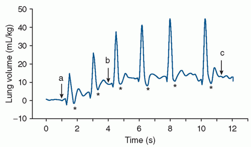
Before birth, the airways are liquid filled, and the lungs take no part in gas exchange (5). At birth, lung liquid has to be cleared from the airways to allow air entry and establishment of a functional residual capacity (FRC) (5). The purpose of the initial postnatal breaths is to clear lung liquid, establish an FRC, and initiate spontaneous breathing while facilitating gas exchange (6). Within minutes of delivery, the newborn’s pulmonary vascular resistance decreases by 10-fold, resulting in an increase in pulmonary blood flow (7). The first breath must generate a high transpulmonary pressure to overcome the viscosity of the lung fluid and the intra-alveolar surface tension. It also helps to drive the alveolar fluid across the alveolar epithelium. Lung expansion and aeration also stimulate surfactant release with the resultant establishment of an air-fluid interface and development of FRC (5,7,8) (Fig. 17.1).


▪ CIRCULATORY ADAPTATION

For the lungs to exchange gas adequately after birth, the airways and the alveoli must be cleared of fetal lung fluid, and an increase in pulmonary blood flow must occur. In utero, fetal pulmonary vascular resistance is high, and the fetal systemic vascular resistance is low; 90% of the cardiac output is shunted away from the lungs and is directed to the placenta where fetoplacental gas exchange occurs (Fig. 17.2). With clamping of the umbilical cord, the infant must immediately switch its site of gas exchange from the placenta to the lungs, and it must redirect cardiac output through the lungs to support the onset of pulmonary gas exchange. At birth, the clamping of the umbilical cord increases the systemic vascular resistance, which results in an increase in left ventricular and aortic pressures. Lung aeration and subsequent gas exchange result in increased PaO2 and pH, which result in pulmonary vasodilation. These physiologic changes increase blood flow to the left atrium via the pulmonary veins, so that left atrial pressure exceeds right atrial pressure, resulting in functional closure of the foramen ovale (Fig. 17.2). When pulmonary vascular resistance decreases to a level lower than the systemic vascular resistance, the ductus arteriosus closes functionally. As a result of the cessation of umbilical venous return, clamping of the umbilical cord also leads to the closure of the ductus venosus.

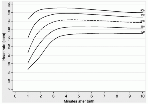
During neonatal transition, heart rate (HR) is the most important, objective clinical indicator of the health of newly born infants. Increasing HR is considered to be a good marker of effective resuscitation (9), and an HR exceeding 100 beats per minute (bpm) is considered normal (4). Recently, Dawson et al. (10) reported a nomogram of HR changes within the first 10 minutes. These data suggest that more than 50% of infants have a HR less than 100 bpm at 1 minute of age and 21% at 2 minutes (Fig. 17.3). It should be noted that an HR less than 60 bpm was measured in these infants with good muscle tone and normal respiratory effort at 1 and 2 minutes, in 17% and 7%, respectively (Fig. 17.3). Moreover, in preterm infants and those born by cesarean section, the HR rose more slowly than in term vaginal births. Therefore, low HRs in the first 2 minutes may be considered “normal” and, in isolation from other signs, should not be an indication for immediate ventilation (10).


▪ HIGH-RISK PREGNANCIES

Certain situations during pregnancy, labor, or delivery carry an increased risk of intrapartum asphyxia. If these high-risk deliveries are identified before birth, their progress during labor and delivery should be closely monitored, and resuscitation can be initiated at birth. Tables 17.1 and 17.2 list some of the factors that alert the physician to a high-risk delivery. Optimal management of these cases requires good communication between the obstetrician, anesthesiologist, and pediatrician.


▪ RESUSCITATION OF THE ASPHYXIATED INFANT

If a severely asphyxiated infant is expected, a resuscitation team must be present at delivery. In the majority of instances, good communication between the obstetrician and neonatal personnel will provide timely notice of the impending delivery of an asphyxiated infant. The actual extent of resuscitation needed can be determined only after someone with considerable clinical experience evaluates the infant’s condition. It is helpful to assign the responsibility of each member of the resuscitation team before delivery (Table 17.3).
Although knowledge is increasing from randomized trials, the majority of these recommendations are based on observational human and animal data but have not been rigorously tested. It is expected that in the future, some of these recommendations will undergo close scrutiny and possible change.
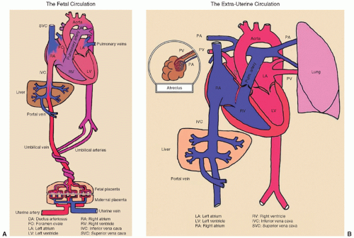


FIGURE 17.1 Plethysmography recording of breathing activity and the increase in end-expiratory lung gas volumes from birth in a spontaneously breathing newborn rabbit pup delivered near term. The recording demonstrates that pups can rapidly generate an end-expiratory lung air volume of approximately 16 mL/kg within 10 to 12 seconds of the onset of breathing. Note that the end-expiratory lung air volume increases with each breath. Phase-contrast x-ray images were acquired at the times indicated by the arrows in the plethysmography recording and demonstrate the increase in lung aeration associated with each breath (a, b, c). Reductions in gas volume immediately following inspiration (asterisk) are recording artifacts. From Hooper SB, Siew M, Kitchen MJ, et al. Imaging lung aeration and lung liquid clearance at birth. FASEB J 2007;21:3329, with permission.


FIGURE 17.2 A and B: Fetal and neonatal circulation. From David Atkinson, MD, David Geffen School of Medicine at UCLA, with permission.


Initial Steps in Transitioning and Resuscitation

Initial assessment, performed immediately after delivery, dictates the extent of resuscitation needed. Initial assessment includes determining whether or not the infant is breathing, if there is good muscle tone, and whether the infant looks term or preterm (4). If the infant is term, vigorous, without any known risk factors, and born through clear amniotic fluid, the infant can stay with the mother to continue care. Thermal care can be given by putting the infant on the mother’s chest (direct skin-to-skin contact), drying, and covering the infant with dry linen (4).

If the infant is apneic, gasping, and has decreased muscle tone, immediate resuscitation is needed. Place the infant under a radiant warmer; quickly towel dry the infant if greater than 28 weeks of gestation or wrap in plastic if less than 28 weeks of gestation (4); open the airway by positioning the infant in the sniffing position (11); provide tactile stimulation (e.g., by gently rubbing the back) (4). In the majority of cases, with these initial steps, the infant will start breathing adequately. However, if the infant fails to initiate breathing or HR is less than 100 bpm, positive pressure ventilation (PPV) should be started.

There is a large body of evidence that blood oxygen levels in uncompromised babies generally do not reach extrauterine values until approximately 10 minutes following birth. Oxygen saturation may normally remain in the 70% to 80% range for several minutes following birth, thus resulting in the appearance of cyanosis during that time (4). An observational study in the DR reported that there is substantial interobserver and intraobserver variability in clinical assessment of skin color (12). Therefore, experts have recommended the use of pulse oximetry to measure oxygenation in this setting (13). Optimal management of oxygen during neonatal resuscitation becomes particularly important because of the evidence that either
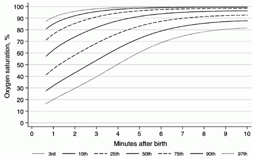
insufficient or excessive oxygenation can be harmful to the newborn infant (14,15). Percentiles of oxygen saturation as a function of time from birth in uncompromised babies born at term have been recently published (Fig. 17.4) (16). To appropriately compare oxygen saturations to similar published data, the probe should be attached to a preductal location (i.e., the right upper extremity, usually the medial surface of the palm). During respiratory support, a special emphasis should be given to prevent oxygen-induced lung injury by providing the lowest level of oxygen supplementation that maintains adequate delivery of oxygen to tissues (17,18,19). Studies suggest that resuscitation with 100% oxygen may generate oxygen free radicals, which may cause tissue damage, particularly to the brain (15,20,21). Therefore, continuous monitoring of oxygen saturation should start in the DR by using recently published nomograms (Fig. 17.4) to reduce overall oxygen exposures (16). The current resuscitation guidelines recommend to start resuscitation in room air in all term infants (4). Although, for preterm infants, no definitive recommendation has been made (4), there is increasing evidence that DR resuscitation of preterm neonates with an initial oxygen of less than 30% is feasible, decreases oxygen exposure without increasing need for additional resuscitation, and decreases oxidative stress (18,19,22). In the absence of DR studies comparing important clinical outcomes such as bronchopulmonary dysplasia and long-term neurodevelopment, neonatal resuscitation of preterm infants should be initiated with air or a blended oxygen and titrating the oxygen concentration to achieve an oxygen saturation value in the interquartile range of preductal saturations measured by pulse oximetry (Fig. 17.4) (4,16).


FIGURE 17.3 The 10th, 25th, 50th, 75th, and 90th HR centiles for all infants with no medical intervention after birth. bpm, beats per minute. Dawson J, Kamlin O, Wong C, et al. Changes in heart rate in the first minutes after birth. Arch Dis Child Fetal Neonatal Ed 2010;95:F177.


TABLE 17.1 Some Factors That Place the Newborn at High Risk for Asphyxia


Maternal Conditions


Labor and Delivery Conditions


Fetal Conditions


Diabetes mellitus


Forceps delivery other than low-elective or vacuum-extraction delivery


Premature delivery


Preeclampsia, hypertension, chronic renal disease


Breech or other abnormal presentation and delivery


Postmature delivery


Anemia (i.e., hemoglobin <10 g/dL)


Cephalopelvic disproportion: shoulder dystocia, prolonged second stage


Acidosis determined by fetal scalp capillary blood


Blood type or group alloimmunization


Cesarean section


Abnormal HR pattern or dysrhythmia


Abruptio placentae, placenta previa, or other antepartum hemorrhage


Prolapsed umbilical cord


Meconium-stained amniotic fluid


Oligohydramnios


Polyhydramnios


Narcotic, barbiturate, tranquilizer, psychedelic drug use or alcohol intoxication


Cord compression (e.g., nuchal cord, cord knot, compression by aftercoming head in breech delivery)


Decreased rate of growth: uterine size or fetal size determined by ultrasonography


History of previous perinatal loss


Maternal hypotension or hemorrhage


Macrosomia


Prolonged rupture of membranes


Immaturity of pulmonary surfactant system


Lupus


Fetal malformations determined by sonography


Maternal heart disease


Hydrops fetalis


Maternal fever or other evidence of amnionitis


Low biophysical profile


Abnormal umbilical artery


Doppler velocity


Multiple birth; in particular, discordant, stuck, or monoamniotic


TABLE 17.2 Fetal Heart Rate Patterns Associated with Fetal and Neonatal Distress


Heart Rate Pattern


Fetal or Neonatal Problems


Severe (i.e., <80 beats/min), sustained bradycardia, with loss of variability


Fetal hemorrhage, fetal asphyxia


Sustained tachycardia, uncomplicated by other abnormal patterns


Infection, often with apnea


Late decelerations with loss of variability


Asphyxia


Severe, recurrent variable decelerations, with loss of variability


Asphyxia and possible hypovolemia


Sinusoidal


Severe anemia with asphyxia


Respiratory Support


PEEP and CPAP

Positive end expiratory pressure (PEEP) is used in the Neonatal Intensive Care Unit (NICU) during mechanical ventilation to help maintain end expiratory lung volume, and continuous positive airway pressure (CPAP) supports lung volume and improves gas exchange in nonintubated neonates with respiratory failure. Neither of these has been mandated in neonatal resuscitation guidelines (4). The infant who is born very preterm has difficulty maintaining FRC and upper airway patency for many reasons. CPAP or PEEP can reduce the possibility
of atelectrauma and improve respiratory function in different ways: (a) CPAP reduces upper airway obstruction by decreasing upper airway resistance and increasing the pharyngeal cross-sectional area; (b) both CPAP and PEEP increase FRC; (c) CPAP and PEEP reduce inspiratory resistance by dilating the airways and allow a larger tidal volume for a given pressure with a reduction in the work of breathing; (d) CPAP and PEEP increase the compliance and tidal volume of stiff lungs with a low FRC by stabilizing the chest wall; (e) CPAP and PEEP increase the mean airway pressure and improve ventilation-perfusion mismatch; (f) PEEP conserves surfactant on the alveolar surface; (g) as CPAP and PEEP increase lung volume, oxygenation is also improved (23).


TABLE 17.3 Team Assignment During Neonatal Resuscitation


Member A


Member B


Member C


Responsible for respiratory


Responsible for cardiovascular


Responsible for support


Initial assessment of infant


Check HR and, if needed, give CC


Dries baby; radiant monitor servo control, apply electrocardiograph leads and transcutaneous pulse oximetry sensor


Airway management


Catheterize umbilical vessels and maintain patency of catheter


Keep timed, written record of resuscitation and vital signs and assign the Apgar scores at 1 and 5 min and every 5 min thereafter until the score is 7 or greater; time and record the rate and volume of infusions


Provide PP or CPAP


Administer fluids and drugs


Assist member A by providing ET suction, adjusting FiO2, and helping to secure ET


Intubate if necessary


Measure arterial pressures, assess perfusion, sample blood for blood gas and draw blood cultures


Help member B by providing medications and blood volume expanders in sterile syringes; B is working in a sterile field early in resuscitation


Secure ETT


Auscultate the chest to be sure ET is in proper position and gas exchange is good


Monitor baby’s temperature and capillary blood glucose


Continue assessment of the infant


Studies using animal models of resuscitation support the use of PEEP in the DR (24,25). Polglase et al. (25) showed that 4 to 8 cm H2O PEEP maintains arterial oxygenation and decreases pulmonary vascular blood flow with no adverse effects on the cardiovascular system. Probyn et al. (24) ventilated preterm lambs at PEEP levels of 0, 4, 8, and 12 cm H2O. All lambs ventilated with a PEEP above zero showed an improvement in oxygenation, but increasing the PEEP to 12 cm H2O resulted in pneumothoraces (24). PEEP levels up to 8 cm H2O should be considered for resuscitation of preterm infants. However, randomized controlled trials of different levels of PEEP during resuscitation of human infants at birth are required.

Although the majority of infants breathe and cry immediately after birth (26), PPV is required if infants fail to initiate spontaneous breathing after birth. PPV is commonly used in the DR and is the cornerstone of respiratory support after birth (4). An international consensus statement recommends that infants with inadequate breathing or bradycardia be given PPV via face mask with a self-inflating bag, flow-inflating bag (also called an anesthesia bag), or T-piece device (4). Each type has its own advantages and disadvantages, and the resuscitators should be familiar with the type of device used in their institution (27,28,29). The purpose of PPV is to establish an FRC and deliver an adequate tidal volume to achieve effective gas exchange (5). Adequacy of ventilation is then judged by assessing HR (4). However, if HR does not increase, chest wall movements should be assessed to gauge the adequacy of ventilation (4,5).


FIGURE 17.4 Third, 10th, 25th, 50th, 75th, 90th, and 97th SpO2 percentiles for all infants with no medical intervention after birth. Dawson J, Vento M, Kamlin O, et al. Defining the reference range for oxygen saturation for infants after birth. Pediatrics 2010;125:e1340.

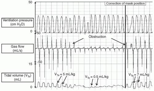
Manikin and DR studies have shown that PPV is difficult, and mask leak and airway obstruction are common problems during PPV (Figs. 17.5 and 17.6) (30,31,32,33,34). However, the delivery of adequate PPV in the DR is dependent on good face mask technique. Several factors can reduce the effectiveness of PPV. These include poor face mask application resulting in leak or airway obstruction (7,12,17,18), spontaneous movements of the baby, movements by or distraction of the resuscitator (34), and procedures such as changing the wet towels or fitting a hat (7,17,18). In addition, correct positioning of the infant’s head and neck with airway maneuvers (e.g., jaw thrust or chin lift) are crucial steps during mask ventilation (4,11).

In term and preterm infants, PPV is provided at a rate of 40 to 60 breaths/minute with an initial peak inflation pressure of 20 cm H2O (4). However, higher peak inflation pressure might be required, particularly in infants without spontaneous ventilation (4). In the majority of instances, provision of appropriate PPV is followed by an increase in HR and spontaneous breathing (9,35). Rate and pressures of PPV should be gradually reduced before deciding to see if the infant will tolerate its discontinuation. Some of the factors determining the success of mask PPV include choosing the correct size mask, proper positioning of the infant, achieving tight seal between the face and the mask, and using adequate inspiratory pressure (36). If despite correct PPV no increase in HR to greater than 60 per minute is observed or it continues to deteriorate, consider beginning CCs and endotracheal intubation.

It is important to realize that infants respond to initial lung inflation by eliciting a variety of physiologic responses. The infant may respond by the “rejection response,” in which the infant responds to PPV with a positive intraesophageal pressure to resist the inflation, that is, the infant actively resists attempts to inflate the lungs by generating an active exhalation. This response not only acts to reduce lung inflation but also may cause high transient inflation pressures. Another response is “Head’s paradoxical response” in which the neonate responds to PPV with an inspiratory effort, causing a negative intraesophageal pressure. This inspiratory effort, with the resultant
negative pressure, produces a fall in inflation pressures but results in a transient increase in tidal volume. Of course, the neonate may demonstrate no response to the inflation attempt, that is, not generating any change in intraesophageal pressure during PPV, and passive inflation subsequently results. It is important to recognize that these physiologic responses to PPV in the DR may cause large variability in the tidal volume and intrapulmonary pressures, despite delivery of constant inflation pressure.


FIGURE 17.5 Airway obstruction during mask PPV in a very preterm infant with a self-inflating bag. Initially, PPV delivered an expired tidal volume (VTe) of 5 mL/kg. Both inflation and expiratory flow waves rapidly reduced in size. This is reflected in the VT curve, which displays a 90% reduction in VTe. By correcting the face mask position, the tidal volume is restored. Throughout PPV, the peak inflation pressure is achieved. From Schmölzer GM, Dawson J, Kamlin O, et al. Airway obstruction and gas leak during mask ventilation of preterm infants in the delivery room. Arch Dis Child Fetal Neonatal Ed 2011;96:F254, with permission.


Chest Compressions

Although CCs are an infrequent event in newly born infants, outcome studies of DR resuscitations have reported high rates of mortality and neurodevelopmental impairment in those infants receiving CC or epinephrine (2,3). Current resuscitation guidelines recommend to deliver coordinated CC and ventilation using a 3:1 compression:ventilation (C:V) ratio (Fig. 17.7) (4). In addition, current resuscitation guidelines recommend 120 events per minute, which compromises 90 CC and 30 inflations (one & two & three & breathe & one & two & three & breathe & …) (4

Only gold members can continue reading. Log In or Register to continue

Stay updated, free articles. Join our Telegram channel

May 30, 2016 | Posted by in PEDIATRICS | Comments Off on Delivery Room Management

Full access? Get Clinical Tree

Get Clinical Tree app for offline access