Cardiac Disease


Cardiac Disease


Michael F. Flanagan

Scott B. Yeager

Steven N. Weindling


▪ INCIDENCE

The incidence of congenital heart disease detectable by routine clinical examination has been about 8 per 1,000 live births (1). The incidence of congenital heart anomalies in neonates seen with detailed echocardiographic examination is 4- to 10-fold higher, with most of the difference being clinically insignificant ventricular septal defects (20 to 50 per 1,000) and nonstenotic bicommissural aortic valves (2). Severe forms of cardiac anomalies requiring cardiac catheterization or surgery, or resulting in death, occur in 2.5 to 3 infants per 1,000 births (2,3). Almost one-half of these are diagnosed during the first week of life. Additionally, moderately severe forms of cardiac anomalies occur in another 3 per 1,000 live births, and another 13 of 1,000 live births have a bicommissural aortic valve that may require care eventually (2). The distribution of congenital heart anomalies in newborns seen at a primary pediatric cardiac center is shown in Table 30.1.


▪ INFANT MORTALITY

Before aggressive intervention, Mitchesll found that 2.3 of 1,000 live births died with cardiac problems in infancy (1). Infant cardiac mortality in developed countries has progressively declined over the last several decades with better pre- and postnatal recognition and with the development and refinement of definitive interventions and periprocedural management. The infant cardiac fatality rate in the United States was 0.15 per 1,000 births in 2000, ranking 10th among leading causes of infant death (4). At high-volume surgical centers, more commonly occurring cyanotic cardiac anomalies such as transposition of the great arteries and tetralogy of Fallot have surgical mortality rates of 1% to 5% or less. Complex anomalies with the highest risk have also had significant improvements in neonatal survival in developed countries. For example, surgical survival of neonates with “uncomplicated” hypoplastic left heart syndrome increased from 40% to 60% to 74% to 93% at specialized surgical centers (5), with overall mortality in infancy approximately twice this. Although surgical outcomes are much improved, including in babies with prematurity and multiple anomalies, prematurity and associated noncardiac anomalies still strongly influence survival for infants with cardiac disease (Table 30.2) (4).


▪ LONG-TERM SURVIVAL

This chapter focuses on infancy; however, discussions with parents concerning their newborn with a cardiac anomaly often quickly, and appropriately, move to the length and quality of life anticipated in later childhood and adulthood. It is important that parents are accurately advised by cardiologists or other practitioners are aware of recent advances and outcome findings of the potential long-term outcomes with current therapies. In general, those with common acyanotic anomalies such as uncomplicated septal defects or pulmonary valve stenosis, and most of those with cyanotic anomalies such as simple d-transposition of the great arteries may have an essentially near-normal survival and physical activity after appropriate intervention. Even with many complicated anomalies, most can expect to survive to at least midadulthood, although long-term survival is highly dependent on the specific diagnosis, with the highest mortality now generally occurring in infancy (see Table 30.2). With few exceptions, a cardiac operation or catheter intervention can lengthen and improve the quality of life of a child with heart disease. Even when the nature of the long-term management is unclear, care has proceeded with the conviction that childhood survival often allows yet to be planned later interventions, resulting from future progress in the field, to provide even longer and better survival. The palliative shunt operations of 20 to 40 years ago unexpectedly produced candidates for later Fontan procedures. The central principle continues to be “where there is life, there is hope” (see Ecclesiastes 9:4, Theocritus, Cicero in Epistle to Atticus).

With reductions in mortality and new generations of adults and older children with repaired and palliated cardiac anomalies, it has become evident that cardiac anomalies and procedures utilized to treat them sometimes have late residuae and sequelae, including neurologic and cognitive morbidity not evident until preschool or school-age (6 to 8 years) and arrhythmias and ventricular dysfunction developing in adolescence and adulthood. Most difficult and not yet fully delineated has been the neurologic and cognitive morbidity. These abnormalities appear rarely with common septal and valvar malformations and tend to be more frequent and severe with more complicated anomalies and repairs. Cardiac, neurologic, and cognitive outcomes after repair of septal defects have appeared generally normal (8). While most children with complex anomalies and procedures, such as hypoplastic left heart syndrome and operative circulatory arrest, have neurocognitive outcome within the normal range, on average, they have slower development, lower IQ scores, and higher rates of learning disabilities and special needs. Moreover, a significant number have major impairments (8). The etiologies are complex and include possible genetic issues, coexistent brain anomaly, diminished fetal cerebral oxygenation (e.g., d-transposition of great arteries, tricuspid and pulmonary atresia) and, with some anomalies, decreased cerebral perfusion (e.g., hypoplastic left heart syndrome—see section on fetal circulation), pre- and/or perioperative low cardiac output, cyanosis, thromboembolism, or intracerebral hemorrhage, and operative use of hypothermic circulatory arrest (6,7,8). The possibility that brain injury or other injury may be acquired prenatally, pretreatment, or in the process of treatment should be understood.

The goal is to provide a satisfying life through childhood and adulthood. The long-range future of patients undergoing intracardiac repair, arterial switch operations, staged multiple complex palliative operations, Fontan operations, or cardiac transplantation requires detailed discussion. The ramifications of treating a child who has major anomalies must be discussed with the parents in understandable language. The expected physical capabilities of the child after treatment should be delineated. After the physician is confident that the parents thoroughly understand the known facts, he or she is free to express an opinion about what may be best for the child.


▪ ETIOLOGY

Heart formation is a fantastic metamorphosis regulated by many sequences of genes. Given the remarkably complex orchestration of molecular and morphologic processes in formation of the heart, even small genetic and/or environmental changes in the control of these processes can have major and variable consequences. Truly, it is wonderful that development occurs as well as it does, as often as it does. Nevertheless, congenital cardiac disease is the most common type of birth defect (9,10). Understandably, parents ask why their baby was born with a cardiac abnormality and whether it is likely to recur with a subsequent pregnancy.

Cardiovascular malformations often appear to arise from altered number or function of specific genes (10,11). However, while some individuals with an isolated cardiac anomaly have a parent or
another family member who has survived with a cardiac anomaly, more often, there is no family history. Inheritable or even acquired defects in a single gene (e.g., Marfan syndrome) or a chromosomal aneuploidy (e.g., trisomy) is not yet identifiable in most patients with cardiac malformations. More commonly, there may be susceptibility from inherited or acquired mutations in two or more genes, perhaps with additional alterations in gene transcription or posttranscriptional processes from maternal-fetal folate metabolism or fetal exposure to specific pharmacologic, biochemical, infectious, and environmental factors that cumulatively surpass a threshold of liability (9,10,11,12). These result in pathogenic changes in embryonic development, including the following:


TABLE 30.1 Neonatal (First Month) Cardiac Diagnostic Distributiona


Percent


Ventricular septal defect


41


Atrial septal defect secundum


12


Valvar pulmonary stenosis


11


Coarctation of the aorta


6


Tetralogy of Fallot


5


Cardiomyopathy


4


Transposition of the great arteries


3


Endocardial cushion defects


3


Hypoplastic left heart syndrome


2


Aortic stenosis


2


Tricuspid atresia


1


Malposition


1


Total anomalous pulmonary venous connection


1


Truncus arteriosus


1


Aortic-pulmonary window


1


Hemitruncus


<1


Interrupted aortic arch


<1


l-Transposition of the great arteries


<1


Tricuspid valve diseases


<1


Pulmonary atresia and intact interventricular septum


<1


Single ventricle


<1


aBased on 361 patients diagnosed by echocardiography by age 1 mo at Children’s Hospital at Dartmouth. Neonates with patent ductus arteriosus, persistent fetal circulation, arrhythmia and normal heart exams are not included.


  • Defects in mesenchymal tissue migration (tetralogy of Fallot, truncus arteriosus, interrupted aortic arch, malalignment conal-septal ventricular septal defects, transposition of great arteries)


  • Extracellular matrix defects (endocardial cushion defects)


  • Abnormal cell death (muscular ventricular septal defect, Ebstein anomaly)


  • Targeted growth (anomalous pulmonary vein connection, single atrium)


  • Defective situs and cardiac looping (heterotaxy syndromes, l-transposition)


  • Secondary effects from alterations in blood flow in the right heart (secundum atrial septal defect, pulmonary valve stenosis, and atresia) or left heart (hypoplastic left heart syndrome, coarctation of aorta, aortic valve stenosis, patent ductus arteriosus) (9,10,11,12)

Loss of function of specific genes from genomic copy number variations associated with inappropriate recombination of DNA segments or chromosomal aneuploidy is responsible for a significant proportion of cardiovascular anomalies. When multiple contiguous genes are involved, cardiovascular anomalies with syndromes can result (10). Approximately 13% of children with cardiac anomalies have chromosomal aneuploidy associated with cardiovascular malformation. Another approximately 8% to 13% of children have inheritable syndromes with associated cardiovascular abnormalities (11,12,13). The genes affected in many of these syndromes have been identified (9,10,11,14,15,16) (Table 30.3). The most common human mutations with copy number variants are in a critical 30-gene region of chromosome 22q11 involved in neural crest and cardiac development that cause DiGeorge and velocardiofacial syndromes and associated conotruncal and aortic arch malformations (15). Altered function of one of the genes in this region, TBX1, a transcription factor that promotes cell proliferation in progenitors of the right ventricle and conal outflow, accounts for most of the cardiovascular phenotype (10).


TABLE 30.2 Infant (First-Year) Cardiac Diagnostic Distribution and Mortalitya


Diagnosis


Frequency (%)


Mortality (%)


Ventricular septal defect


31


0.7


Pulmonary valve stenosis


19


0.5


Atrial septal defect


12


0.2


Aortic valve stenosis


8


4


Atrio-ventricular canal septal Defects


5


1.8


Coarctation of aorta


4


3.5


d-Transposition of great arteries


4


1.0


Tetralogy of Fallot with pulmonary outflow stenosis


4


2.2


Malposition


3


9


Hypoplastic left heart syndrome


2


22


Pulmonary valve atresia with intact ventricular septum


2


4


Pulmonary valve atresia with tetralogy of Fallot


1


11


Double outlet right ventricle


0.9


6


Total anomalous pulmonary venous connection


0.8


7


Truncus arteriosus


0.7


5


Single ventricle


0.7


<0.1


Tricuspid atresia


0.5


<0.1


Interrupted aortic arch


0.3


<0.1


Aortic-pulmonary window


0.2


<0.1


Hemitruncus


0.1


<0.1


l-Transposition of great arteries


0.1


<0.1


Total


2.2


aData is based on 5,182 infants diagnosed at Childrens Hospital, Boston between January 1, 1998 and January 1, 2002, and their overall mortality by age 1 y. Babies with patent ductus arteriosus and primary cardiomyopathy were not included. Mortality by age 1 y was significantly influenced by coexistent prematurity and non-cardiac disease.


Point mutations within a specific gene may result in transcription of dysfunctional proteins in structural or regulatory protein (missense mutation) or gene inactivation (frameshift or nonsense mutations). These may alter transcriptional regulation, signal transduction, or structural proteins, resulting in a phenotypic constellation with particular cardiovascular anomalies. For example, mutations in the genes encoding the extracellular matrix proteins fibrillin-1 and elastin are responsible, respectively, for Marfan and William syndrome. Point mutations in specific genes (see Table 30.3) are also responsible for Noonan syndrome, Alagille arteriohepatic dysplasia syndrome, and Holt-Oram syndromes (10).

Whether from copy number variation or point mutation, genetic syndromes associated with abnormal cardiac development are generally associated with specific cardiac malformations, for instance,


Down syndrome and endocardial cushion defects, Williams syndrome and supravalvar aortic stenosis, DiGeorge syndrome and tetralogy of Fallot, truncus arteriosus, and interrupted aortic arch (Table 30.3). Recognition that a child has a syndrome associated with a cardiac anomaly or vice versa should prompt an investigation for possible associated anomalies (16,17,18,19,20).


TABLE 30.3 Congenital Disorders Associated with Cardiac Disease


Selected Disorders


Identified Gene(s)


Chromosome Location


% Heart Disease


Cardiovascular Anomalies


Autosomal Dominant


Alagille Arteriohepatic dysplasia


Jagged 1, Notch 2


20p12, 1p11-12


100


Multiple PA stenosis and hypoplasia, PDA, ASD, VSD,renal artery stenosis, carotid aneurysm


Apert acrocephalosyndactyly


FGFR-2


10q26


VSD, hypoplastic PA


Beckwith-Wiedemann syndrome


H19, KCNQ10T1, CDK1C


11p15.5, 5q35.2-3


15 ?


HCM, ASD, VSD, TF, PDA


Cardiofacial cutaneous syndrome


KRAS, BRAF, MAP2K1, MAP2K1


multiple


ASD, PS, HCM


CHARGE syndrome


CHD7


8q12.1


44


TF, ASD, VSD, DORV, PDA, PS


Costello syndrome


HRAS


11p15.5


HCM, PS, MVP, ASD, VSD, arrhythmia


de Lange syndrome CLLS1


N1PBL


5p13.2


30


VSD, ASD, PDA, AS, EFE


DiGeorge/velocardiofacial syndromes


Multiple in DGCR including TBX1


22q11 DGCR


>50


TF, interrupted Ao arch, truncus arteriosus, right Ao arch


Goldenhar hemifacial microsomia/OAVS


multiple


multiple


15


TF, VSD, PDA, COARC


Hereditary hemorrhagic telangiectasia


Osler-Weber-Rendu


endoglin


9q34.1


100


pulmonary and systemic AVM, arterial aneurysm telangiectasia


Holt-Oram Heart-hand syndrome


TBX5


12q24.1


100


ASD-2, VSD or PDA in 2/3, HLHS, conduc. block, HLHS, TAPVC, truncus art.


Juvenile Polyposis Hereditary


Telangiectasia JPHT


SMAD4


18q21.2


?


Ao aneurysm, MVP, MR, AVM


Leopard syndrome


PTPN11


12q24.13


Loeys-Dietz syndrome 1-4


TGFBR1, TGFBR2, SMAD3, TGF


multiple


100


Ao aneurysm/dissection, cerebral and arterial aneurysm and tortuosity, MVP, MR, BAV, PS


Marfan syndrome


fibrillin-1


15q21.1


up to 100%


Ao aneurysm; AR, MR, TR & prolapse


Myhre syndrome


SMAD4


18q21.2


ASD, VSD, PDA, AS, COARC, pericardial fibrosis


Neurofibromatosis-type 1


NFI


17q11.2


rare


PS, COARC, renal artery stenosis


Noonan syndrome types1,4,8, NF type


PTPN11 (50%), SOS1 (28%), KRAS, NRAS, BRAS, RAF1, RITI, NF1


12q24.13 (PTPN11)


?


PS/dysplasia, PDA, HCM, COARC


Rubinstein-Taybi type 1


CREBBP


16p13.3


35


VSD, PDA, ASD, COARC, PS, hemangioma, BAV


Saethre-Chotzen syndrome


TWIST, FGFR-3&2


7p21, 10q26


?


various, subvalvar AS


Shprintzen-Goldberg Craniosynostosis syndrome


SKI


1p36.33


Ao aneurysm, carotid and vertebral arterial tortuosity, MVP


Treacher-Collins syndrome


TCOF1


5q32-q33.1


10


ASD, VSD, PDA, Ao aneurysm,


Tuberous sclerosis 1, 2


TSC1 (hemartin), TSC2 (tuberin)


9q34.13, 16p13.3


30


rhabdomyomas, WPW, rarely Ao aneurysm


Williams-Beuren syndrome


elastin


7q11.2


50-80


supravalvar AS, small aorta, BAV, AS, PS, stenoses of LCA, multiple PAs, cerebral & renal arteries, MVP, MR, ASD, VSD


Autosomal Recessive


Carpenter acrocephalopolysyndactyly 1,2


Type 1MEGF8


19q13.2


ASD, VSD, PDA, PS, VSD, TF, TGA, dextrocardia


Type 2 RAB23


6p11.2


Coffin-Siris fifth digit syndrome


?


?


33


PDA, ASD, VSD, TF


Ellis van Creveld syndrome


ECC


4p16.2


?


ASD


Klippel-Feil syndrome-2


MEOX1


17q21.31


?


VSD, dextrocardia


Mucopolysaccharidosis type1


EVC


4p16


50-60


single atrium, primum ASD, COARC, HLHS


type 2


iduronidase


4p16.3


>50


all types have valvular disease,


type 3D


iduronate 2-sulfatase


Xq28


coronary disease (type 2)


type 6


GNS


12q14


Pierre Robin syndrome


arylsulfatase B


5q11-q13


Smith-Lemli-Opitz syndrome 1&2


?


?


2q32.3-q33.2


TF, COARC, pulmonary hypertension


Thrombocytopenia absent radius


SLOS


7q32.1


20/100


VSD, PDA, ASD, TF, AV canal, COARC


Zellweger cerebrohepatorenal syndrome


Multiple PEX genes


multiple peroxin-5,2, 6,12


?


VSD, ASD, PDA


Selected Chromosomal Disordersa


Trisomy 13 Patau syndrome


Trisomy 18 Edwards syndrome


13


80


PDA, VSD, ASD, COARC,AS, PS


Trisomy 21 Down syndrome


18


90-100


VSD, polyvalvular, ASD, PDA


+8 Mosaicism


21


40-50


AV canal, VSD, ASD1&2, PDA, TF


+9 Mosaicism


8


25


VSD, PDA, CoAo, PS


XO Turners syndrome


9


70


VSD, PDA, LSVC


4p-Wolfe syndrome


X


>50


bicusp AV, COARC, Ao anuerysm, AS, VSD


5p- Cri-du-Chat syndrome


4p


33


VSD, ASD, COARC


7q-


5p


20


VSD, PDA, ASD, PS


13q-


7q


20


18q-


13q-


common


ring 18


18q


25


VSD, ASD, PDA, PS


10p trisomy


18


20


COARC, PA hypoplasia, HLHS, LSVC


10q24 trisomy


10p


30


AV canal, VSD, ToF


22+ Cat eye syndrome


10q24


50


Fragile X


22


40


TAPVC, TF


Syndromes with unknown etiology


x


50


Asymmetric crying facies


65-75


TF, DORV, ASD, VSD, PDA, COARC, AV canal


VACTERL Association


Non-Random Associations


10


VSD, ASD, TF


Cleft lip and palate


diaphragmatic hernia


25


VSD, PDA, TGA, TF, SV


lung agenesis


25


TF


omphalocele


20


PDA, VSD, TF, TAPVC


intestinal atresia


20


TF, ASD


renal agenesis unilateral/bilateral


10


VSD


VSD


17/75


VSD


Ao, aortic; AR, aortic regurgitation; AS, aortic stenosis; ASD, atrial septal defect; ASD-1, primum atrial septal defect; ASD-2, secundum atrial septal defect; AV, aortic valve; AV canal, atrioventricular canal defect; AVM, arteriovenous malformation; BAV, bicuspid aortic valve; COARC, coarctation of the aorta; DORV, double-outlet right ventricle; EFE, endocardial fibroelastosis; HLHS, hypoplastic left heart syndrome; LCA, left coronary artery; LSVC, left superior vena cava; MR, mitral regurgitation; MVP, mitral valve prolapse, PAs, pulmonary arteries; PDA, patent ductus arteriosus; PS, pulmonary valve stenosis; TAPVC, totally anomalous pulmonary venous connection; TF, tetralogy of Fallot; TGA, transposition of great arteries; TR, tricuspid regurgitation; truncus art., truncus arteriosus; VSD, ventricular septal defect.


References (10,12,16,17,18,19,20).


a See omim.org and rarecrhromo.org for many additional chromosomal deletion and duplication syndromes.


TABLE 30.4 Incidence of Severe Associated Noncardiac Anomalies Among 2,220 Infants with Heart Disease


Diagnosis


Incidence (%)


Endocardial cushion defect


43


Patent ductus arteriosus


31


Ventricular septal defect


24


Malpositions


13


Tetralogy of Fallot


10


Coarctation of aorta


9


Pulmonary atresia with intact septum


1


d-Transposition of the great arteries


1


Most children with cardiac malformations, even those with tetralogy of Fallot, have isolated cardiac anomalies, without generalized syndrome or other apparent abnormality. Many specific cardiac anomalies are rarely associated with a noncardiac syndrome, for example, transposition of the great arteries, and pulmonary atresia with intact ventricular septum (Table 30.4). Although the molecular biology of cardiovascular development is being unraveled and dozens of genes responsible for isolated nonsyndromic cardiac anomalies have been identified, of the more than three dozen identified, the majority involve transcription factors and cofactors (e.g., NKX2, TBX1, TBX5, GATA4, GATA6, ZIC3, and others) or signal ligands and receptors (e.g., JAG1, SMAD6, and others), and relatively few encode structural proteins (10,11). There appears to be a causative convergence of environmental risk factors for congenital heart disease with a relatively small set of genes identified as genetic risk factors for congenital heart disease (e.g., ACTC1, JAG1, MYH6, NOTCH1, RAF1, RARG, RARA, TBX5, TMOD1, TWIST1) known to act upon protein networks driving heart development (11). However, the specific genetic and/or environmental causes of isolated cardiac anomalies remain unknown in most individual cases (9,10,11,14,15,21).

Although the occurrence of cardiac malformation has varied little year by year and by location, there are exceptions. Some of these variations may be from regional variations in exposure to biochemical risks during fetal development that play roles in causation of new genetic mutations and in transcriptional and posttranscriptional processes (9,10,11,12,14,15,22). For instance, the risk for development of conotruncal anomalies is significantly influenced by maternal-fetal intake and metabolism of folic acid and homocysteine (24,25,28). Additionally, maternal obesity and associated diabetes mellitus increase the likelihood for development of cardiovascular anomaly (10). Some variations may be from regional variations in parental consanguinity (10). Although fetal exposure to specific biochemical, pharmacologic, infectious, and environmental factors may increase the risk for developing a cardiac abnormality (Table 30.5) (9,10,11,12,29,30), these factors alone do not appear to explain most cases. In individual cases, it is usually difficult or impossible to identify specific extrinsic factors that may have modified the baby’s genotype or genotypic expression.

A singular abnormality results in a characteristic complex cardiac malformation by altering migration or function of embryonic primordial cells, such as in the neural crest or endocardial cushion, before formation of cardiac structures in the conotruncus or atrioventricular valves, respectively. Animal studies have demonstrated that embryonic cervical neural crest cells migrate into the thorax and contribute to formation of the aortic arch and conotruncal outflow region of the heart. Blockage of the normal function of these embryonic neural crest cells results in aortic arch anomalies including aortic interruption; conotruncal abnormalities including tetralogy of Fallot, truncus arteriosus, and transposition; and ventricular inlet anomalies including tricuspid atresia and double-inlet single left ventricle (11,14). Cells in the embryonic endocardial tissue undergo a different developmental sequential process controlled by a large number of factors. Perturbation of specific steps in these embryonic cell process changes developmental sequences in characteristic ways and alters blood flow patterns affecting vascular growth downstream in characteristic ways (9,11). Because growth of specific cardiovascular structures is flow dependent, limitation of flow can cause additional hypoplasia of downstream structures (11). For example, a mildly stenotic bicommissural aortic valve may decrease blood flow through the aortic isthmus and result in coarctation.


TABLE 30.5 Possible Teratogens for Congenital Heart Disease


Vitamin deficiency


Folate deficiencya


Environmental agents


High altitude,a organic solventsa, dioxinsa, pesticidesa, irradiation


Drugs


Ethanol,a folic acid antagonists (a trimethoprim,a sulfasalazine,a triamterene,a trimethadione,a phenytoin,a primidone,a phenobarbitol,a carbamazepine,a methotrexatea), valproic acid,a lithium,a thalidomide,a retinoic acid,a antineoplastic agents (?), coumadin,a amphetamine, cocaine


Metabolic factors


Maternal pregestational diabetes,a maternal phenylketonuria,a maternal obesity, homocysteine


Immune factors


Maternal autoimmune disease with anti-Ra anti-LA antibodiesb


Infectious agents


Rubella,a influenza,a febrile illness,a mumps (?), cytomegalovirus (?)


a It is generally accepted that these prenatal factors increase the risk for congenital heart disease.

b Immune-mediated conduction block and myocarditis.


?, uncertain.


From refs. (5,11,12,16,18,114).


Certain cardiac anomalies are associated with prematurity or low birth weight. Because closure of the ventricular septum may be delayed until the first months of life, it is not surprising that there is a somewhat greater incidence of ventricular septal defect among premature infants. The increased incidence of patent ductus arteriosus in prematurely born infants can be viewed as the result of birth long before the programmed time for closure of the ductus. Hypoxemia of pulmonary origin also promotes ductal patency.

As with gross anatomic cardiac anomalies, specific causes of cardiac muscle diseases are being identified. Most hypertrophic and many dilated cardiomyopathies previously known as idiopathic are now known to be caused by specific gene mutations and pathogenic mechanisms (16,27,28,29,30,31,32). Most patients with isolated hypertrophic cardiomyopathy have newly acquired or autosomal dominant inherited mutations in the genes encoding sarcomeric contractile proteins, most commonly cardiac β-myosin heavy chain, cardiac myosin-binding protein C, or troponin T2 (27,28,29). Isolated dilated cardiomyopathy has been associated with dozens of genetic loci, involving contractile, cytoskeletal, and other proteins, and identification of more is likely (16,28,29,30,31,32). Dilated and hypertrophic cardiomyopathies also occur in association with a large number of more generalized neuromuscular and metabolic disorders occurring from specific nuclear and mitochondrial genetic mutations (see Cardiomyopathy and Tables 30.14 and 30.15) (16,34).


Some specific arrhythmia syndromes are caused by specific genetic mutations. These include patients with ventricular tachycardia associated with prolonged QT syndrome and arrhythmogenic right ventricular dysplasia and rare forms of Wolff-Parkinson-White (WPW) syndrome with supraventricular tachycardia (SVT). Long QT syndrome results from mutations in genes encoding specific cardiac potassium and sodium ion channels that regulate repolarization (33); the ensuing prolongation of repolarization results in ventricular tachycardia.


▪ FETAL CARDIOLOGY


Fetal Circulation

Extensive information about the circulatory physiology of the fetus and newborn has accumulated. The work of Rudolph (34) should be consulted for details, but the central features are discussed here. The circulation before birth consists of parallel circuits (Fig. 30.1). Blood in the aorta may follow several routes to a capillary bed in the fetus or the placenta, back to the heart, passing through either ventricle, and out again to the aorta. The stream of newly oxygenated blood from the placenta passes through the umbilical vein, the ductus venosus, the inferior vena cava, and the right atrium. Unlike the circulation after birth, the streams of oxygenated and unoxygenated blood are not completely separated, although the more oxygenated blood from the inferior vena cava is mostly diverted through the foramen ovale into the left atrium. Consequently, in the normal heart blood from the left ventricle entering the ascending aorta and coronary and carotid circulations is somewhat higher in oxygen than that entering the descending aorta from the right ventricle by way of the ductus arteriosus.


FIGURE 30.1 Fetal circulation is in parallel, and the amount of blood handled by the left and right ventricles is 125 and 90 mL, respectively. Only 40 mL passes through the aortic arch to the descending aorta, and only a small fraction passes through the lungs. The numbers inside the diagram represent relative blood flow (mL); the numbers in italics are pressure measurements. Modified from McElhinney DB, Tworetzky W, Lock JE. Current status of fetal cardiac intervention. Circulation 2010;121:1256.

The volume pumped by the right ventricle is normally about 55% of the combined output of both ventricles. Because both ventricles pump against the systemic resistance, pressures in the two ventricles are comparable. The resistance to blood flow through the lungs is relatively great; only minimal flow through the lungs occurs in utero, and almost all of the right ventricular output into the pulmonary artery passes through the ductus arteriosus to the descending aorta. The parallel arrangement of the ventricles allows fetal survival despite a wide variety of cardiac lesions. With total obstruction of either ventricle, the other ventricle assumes the entire cardiac output. Reversal of the pulmonary arterial and aortic streams of blood, as occurs in transposition of the great arteries, produces no obvious deleterious effect on the somatic growth in the fetus. However, while these alterations of fetal circulation permit adequate cardiac output for normal fetal somatic growth, there are important alterations in regional oxygenation. In the fetus with transposition of the great arteries, the streaming of more oxygenated umbilical venous flow across the atrial septum results in lower than normal oxygen levels in the ascending aorta and coronary and cerebral circulations. Cerebral oxygenation is decreased in other cardiac anomalies. For instance, anomalies with right-heart obstruction result in fetal shunting of relatively deoxygenated right heart blood to the coronary and cerebral circulations, across the atrial septal defect in tricuspid atresia and across the ventricular septal defect in tetralogy. In hypoplastic left heart syndrome, all venous return mixes in the right heart, and this less oxygenated blood enters the aorta via the ductus arteriosus. Cerebral and coronary blood flow occurs by flow backward around the aortic arch and is compromised in the frequent situation of coexisting aortic arch obstruction (5,6,7,8).

The impact of these defects on broader fetal development is increasingly recognized. It appears that well over 50% of neonates with cyanotic congenital heart abnormalities demonstrate anatomic and clinical manifestations of central neurologic injury, including focal and diffuse white matter injury, ischemic lesions, periventricular leukomalacia, hemorrhage, and a variety of neurobehavioral abnormalities. Additionally, there is evidence that the presence of serious heart disease in the fetus is associated with delayed brain maturation, increasing neonatal vulnerability (5,6,7,8).

Despite this remarkable ability, while fetal cardiovasculature adaptations maintain adequate overall cardiac output with many anomalies, the cardiac performance and survival of the fetus are affected by limitations in myocardial contractility. Prolonged, severe volume loading of the heart or sustained tachyarrhythmia or primary myocardial disease may result in congestive heart failure, manifested by hydrops fetalis, with substantial fetal and neonatal mortality. The interplay between the metabolic effects of congestion in the fetus and the possible compensatory role of the placenta is not understood. Because lesions that may be expected to cause gross intrauterine difficulty are tolerated surprisingly well, the postulate that the placenta helps compensate for the metabolic abnormalities resulting from congestive heart failure is tenable.


Circulatory Adjustments at Birth


Changes in the Source of Oxygenated Blood and in the Ductus Venosus and Ductus Arteriosus

With the first breath, the resistance to pulmonary blood flow drops sharply. The oxygen content of the left heart and systemic circulation rapidly reaches levels well above that of the fetal
circulation. Oxygen saturation in the ascending aorta rises from about 65% in the fetus, to about 93% immediately after birth. The ductus venosus functionally closes, establishing the portal circulation as an independent loop between two capillary beds. With removal of the low-resistance placenta, systemic vascular resistance increases. The relative fall in the pulmonary and rise in the systemic vascular resistance result in a transitory left-toright shunt through the ductus arteriosus. In 50% of term babies, the ductus is completely constricted by the end of the first day of life and normally becomes anatomically obliterated at about 10 days of age. Even among cyanotic newborns who are duct dependent, the ductus may inexorably close, often severing the infant’s only source of pulmonary or systemic blood flow. The mechanisms causing closure of the ductus arteriosus are not completely understood but involve decreased prostaglandins and increased blood oxygen. Prostaglandin levels in the blood decrease at birth as a result of removal of their placental source of production and increase in perfusion of the lungs, where prostaglandins are metabolized.


Foramen Ovale

Functional closure of the foramen ovale occurs soon after birth, largely as a result of increased left atrial volume and pressure secondary to the increased pulmonary venous return, the ductal lefttoright shunt, and the developing differences in diastolic pressure of the two ventricles. Anatomic closure normally is delayed for months or years. Among infants with cardiac defects, lesions with increased right atrial pressure favor indefinite patency of the foramen ovale (e.g., pulmonary stenosis), but abnormally increased left atrial pressure promotes early anatomic closure (e.g., ventricular septal defect). Before birth, the pulmonary arterioles are relatively muscular and constricted.


Pulmonary Vasculature

With the first breath, total pulmonary resistance falls rapidly because of the unkinking of the vessels with expansion of the lungs and because of the vasodilatory effect of inspired oxygen. The muscular constriction relaxes, and gradually, during the subsequent days and weeks, the muscular wall of the pulmonary arterioles thins. During the first weeks of life, the muscular arterioles retain a significant capacity for constriction. Pulmonary alveolar hypoxia normally produces an increase in pulmonary artery pressure at all ages, but in the young infant, the response is more profound and occurs more rapidly. Therefore, pulmonary hypertension equal to or greater than systemic pressure occurs commonly in neonates with severe respiratory disease.


Ventricular Work

Before birth, the two ventricles share in supplying systemic blood flow and placental flow, and after birth, the two ventricles sequentially and independently handle the entire cardiac output. At birth, the volume of blood to be pumped by the right ventricle decreases to the level of the systemic blood flow; right ventricular pressure falls as a result of the decrease in pulmonary vascular resistance and closure of the ductus arteriosus. Although right ventricular work decreases, left ventricular work increases (Fig. 30.2). At birth, the left ventricle abruptly becomes the sole supplier of systemic blood flow, and the volume that it pumps is fractionally increased. The left-to-right shunt through the ductus arteriosus adds further volume work, and the elevated systemic vascular resistance must be overcome. Although this is a stressful time for the left ventricle, the magnitude of these suddenly acquired burdens generally does not result in any detectable left ventricular difficulties, but any impairment of myocardial function may be magnified as a consequence. Myocardial disease, as a cause of symptoms, is more common in the first days of life than at any other time during infancy; 25% of infants with myocardial disease presented in the first week of life (4).


FIGURE 30.2 Mature circulation is in series, and the amount of blood carried by the two ventricles is approximately the same as before birth. The lungs carry an amount equivalent to the cardiac output, as does the ascending aorta. The numbers inside the diagram represent relative blood flow (mL); the numbers in italics are pressure measurements.


Myocardial Function

Important changes occur in the fetus and neonate in many aspects of myocardial biochemistry and structure. These include myocyte size and number, microvascular structure, myocyte utilization of lactate and fatty acids, and antioxidant systems. Many structures and proteins involved in calcium handling within the myocyte, such as t-tubules, sarcoplasmic reticulum, Na+-Ca2+ exchange, Ca2+-ATPase, and phospholamban, have important developmental changes. These changes influence the effects on ventricular rhythm and function of normal development, prematurity, ischemia, cardioplegia, and various inborn errors of metabolism.


Fetal Echocardiography

High-resolution two-dimensional ultrasound evaluation of the fetal heart is a useful and accurate technique in the diagnosis and management of the fetus at risk for structural or functional cardiac abnormalities. Indications for prenatal echocardiography may include maternal, fetal, and genetic considerations (25) (Table 30.6). The optimal time for performing fetal echocardiography is 18 to 24 weeks of gestation. At this age, the fetal heart is usually large enough for detailed anatomic evaluation, and the images are unimpaired by dense rib or spine calcification. There is also a relatively large volume of amniotic fluid, which facilitates imaging from a variety of angles. For accurate diagnosis, the examiner must be experienced in the technical aspects of fetal ultrasonography and knowledgeable in the anatomic patterns and physiologic consequences of congenital heart defects (36). Transvaginal ultrasound can provide diagnostic information as early as 10 to 12 weeks’ gestation and may be useful when there is a high suspicion of major cardiac anomalies. Three-dimensional echocardiography and magnetic resonance imaging have been applied to fetal cardiac evaluation and may improve diagnostic accuracy, although difficulty gating to the fetal heart rate has limited their utility to date (37).


TABLE 30.6 Indications for Fetal Echocardiography


Suspected cardiac malformation on general ultrasound


Other malformations noted on general ultrasound


Oligo- or polyhydramnios


Fetal dysrhythmia


Suspected or known chromosomal abnormality


Family history of congenital heart disease


Family history of chromosomal abnormality


Maternal diabetes


Maternal collagen vascular disease


Rubella exposure


Evidence of hydrops fetalis


Intrauterine growth restriction


Maternal drug exposure, including:


Lithium


Hormones


Anticonvulsants


Chemotherapy


Alcohol


Cardiac Anatomy

Virtually, all major cardiac malformations can be detected prenatally using high-resolution two-dimensional, real-time sector scanning by an experienced examiner. The details of systemic and pulmonary venous connections, arterial alignment, chamber size and orientation, and valve position and function can be determined (Fig. 30.3) and abnormal structures demonstrated (Figs. 30.4 and 30.5).


Cardiac Physiology

Color Doppler provides a quick and sensitive means of evaluating the function of atrioventricular and semilunar valves, the direction of flow in fetal vessels, and the presence of normal and abnormal connections (Fig. 30.5). If abnormal flow is detected, it can be evaluated further using the quantitative capabilities of pulsed- or continuous-wave Doppler.


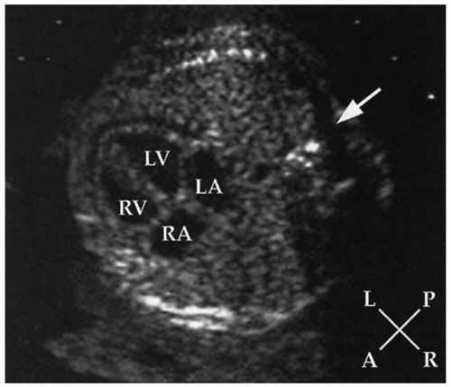
FIGURE 30.3 Echocardiogram of the normal fetal heart in a four-chamber view demonstrating the position of the heart and the cardiac chambers in a cross-section of the chest. A, anterior; L, left; LA, left atrium; LV, left ventricle; P, posterior; R, right; RA, right atrium; RV, rightventricle; arrowdenotes the spine.


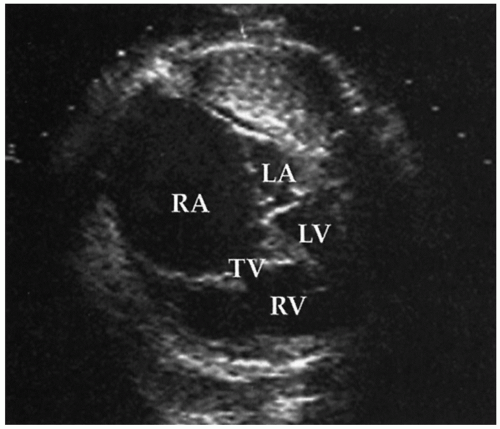
FIGURE 30.4 Echocardiographic cross-sectional views of the fetal chest in an infant with Ebstein anomaly of the tricuspid valve. The right atrium (RA) is markedly dilated and fills much of the thorax. The left atrium (LA) and left ventricle (LV) are of normal size but are dwarfed by the right-sided structures. The severely regurgitant tricuspid valve has apical displacement of the septal leaflet into the right ventricle (RV).


Cardiac Function

A qualitative assessment of cardiac function is obtained by visual inspection of ventricular motion during real-time sector scanning. When more quantitative information is desired, M-mode recording can provide precise dimensions and an accurate measure of ventricular shortening (Fig. 30.6). Doppler-derived indices of cardiac function may also provide insight into fetal cardiac performance, and three-dimensional imaging and fetal MRI may ultimately prove useful as well but remain hampered by the relatively
small fetal cardiac size, rapid heart rate, and lack of ECG gating (38,39). Severe ventricular dysfunction may manifest as generalized hydrops fetalis, which is readily recognized by ultrasound as pleural and peritoneal fluid accumulation and cutaneous edema.


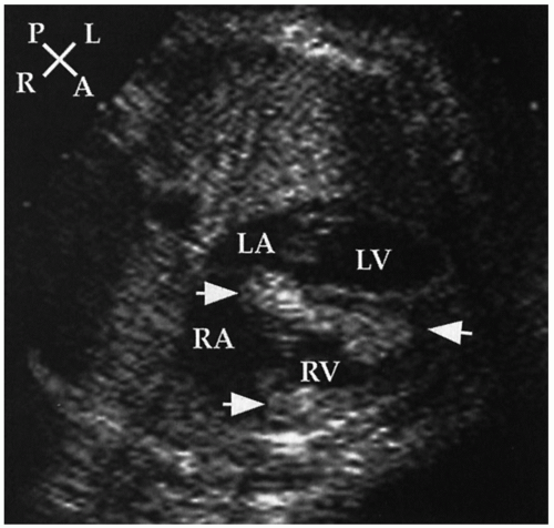
FIGURE 30.5 Echocardiographic cross-sectional view of a fetus with multiple intramyocardial rhabdomyomas ( arrows). The infant was subsequently diagnosed with tuberous sclerosis. A, anterior; L, left; LA, left atrium; LV, left ventricle; P, posterior; R, right; RA, right atrium; RV, right ventricle.


FIGURE 30.6 Echocardiogram of a fetus with a premature atrial beat. The upper left image is a two-dimensional view with a cursor (line) through the fetal right atrium (RA) and aortic valve (AOV), demonstrating the axis of the simultaneous M-mode echocardiogram seen in the lower panel. The M-mode tracing depicts the motion of the fetal right atrial wall and aortic valve in the cursor line over a time frame of 3 seconds. A series of normal atrial wall contractions are interrupted by a premature contraction (large arrow) followed by opening of the aortic valve (small arrow), demonstrating a premature atrial beat conducted to the ventricle. LV, left ventricle; RV, right ventricle.


Arrhythmias

Tachyarrhythmias, bradyarrhythmias, and irregular cardiac rhythms are common reasons for referral for evaluation. Structural and functional abnormalities should be excluded, as described above. The mechanism of the rhythm disturbance can usually be elucidated by determining the timing of atrial and ventricular contraction using an M-mode recording (Fig. 30.6), as well as two-dimensional and Doppler echocardiography, which simultaneously display atrial and ventricular wall and valve motions and flows. By this means, the timing and sequence of atrial and ventricular activation can be deduced. The most common rhythm disturbance is isolated premature atrial contractions in a structurally normal heart or in association with an atrial septal aneurysm. Sustained tachyarrhythmia usually represents a reentrant or ectopic atrial tachycardia. These infants must be monitored closely for the development of congestive heart failure and hydrops fetalis, which would be an indication for induced delivery of the mature fetus or maternal antidysrhythmic therapy in the immature fetus. Sustained bradyarrhythmias may be secondary to heart block, nonconducted premature atrial contractions, or noncardiac sources of fetal distress. The mechanism can be inferred as described above and appropriate therapy initiated if indicated (see Arrhythmias).


▪ PREMATURITY

The circulatory adjustments and myocardial biochemical changes at birth and in the neonatal period are modified in direct relation to the degree of prematurity. The muscular coat of the pulmonary arterioles develops late in gestation; the more premature the infant, the less muscular are the pulmonary arterioles at birth. The most notable consequence of this is that the difference between systemic and pulmonary vascular resistance after birth is greater among premature than term infants. Shunting through a ductus arteriosus is often audible. Developmental biologic factors in the ductus arteriosus and hypoxia, so common among premature infants, may be factors that contribute to the delay in closure of the ductus in premature infants. The propensity of the ductus to close at around 41 weeks after conception is clinically recognized. Developmental changes in myocardial structure and biochemistry may influence the function of the left ventricle in response to stress such as volume overload associated with the left-to-right shunt through a patent ductus arteriosus.


▪ RECOGNITION OF CLINICAL FEATURES

Despite increased prenatal recognition by advances in fetal echocardiography, most babies with congenital cardiac anomalies, including many with critical ductal-dependent anomalies, are not recognized prenatally. Only a few such infants are born in hospitals equipped for all eventualities. Infants with serious heart anomalies require transportation to a specialized pediatric cardiac center for detailed diagnostic assessment with echocardiography, CT-A or MR-A imaging, and treatment that may include cardiac catheterization and/or surgery. Timely clinical recognition of the likelihood of a specific cardiac anomaly, that without intervention will result in serious deterioration of the baby’s condition (e.g., critical coarctation of the aorta, pulmonary valve atresia), is necessary, allowing for initiation of medical therapy to prevent and/or reverse clinical deterioration (e.g., administration of prostaglandin, inotropic agents, oxygen, and ventilation). This results in timely appropriate management and early transfer to a centre for full evaluation and treatment. Initial evaluation includes assessment for cyanosis, measurement of transcutaneous oxygen saturation, perfusion, pulses and blood pressure, respiratory work and rate, precordial activity, second heart sound splitting, and murmur intensity, quality, pitch, and timing. Chest radiograph and electrocardiogram (ECG) remain cost- and time-efficient tests that aid in
the initial evaluation. Any one of these alone is rarely diagnostic. A number of lesions result in cyanosis; quite a number of lesions are also associated with loud murmurs; others are associated with little or no murmur; some cause shock (see Figs. 30.7 and 30.8). Others have chest radiographs with increased pulmonary arterial or venous markings; others have diminished pulmonary vascular markings. Most have an undistinguished ECG at birth; while some have left-axis deviation on ECG (see Figs. 30.9 and 30.10). Most cardiac anomalies vary in their characteristics at presentation. Furthermore, often it is not possible to determine with certainty if the second heart sound is split or not, or if the pulmonary vascular markings on chest radiograph are normal versus increased or normal versus decreased. Clinical analysis requires weighting of the categories of evidence as to its certainty and other possibilities. A classical diagnostic approach based on sequential analysis of data categories is limited by these types of weaknesses in the clinical information and is no stronger than the weakest link in the chain of information. However, interweaving of the findings provides a matrix of diagnostic information that remains intact even when one category of findings is weak. Overlapping possible anomalies suggested from history, physical examination, chest radiograph, and ECG, as if with a series of Venn diagrams (see Figs. 30.7, 30.8, 30.9, and 30.10), provides information that allows a careful observer to quickly determine which anomaly, or which two or three possible anomalies, may be present. Comparing the anomalies consistent with the clinical presentation with the anomalies consistent with the murmur findings, other physical exam findings, chest radiograph findings, and electrocardiographic findings usually focuses the list of possible anomalies on one or two primary choices (see Fig. 30.11). This may provide an important advantage in the timely and efficient management of potentially lifethreatening anomalies. For example, the combination of cyanosis, soft or no murmur, single S2, chest radiograph with decreased pulmonary vascular markings and normal heart size, and ECG R axis of 50 degrees suggests pulmonary atresia, which is an anomaly in which life depends upon maintaining ductal patency (see Figs. 30.8 and 30.10). Two-dimensional echocardiogram should be obtained promptly if significant cardiac disease is suspected. This technique when done by personnel trained for evaluation of congenital cardiac anomalies in neonates accurately demonstrates the
anatomy, occasionally uncovering a potentially lethal lesion before symptoms. Appropriate initial management (e.g., infusion of PGE1 in a cyanotic infant suspected to have pulmonary atresia) need not await availability of echocardiography (see Fig. 30.12 and Management Procedures for Severe Cardiac Disease).


FIGURE 30.7 The differential diagnosis of cardiac exam findings in acyanotic neonates. Anomalies in larger print are more common. +/-, sometimes; up arrow, increased; down arrow, decreased; AP window, aorticopulmonary window; AS, aortic stenosis; ASD, atrial septal defect; CAVC, complete atrioventricular canal defect; CoAo, coarctation of the aorta; DCM, dilated cardiomyopathy; HCM, hypertrophic cardiomopathy; Interrup. Ao, interrupted aortic arch; MS, mitral stenosis; PDA, patent ductus arteriosus; PS, pulmonary stenosis; VSD, ventricular septal defect.


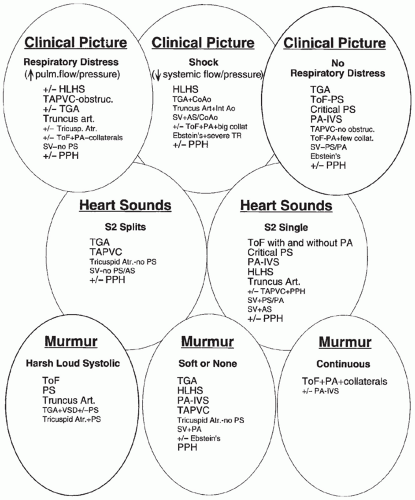
FIGURE 30.8 The differential diagnosis of cardiac exam findings in cyanotic neonates. +/-, sometimes; up arrow, increased; down arrow, decreased; AS, aortic stenosis; CoAo, coarctation of aorta; collat., systemic to pulmonary artery collateral vessels; Ebstein’s, Ebstein anomaly of the tricuspid valve; HLHS, hypoplastic left heart syndrome; Int Ao, interrupted aorta; PA, pulmonary atresia; PA-IVS, pulmonary atresia with intact interventricular septum; PPH, persistent pulmonary hypertension syndrome; PS, pulmonary stenosis; SV, single ventricle; TAPVC, totally anomalous pulmonary venous connection; TGA, transposition of great arteries; ToF, tetralogy of Fallot; tricuspid atr., tricuspid atresia.


Age of Presentation

Most children with a critical congenital cardiac anomaly develop symptoms within the first weeks of life (8). The age when infants develop cardiac symptoms is diagnostically useful. For instance, while ventricular septal defect is far more common (Table 30.2), transposition of the great arteries, coarctation of the aorta, and the hypoplastic left heart syndrome are the most common life-threatening anomalies presenting in the first days of life (see Table 30.7). Isolated ventricular septal defect is not associated with cyanosis, the associated murmur generally develops after several days or more, and respiratory symptoms usually do not develop until after the first week of life. Among those whose problem is cyanosis, transposition of the great arteries is the leading cause through the 3rd week of life; after that time, tetralogy of Fallot becomes the dominant cause of cyanosis. Among neonatal cardiac patients admitted because of respiratory symptoms, the hypoplastic left heart syndrome is the leading cause in the first week, complex coarctation leads in the second week, and thereafter, ventricular septal defect becomes the main cause (see Table 30.6).


Physical Examination


Systemic Perfusion and Pressure

Decreased systemic cardiac output is an ominous sign that requires rapid assessment and rapid appropriate management for the infant to survive. There are many potential noncardiac causes, the most common being sepsis. Signs of diminished systemic cardiac output include poorly perfused, cool, and/or mottled skin, listlessness, diminished peripheral pulse intensity, diminished systolic and pulse pressure, decreased urine output, and metabolic acidosis. Blood pressure should be measured in all four extremities in an infant who appears severely ill with these signs, particularly with coexistence of a murmur or cyanosis. Oscillometric devices can quickly and noninvasively measure blood pressure, although their correlation with centrally measured blood pressure declines in the presence of very low perfusion and pressure. It is very important to establish if the pulse amplitude and systolic blood pressure are diminished only in the postductal arterial distribution or throughout the body; that is, if the right arm systolic blood pressure is similar to or higher than that in the other extremities. Significant differential in the systolic blood pressure between the arms (usually right) and the legs (and umbilical artery) is diagnostic of an aortic obstruction. The additional presence of a murmur, gallop, hepatomegaly, or cyanosis further strongly suggests that a cardiac anomaly is causative of poor perfusion.


Murmur

Hearing a murmur is the most common means of recognizing the presence of heart disease in an infant. The diagnosis requires ascertaining the characteristics of the murmur. These include the history of the baby’s age when the murmur was first audible and examination findings of murmur timing in systole versus diastole, loudness, pitch, and the association of a thrill. The murmurs of valvar regurgitation and stenosis are audible immediately after birth, and the murmurs of septal defects are usually delayed days to weeks, or as long as several months in the case of atrial septal defects. Diastolic murmurs are rare but indicative of cardiac pathology. A prominent continuous murmur in a cyanotic neonate, particularly in the back or axilla, is rare but is very characteristic of tetralogy of Fallot with pulmonary atresia and systemic-to-pulmonary artery collateral vessels (the latter being the cause of the murmur). The loudness of a murmur, in combination with other findings, may suggest the likelihood of
various anomalies but is often not proportional to the severity of the lesion. The absence of a murmur does not preclude serious heart disease. To the contrary, many life-threatening cardiac anomalies may be associated with little or no murmur. In a neonate with cyanosis and/or shock and suspected cardiac anomaly, the presence of little or no murmur provides a diagnostic clue (see Figs. 30.7 and 30.8). The pitch of a murmur is associated with the pressure gradient across the abnormality causing the murmur. Tiny ventricular septal defects develop a characteristic fairly high-pitched murmur when the right ventricular pressure decreases to much less than the left ventricular pressure. Severe pulmonary or aortic stenosis can sometimes be distinguished from mild stenosis by a high-pitched harsh loud murmur and an associated thrill.


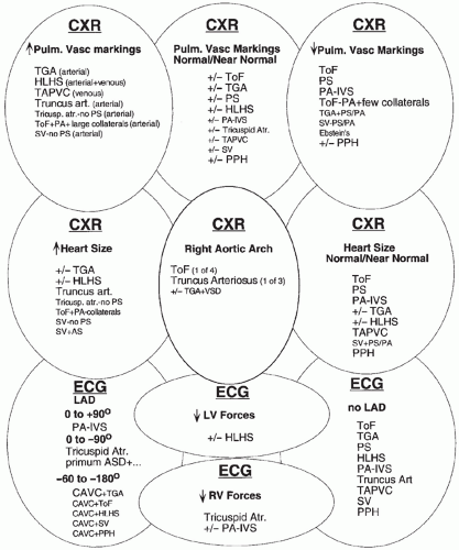
FIGURE 30.10 The differential diagnosis of chest radiographic and electrocardiographic findings in cyanotic neonates. Abbreviations, see Figs. 30.8 and 30.9.


Heart Sounds

Auscultation of the heart sound splitting is the most difficult part of the cardiac examination in neonates because of the relatively rapid heart and respiratory rates in neonates. However, when abnormalities of the first and second heart sounds are strongly suspected or excluded, it provides important information. Detection of splitting of the heart sounds requires practice and a minute or so of focused attention on just that sound, using a quality stethoscope, in a quieted baby. The absence of splitting may result from a heart rate too fast to discern splitting or a truly singlecomponent sound. A split first heart sound in a neonate suggests a click. The second heart sound emanates from closure of the aortic and pulmonary valves. Determining that the second heart sound is split (i.e., has two components) suggests that both aortic and pulmonary valves are not severely abnormal; that is, it is against the presence of aortic or pulmonary valve atresia or severe stenosis. However, other serious anomalies with two semilunar valves may still be present, for example, simple transposition of the great arteries. A second heart sound that always appears single, particularly at heart rates less than 120 per minute, may be caused by relatively early pulmonary valve closure associated with elevation of pulmonary artery pressure, but it suggests that the pulmonary or aortic valve may be abnormal (as in pulmonary atresia or critical stenosis, hypoplastic left heart syndrome, truncus arteriosus). Although difficult to detect, constant splitting of the second heart sound, as opposed to the usual intermittent splitting, suggests an atrial septal defect.


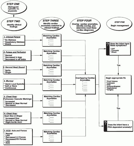
FIGURE 30.11 A process for diagnosing cardiac anomalies from findings on cardiac exam, chest radiograph, and electrocardiogram. ASAP, as soon as possible; c/w, consistent with; FiO2, fractional percentage of inspired oxygen; PDA, patent ductus arteriosus.


Cyanosis

Much more threatening than a murmur is the presence of cyanosis. Cyanosis without pulmonary disease is almost invariably the result of a serious cardiac abnormality. Generalized cyanosis may result from the following:


  • Poor mixing of separate parallel circulations (e.g., transposition of the great arteries


  • Other anomalies with transposition physiology such as Taussig-Bing-type double-outlet right ventricle


  • Restricted pulmonary blood flow and right-to-left shunting of unoxygenated systemic venous blood to the systemic arterial circulation (e.g., tetralogy of Fallot, critical pulmonary stenosis, tricuspid atresia)


  • Right-to-left shunting from intracardiac mixing with normal or increased pulmonary blood flow (e.g., total anomalous pulmonary venous connection without obstruction, truncus arteriosus, single ventricle without pulmonary stenosis, hypoplastic left heart syndrome)


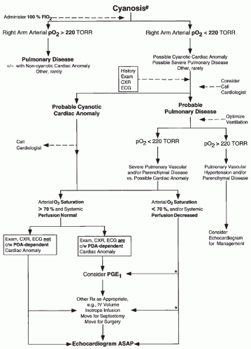
FIGURE 30.12 An approach to the diagnosis and management of cyanotic infants. #, cyanosis; see text for additional details of assessment of cyanosis; +/-, possibly; *, see text concerning management of specific anomalies; ASAP, as soon as possible; c/w, consistent with; FiO2, fraction inspired oxygen; Rx, treatment.

Differential cyanosis, most frequently in the postductal lower body circulation, may result from ductal-dependent severe aortic and/or left ventricular outflow or inflow obstructions (critical aortic coarctation or aortic valve stenosis) or severely elevated pulmonary vascular resistance.

The clinical recognition of cyanosis is dependent on the amount of arterial hemoglobin and therefore is influenced by the total blood hemoglobin concentration. An anemic infant may have severe arterial oxygen unsaturation without obvious cyanosis, and infants with polycythemia may appear cyanotic with near-normal arterial oxygen levels. Cyanosis is particularly evident in the lips. Perioral or nailbed cyanosis without lip cyanosis is usually not caused by cyanotic heart disease. Hypothermic infants may seem blue; babies viewed in fluorescent lighting or in blue surroundings may make the estimation of cyanosis more difficult. Methemoglobinemia is a rare cause of cyanosis. When cyanosis is suspected, indirect assessment of arterial oxygen saturation by the transcutaneous pulse oximetry can provide a rapid noninvasive measurement.


Noncardiac Anomalies

It is useful to know the relative frequency of the cardiac diagnostic possibilities when there are associated noncardiac anomalies (see Tables 30.3 and 30.4) or prematurity. Among premature infants, patent ductus arteriosus, coarctation of the aorta, and ventricular septal defect occur more often. Chromosomal abnormalities and congenital syndromes are also associated with lower birth weight and specific cardiac malformations (e.g., Down syndrome).


TABLE 30.7 Top Five Diagnoses Presenting at Different Ages


Percentage of Diagnosis


Patients


Age on Admission: 0-6 days (n = 537)


d-Transposition of great arteries


19


Hypoplastic left ventricle


14


Tetralogy of Fallot


8


Coarctation of aorta


7


Ventricular septal defect


3


Others


49


Age on admission: 7-13 days (n = 195)


Coarctation of aorta


16


Ventricular septal defect


14


Hypoplastic left ventricle


8


d-Transposition of great arteries


7


Tetralogy of Fallot


7


Others


48


Age on admission: 14-28 days (n = 177)


Ventricular septal defect


16


Coarctation of aorta


12


Tetralogy of Fallot


7


d-Transposition of great arteries


7


Patent ductus arteriosus


5


Others


53


▪ DIAGNOSTIC TOOLS


Transcutaneous-Pulsed Oximetry

Especially in the first week of life, generalized or differential oxygen desaturation may be the sole evidence of an important cardiac lesion. One-third of infants with potentially lethal congenital heart disease have cyanosis as their major symptom; another one-third have cyanosis associated with respiratory symptoms. At 2 days after birth, when most babies in the United States are sent home from the hospital, approximately 75% with ductus-dependent pulmonary circulation and 60% with ductal-dependent systemic circulation (including 75% with aortic coarctation, 45% with interrupted aortic arch, 25% with hypoplastic left heat syndrome) have not been diagnosed (41). About 30% (13% to 48%) of babies with ductal-dependent congenital cardiac anomalies are sent home from the hospital following birth without cardiac diagnosis (41). Prompt infusion of prostaglandin E1 to open the ductus arteriosus, and sometimes prompt catheter intervention to create an atrial septal defect, may be necessary for survival in those with ductal-dependent congenital cardiac anomalies, and most responsible anomalies are amenable to surgery.

Therefore, universal screening of all newborn babies with right upper and lower extremity transcutaneous oximetry and prompt cardiac evaluation of all babies with generalized oxygen saturation less than 95%, or pre- to postductal more than 3% are recommended and in many places mandated (41,42).


Hyperoxia Test: Acute Lung Disease and Cardiac Disease

Rapid diagnosis and initiation of appropriate management are most pressing when the infant is dyspneic and cyanotic. A chest radiograph may suggest lung disease, particularly if the findings are asymmetric. In the presence of diffuse symmetric changes possibly compatible with pulmonary edema or increased vascular markings, caution is necessary, particularly in the full-term neonate. The differential diagnosis between primary lung disease and heart disease causing pulmonary edema (e.g., total anomalous pulmonary venous connection with obstruction) can be difficult. Persistent pulmonary arterial hypertension with right-to-left shunting may coexist with lung disease and cause severe cyanosis. Although carbon dioxide retention is usually prominent among babies with primary lung disease, some severely cyanotic infants with cardiac anomalies can have marked hypercarbia. It can also be difficult to differentiate cardiac anomalies with diminished pulmonary blood flow and little murmur (e.g., pulmonary valve atresia) from persistent pulmonary arterial hypertension without other, radiographically apparent, lung parenchymal disease. Although the absence of hypercarbia suggests cardiac disease, some severely cyanotic infants with pulmonary vascular disease have normal PCO2 values.

The response of the arterial PO2 to administration of 100% oxygen, with the exceptions noted below, can differentiate cyanotic heart disease from lung disease. The infant who responds to breathing pure oxygen with a rise in arterial PO2 to 220 mm Hg or more does not have cyanotic heart disease, and the infant who does not raise his preductal arterial PO2 above 100 mm Hg is likely to have heart disease. Transcutaneous estimation of arterial oxygen saturation is not an accurate alternative because any arterial PO2 greater than 70 mm Hg will result in arterial oxygen saturation greater than 95%. Because the hyperoxia test is inconclusive when the arterial PO2 is between 100 and 220 mm Hg, these babies should be approached as possibly having cyanotic heart disease (see Fig. 30.12). The arterial PO2 while the baby is breathing 100% oxygen may initially be most rapidly measured from an umbilical artery catheter positioned in the descending aorta. However, a low arterial PO2 measured in the descending aorta may be from righttoleft shunting through a ductus arteriosus from coexisting persistent pulmonary hypertension. Comparison of the PO2 measured in blood from the right radial artery with the PO2 measured in blood from the umbilical arterial catheter may help to differentiate persistent pulmonary hypertension from cyanotic cardiac anomaly. The former may have a high PO2 in the right radial artery. Simultaneous mechanical hyperventilation and administration of oxygen may decrease pulmonary resistance and increase pulmonary flow, increasing the PO2 to greater than 220 mm Hg in the descending aortic and/or right radial arterial blood, allowing differentiation of lung disease or persistent pulmonary hypertension from cyanotic cardiac anomaly. Difficulties arise when pulmonary and cardiac pathology coexist. For example, in the baby with both lung and heart disease or those with coexistence of persistent pulmonary hypertension and predominant right-to-left shunting through the foramen ovale (see Chapters 29 and 31) or the baby with heart disease causing pulmonary venous hypertension and pulmonary edema, results may be confusing. Arterial PO2 may not significantly increase in response to 100% oxygen with severe persistent pulmonary hypertension and predominant right-to-left shunting through the foramen ovale. If doubt persists, the physician can make the diagnosis with two-dimensional echocardiography (see Fig. 30.12).


Chest Radiography

Chest radiography is rarely diagnostic of specific cardiac lesions, but it is a relatively quick and relatively inexpensive method to identify lung disease and screen for suspected cardiac anomaly in symptomatic infants. Chest radiographs often appear normal or near normal in the first day or days of life with many cardiac anomalies and may not be cost-effective in asymptomatic infants with an isolated murmur (i.e., no cyanosis or congestive signs or symptoms). However, in cyanotic or symptomatic infants, the presence or absence of cardiomegaly, increased pulmonary arterial or venous markings, diminished pulmonary arterial markings, or right aortic arch provide important information as to the presence of a cardiac anomaly (see Figs. 30.9 and 30.10). In combination with physical exam and electrocardiographic findings, chest radiographic findings may provide important information concerning the possible presence of specific cardiac anomalies that may aid in early management before an echocardiogram can be obtained. The heart size should be differentiated from the thymic shadow. Cardiomegaly is
cardiothoracic ratio greater than 0.6 in an anterior-posterior projection in the presence of an adequate inspiration. The aortic arch position can be assessed, even in the presence of a large overlying thymus, by deviation of the trachea to the opposite side. Associated noncardiac anomalies that provide clues to the cardiac diagnosis may be discovered by radiographic findings, for example, heterotaxy (asplenia syndrome, malrotation), absence of the thymus gland (DiGeorge syndrome), vertebral anomalies (VACTERL association), and abnormal sternal ossification (Down syndrome).


TABLE 30.8 Normal Maturational ECG Changes


Age


Heart Ratea (beats/min)


R axisa (+ degrees)


R amplitude V1a (mm)


R amplitude V6a (mm)


T amplitude V1a (mm)


0-1 day


93-154 (123)


59-192 (135)


5-26


0-11


-30 to +40


1-3 days


91-159 (123)


64-197 (134)


5-27


0-12


-41 to +41


3-7 days


90-166 (129)


77-187 (132)


3-24


0.5-12


-45 to +25


7-30 days


1-7-182 (149)


65-160 (110)


3-21.5


2.5-16


-10 to -52


1-3 months


121-179 (150)


31-114 (75)


3-18.5


5-21


-12 to -62


a 2-98th percentile (mean).


From Schwartz PJ, Garson A Jr, Paul T, et al. Guidelines for the interpretation of the newborn electrocardiogram. A task force of the European Society of Cardiology. Eur Heart J 2002;23:1329.


Electrocardiography

Electrocardiography can be useful in evaluation for cardiac anomaly and arrhythmogenic disorders and in particular for diagnosis and management of arrhythmia (see Arrhythmia section below). Electrocardiographic interpretation in neonates has several caveats. However, when placed within the context of other physical exam and radiographic findings, electrocardiographic findings can provide a timely advantage in diagnosis and management of suspected cardiac anomaly (see Figs. 30.9 and 30.10). Electrocardiography is also useful for timely recognition of life-threatening arrhythmogenic disorders, particularly within the context of other findings, for example, family history and borderline prolonged QTc.


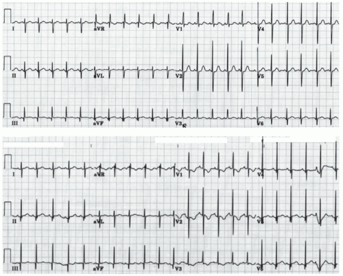
FIGURE 30.13 Electrocardiograms from healthy ½ day old (top) and 5 day old infants (lower) demonstrate normal neonatal repolarization changes. V1 T wave morphology changes from upright to inverted.

Interpretation of neonatal ECGs requires knowledge of maturational changes in heart rate, and electrocardiographic intervals, axes, voltages and repolarization that occur normally during the first days and weeks of life (see Table 30.8 and Fig. 30.13) (43).
Within the first days of life, there are significant changes in repolarization, including T axis and rate-corrected QT interval (QTc), as well as changes in R and S wave amplitudes, that influence interpretation. Compared with a 1 year old, 1 day old babies normally have relatively fast heart rates (93 to 154, average 123/minute), relatively rightward R axis (60 to 195, average 123 degrees) and relative right ventricular hypertrophy (R in V1 5 to 16 mm) (44). During the first 4 days of life a variably longer QTc, an evolution of changes in T wave polarity and voltage (e.g., in V1 from upright to inverted), and nonspecific ST segment changes are common. After age 4 days, QTc is 440 milliseconds or less in 97.5% of newborns but can be much longer with electrolyte abnormality (e.g., hypocalcemia, hypokalemia), drug effect, brain injury and genetic prolonged QT syndromes.

Ventricular hypertrophy in the ECG at birth is a consequence of the hemodynamic abnormalities imposed upon the fetal circulation. The hemodynamic changes and the ventricular hypertrophy associated with various cardiac anomalies are often much different in the fetal circulation than postnatal. For example, neonates with coarctation, and other obstructive anomalies with less than normal blood flow volume through the fetal left ventricle and more than normal flow through the fetal right ventricle, often have right ventricular hypertrophy from increased fetal right ventricular workload. Furthermore, anomalies associated with systemic-level right ventricular hypertension later in infancy often have neonatal electrocardiographic right ventricular forces that are difficult to differentiate unambiguously from normal in the neonate. Therefore, the differential diagnosis of ventricular hypertrophy in the neonate is different from later in infancy (see Figs. 30.9 and 30.10). In term neonates electrocardiographic findings of right ventricular hypertrophy include R amplitude in V1 above the 98th percentile (44) (>26mm in first week, >21mm in 2nd to 4th week), presence of Q wave in V1, or persistence of upright T waves in V1 beyond age 1 week. Findings of left ventricular hypertrophy include elevated R amplitude in V6 (44) (>12mm in first week, >16mm in 2nd to 4th week) and deep Q wave in V6 (>4mm), often accompanied by T wave flattening or inversion in V6.

While most anomalies do not have R-axis deviation at birth, when axis deviation is present, the ECG can be very helpful in diagnosis (see Figs. 30.9 and 30.10). Left axis deviation in acyanotic newborns is most often associated with endocardial cushion anomaly (e.g., primum atrial septal defect, complete atrioventricular canal defect), while in cyanotic newborns left axis deviation occurs with tricuspid atresia and other cyanotic anomalies in combination with an endocardial cushion anomaly (e.g., tetralogy or double outlet right ventricle with complete atrioventricular canal defect).


Echocardiography

Examination of the heart by two-dimensional echocardiography with color Doppler ultrasound allows excellent analysis of the intracardiac anatomy in small infants. Neonates are particularly good candidates for echocardiographic imaging because they are less active and have excellent echocardiographic imaging windows. Detailed segmental examination from subxiphoid, parasternal, apical, suprasternal notch, and additional modified views as necessary delineates almost all relevant cardiac anatomy and anomalies in most neonates. The situs, ventricular relationship, great artery relationships, systemic and pulmonary venous cardiac connections, atrial and ventricular septum, valve structure, great artery anatomy and coronary origins can be accurately determined.

Doppler echocardiography demonstrates the direction and velocity of blood flow within the heart and vessels. Color Doppler visualizes valve regurgitation and blood flow in valvar and subvalvar stenoses, patent ductus arteriosus, septal defects, abnormal coronary arteries, systemic venous anomalies, and arteriovenous malformation. Pulsed and continuous-wave Doppler techniques enable estimation of physiologic measurements such as the pressure gradient across stenotic valves, septal defects, and patent ductus arteriosus (see Fig. 30.14). When physiologic or pathologic tricuspid regurgitation is present, right ventricular peak systolic pressure may be estimated by Doppler measurement of the magnitude of the pressure gradient between the right ventricle and right atrium and the addition of the right atrial V-wave pressure, whether assumed or directly measured through an umbilical vein catheter (usually 3 to 10 mm Hg) (see Fig. 30.15). Right ventricular systolic pressure relative to left ventricular pressure can also be qualitatively assessed by the curvature of the interventricular septum. Contrast echocardiography with injection of agitated saline or albumin into
intravenous or umbilical artery catheters can sometimes serve as a useful adjunct to color Doppler in detection of shunts.


FIGURE 30.14 Echocardiographic parasagittal parasternal view of a patent ductus arteriosus. A: Doppler analysis demonstrates flow away from the transducer within the pulmonary artery and aortic isthmus and, in white, a jet of flow toward the transducer through the patent ductus arteriosus into the pulmonary artery. B: Quantification of the velocity of the flow jet through the ductus arteriosus with a continuous-wave Doppler technique and application of the Bernoulli principle allows the aortic-to-pulmonary-artery systolic pressure gradient to be measured. The pressure gradient by this technique is 4 × (maximum instantaneous velocity)2. The pulmonary artery peak systolic pressure can be estimated by the difference in the arterial systolic pressure and the pressure gradient across the ductus arteriosus. AO, aorta; DAO, descending aorta; LA, left atrium; MPA, main pulmonary artery; PDA, patent ductus arteriosus.


FIGURE 30.15 A: Echocardiographic apical four-chamber view in systole. Color Doppler analysis of the right heart demonstrates a jet of tricuspid regurgitation depicted by the blue flow jet (white arrow). B: Quantification of the velocity of the regurgitant jet by application of continuous-wave Doppler technique along the axis of the dotted line in the upper panel. Application of the measured triscuspid regurgitant velocity within the Bernoulli equation allows the right-ventricle-to-rightatrial peak pressure gradient to be measured and estimation of the right ventricular pressure. LA, left atrium; LV, left ventricle; RA, right atrium; RV, right ventricle; TR tricuspid regurgitation.

The ventricular systolic performance, size, and wall thickness can be assessed. The shortening fraction of the left ventricular internal short-axis dimension is the most commonly used measurement to assess left ventricular systolic function in children. In the sick neonate, right ventricular systolic pressure often is close to left ventricular systolic pressure, resulting in flattening of the ventricular septal curvature; such that shortening fraction may not be indicative of global systolic performance. When regional wall motion abnormalities are present, ventricular systolic performance is assessed by estimation or measurement of the relative change in ventricular volume with contraction, the ejection fraction. This is measured by two-dimensional planimetry in orthogonal planes or by three-dimensional echocardiography. The shortening and ejection fractions measure left ventricular performance, which is a function of contractility, afterload, preload, and heart rate. Contractility can be independently assessed by measuring the relationship of end-systolic wall stress velocity to fiber shortening using directed M-mode echocardiography, indirect central pulse tracing, and phonocardiography. When right ventricular hypertension results in ventricular septal systole flattening, volumetric-based measures are needed (see Chapter 29).

Echocardiography has limitations. Because complete examination of cardiac anatomy in neonates is labor intensive and requires expensive additional technology, the cost is often nearly that of a computerized tomography or magnetic resonance scans. The evaluation has often been unsatisfactory when performed where the use of echocardiography to recognize heart disease in neonates is infrequent and echocardiographic transducers with frequencies appropriate for infants are not available. Training and performance standards for echocardiographic examination of congenital heart disease in fetuses and children have been disseminated (44). Cases of delayed transfer of babies because of erroneous diagnoses of inoperable congenital heart disease and erroneous impression that there is no significant lesion have been encountered. With limited experience in the diagnosis of congenital heart disease in neonates, it may be best to transport the infant to the nearest center for echocardiographic examination. If personnel adequately trained in performing a complete study for congenital heart disease are available, it may be possible with modern technology to transmit the echocardiographic images via the internet or send a CD to a pediatric cardiac center.


Magnetic Resonance Imaging and CT Scanning

The excellent diagnostic quality of echocardiography in the infant, combined with the usual requirements for general anesthesia to obtain adequate MRI and CT images, relegates these other imaging modalities to more specialized roles. Magnetic resonance imaging and CT scanning can supplement assessment of anatomy and function, providing detailed images of intrathoracic structures such as peripheral pulmonary arteries, anomalous pulmonary veins, systemic-to-pulmonary collateral vessels, vascular rings, aortic arch anomalies, and right ventricular function, that may not be adequately displayed by echocardiography (45,46). The choice of imaging modality should recognize that, while CT scanning is less time consuming and provides somewhat greater anatomic detail, infant exposure to ionizing radiation should be minimized. Additionally, MRI may help differentiate cardiac tumors based on tissue characteristics not apparent on echocardiography 47). Use of echocardiography and magnetic resonance imaging or CT scanning, in conjunction with the history and physical examination, enables precise diagnosis without resorting to diagnostic cardiac catheterization in most neonates.


Diagnostic Cardiac Catheterization and Angiography


Anatomy

Catheterization is rarely used to learn the basic anatomy of the heart. The diagnostic information necessary for most cardiac surgical procedures in neonates is now obtained noninvasively by echocardiography. Diagnostic cardiac catheterization is used to provide specific data unavailable through echocardiography, magnetic resonance or CT imaging that are useful in planning management (48). What is the anatomy of the pulmonary arteries and systemic-to-pulmonary collaterals in the patient with tetralogy
of Fallot and pulmonary atresia? Is surgical unifocalization or catheter closure of the specific collaterals preferable? What is the anatomy of the coronary arteries in the patient with pulmonary atresia and intact ventricular septum? What is the anatomy of the coronary arteries with tetralogy of Fallot or transposition where abnormality is suspected and/or echocardiographic imaging is nondiagnostic? In selected patients with cardiomyopathy, light and electron microscopic analysis of ultrastructural anatomy and biochemical analysis of myocardium obtained by biopsy may uniquely provide a diagnosis. In many situations, diagnostic catheterization may be safer or more useful after initial palliative surgery, such as in hypoplastic left heart syndrome or following shunt procedures in those with complex intracardiac anomalies and pulmonary atresia.

The decision to perform a cardiac catheterization should be guided by a careful assessment of the long-term benefits in management versus the risk of the procedure. Before catheterization, the medical condition is optimized for the anomalies present and the rapidity with which catheterization may provide critical information to further stabilize the situation. Infants with a duct-dependent anomaly are best managed with an infusion of prostaglandin E1, begun before and continued throughout the catheterization. The potential for procedural morbidity and mortality is greater in the sick newborn (47). Potential morbidity includes blood loss, hypothermia, metabolic and respiratory acidosis, arrhythmia, electrolyte imbalance, hypoglycemia, thrombosis of femoral arteries, and angiographic myocardial stains.


Hemodynamic Measurements

Hemodynamic data obtained by catheterization can now largely be deduced from noninvasive measurements of blood pressure, oxygen saturation, and echocardiographic Doppler measurements of pressure gradients. Direct measurement does not help preoperative neonatal surgical management of most anomalies. When catheterization is done primarily for delineation of anatomy, hemodynamic measurements can be readily obtained and can facilitate delineating the clinical status and management. Hemodynamic measurements are often used to guide interventional catheterization such as valvuloplasty. Sometimes, particularly postoperatively, information from implanted catheters is useful for management of sick babies in the intensive care unit. Catheters in the right atrium placed through an umbilical vein, systemic vein, or transthoracically in the operating room may be used to obtain central venous pressure and blood oxygen saturation. These data can be used to infer preload and adequacy of cardiac output and, in combination with blood pressure measurements, to infer relative afterload. Catheters in the pulmonary artery, placed transthoracically at surgery or transvenously, can be used to measure left-to-right shunts and to measure pulmonary pressure to titrate pulmonary vasodilators.

The hemodynamic principles for these calculations are based on Ohm’s law and the Fick principle (see Table 30.9) (47). The former, when applied to hemodynamics, is the pressure drop across a vascular bed is equal to the product of the flow and resistance across it. Therefore, the resistance equals the difference of the arterial and venous pressure divided by the flow. The flow can be calculated from the Fick principle, which is based on the premise that oxygen delivery to the body equals oxygen consumption by the body. Oxygen consumption is routinely measured in the catheterization laboratory and, in the intensive care unit, can be assumed to be 200 to 240 mL/min/m2

Only gold members can continue reading. Log In or Register to continue

Stay updated, free articles. Join our Telegram channel

May 30, 2016 | Posted by in PEDIATRICS | Comments Off on Cardiac Disease

Full access? Get Clinical Tree

Get Clinical Tree app for offline access