Fig. 3.1
A clinical photograph showing a peripherally inserted central catheter (PICC)
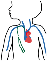
Fig. 3.2
Diagrammatic representation of a tunnelled double lumen central line. Note the proper position of the tip of the catheter in the superior vena cava
Peripheral venous cut-down
Open central venous access:
Open central venous tunnelled catheters
Totally implantable venous access (Port-a-cath)
Peripherally Inserted Central Catheter
The simplest, safest, and most reliable vascular access is PICC (Fig. 3.1).
These catheters are inserted through a peripheral vein and advanced into the heart.
They require local anaesthesia.
They are easy to insert.
They can be done at bedside.
Central Venous Tunnelled Catheter
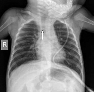
Fig. 3.3
A chest X-ray showing a tunnelled central line. Note the position of the tip of the catheter at the junction of the brachiocephalic vein and the internal jugular vein
It can be single lumen or double lumen.
It is inserted under anaesthesia through a big vein in the neck.
Advantage is it can be inserted in any age group.
It can be kept for longer duration until the treatment is over.
Implantable Ports (Port-A-cath)
Implantable ports are one of the best-suited options for children (Figs. 3.4, 3.5, and 3.6). They are:
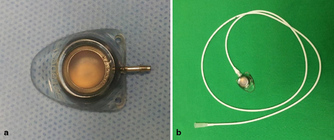
Fig. 3.4
a A photograph showing the single port. b The already connected port. There are also double ports. The length of the catheter to be used is determined intra-operatively and using fluoroscopy to determine the proper position of the catheter
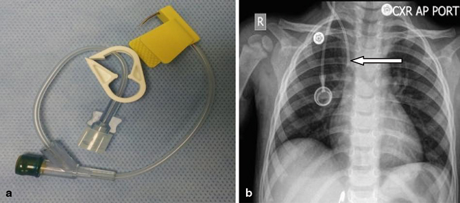
Fig. 3.5
(a) A photograph showing the Huber needle used to infuse fluids and medications via the port. Ordinary needles should not be used and a chest X-ray (b) showing the Port-A-cath. Note the position of the tip of the catheter in the superior vena cava
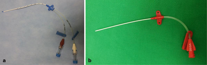
Fig. 3.6


a and b, Two types of percutaneously inserted central lines
Stay updated, free articles. Join our Telegram channel
Full access? Get Clinical Tree
