Fig. 10.1
(a) A catheter is placed in the uterine artery via the femoral artery (Copyright CeloNova Biosciences Inc.). (b) Detail of a. Small particles are injected in the uterine artery obstructing the vessels towards the fibroid (Copyright CeloNova Biosciences Inc.)
Fig. 10.2
(a) Pre-UAE angiogram. (b) Post-UAE angiogram: fibroid blush disappeared
Fig. 10.3
(a) Sagittal MRI demonstrating uterus with fibroids prior to UAE. (b) MRI after UAE will follow later. Sagittal MRI demonstrating same uterus with fibroids from figure a 6 months after UAE
Patient Selection and Prediction of UAE Outcome
Several prognostic factors have been identified to predict effect of UAE on fibroid volume, complaints and need for re-interventions (Table 10.1). Three types of predictive factors can be identified: factors that can be established before UAE, during UAE and after UAE. Before UAE the vascularity of the fibroids, as established by contrast enhanced MRI, predicts outcome: hypervascular fibroids predict a long regrowth free interval [10].
Table 10.1
Predicting UAE outcome
Factor | Outcome | |
|---|---|---|
A. Before UAE | Hypervascular fibroids on MRI | Long regrowth-free interval |
Submucosal location | Larger improvement QOL-score | |
Smaller size of fibroid(s) | Larger improvement QOL-score | |
Heavy menstrual bleeding as presenting symptom | Larger improvement QOL-score | |
Larger size fibroid(s) | Higher chance of symptom recurrence, higher chance of failure | |
B. During UAE | Unilateral UAE | More secondary hysterectomies |
C. After UAE | Less than 90 % infarction (MRI) of fibroids after UAE | More re-interventions |
Post-UAE Dopplers (resistance index) | Fibroid volume decrement | |
No improvement of pain or bleeding at 1 year follow-up | More re-interventions | |
More volume reduction | More satisfaction |
Furthermore, a smaller leiomyoma size, a submucosal location of the fibroids, and heavy menstrual bleeding as the presenting symptom are predictors of a more significant fibroid related symptom change score [11]. Finally, larger fibroids and more numerous fibroids predict higher symptom recurrence and higher failure rates [12, 13]. During the procedure only a unilateral UAE predicts failure, defined as secondary hysterectomy [14]. Directly after the procedure the following factors were of predictive value: when less than 90 % of the fibroids showed signs of infarction on the post-UAE MRI scan there was more chance of re-interventions [15, 16]. Post-UAE fibroid volume at follow up was related to post-UAE dopplers (resistance index) directly after the procedure [17].
When there was no improvement in bleeding or pain at 1 year after UAE, this was indicative for failure [13, 18]. The percentage reduction in dominant tumor volume also predict failure, defined as need for re-intervention [18]. Finally, when volume reduction of fibroids was in the first tertile, patients more likely to be dissatisfied with outcome compared with women in the third tertile [13].
Although all these predictors have not been prospectively evaluated, the somewhat smaller and well vascularized fibroids seem to have the best results. Also it is important to ensure that the whole volume of the fibroid is embolized. Finally, when symptoms have not or only marginally improved after 1 year, the re-intervention rate seems to increase.
Being able to predict the outcome of UAE is very important in selecting patients for this procedure. Especially the first category (factors known before UAE) needs to be further explored, since this differentiates between patients that are good candidates for UAE and the group of patients that might be better off with another treatment.
Indications and Contra-Indications
Uterine Artery Embolization is indicated primarily for uterine fibroids. Another indication is severe post-partum hemorrhage as a last resort before a post-partum hysterectomy needs to be performed [19]. This procedure will not be discussed in this chapter. Although some literature is available on UAE for adenomyosis, the vast body of evidence concerns uterine fibroids. The evidence on UAE for symptomatic adenomyosis is summarized in a review by Popovic et al. [20]. In total 511 women have been reported in this review with a positive effect on symptoms for 75.7 % of patients. No randomized data are available on this topic: patients were treated by UAE for adenomyosis without a control group. Since adenomyosis patients have no other option than radical surgery when conservative treatment fails, UAE could be an option. Randomized data are needed though for appropriate counseling on effectiveness and safety.
UAE for symptomatic fibroids has been performed for a variety of symptoms. Good results have been shown on associated heavy menstrual bleeding (HMB), dysmenorrhea and bulk-symptoms, although performing UAE only for the latter is questionable.
Absolute contraindications are considered to be: current pregnancy, presence of pelvic inflammatory disease, presence of uterine malignancy and asymptomatic fibroids.
Several relative contraindications have been proposed.
Narrow stalked (arguably when the stalk is <50 % of the largest diameter of the fibroid) pedunculated subserosal or submucosal fibroids are considered relative contraindications, since these might cause fibroid expulsion with concurrent intrauterine infection (submucosal fibroids) or sterile peritonitis due to detachment of fibroids (subserosal fibroids). Pedunculated fibroids are considered relatively contraindicated since it depends on the size (with very small fibroids most probably less of a problem than large ones), the possibility to subsequently remove the fibroids by hysteroscopy or laparoscopy.
Since there might be effect on ovarian reserve and uterine function UAE is usually not recommended as a standard treatment when a woman still wishes to get pregnant in the future (see also below). Another relative contraindication might be a very large uterus with multiple very large fibroids. The volume of necrosis can be substantive, with the related post-procedural pain and risk of infection. However, no clear cut-off can be given for size related to problems. No studies are available that provide evidence on a cut-off for size. There are several series though, that demonstrate good results even on very large uteri [21–24]. In these publications there appears to be no relation between the size of the uterus and post-embolization symptomatology or satisfaction. Also, failure rates defined as secondary hysterectomies were similar as in other series. Also volume reduction was similar to patients with smaller fibroids, although a large volume was still present after the procedure because of a large volume before UAE.
The procedure is indicated primarily for premenopausal women, since fibroids tend to decrease in size and symptoms after menopause. A growing uterus after menopause should raise the suspicion of a malignancy and careful imaging and follow up is warranted in that case.
Pre-procedural Imaging and Workup
Most protocols agree on the need of MRI before UAE in order to properly diagnose uterine fibroids. In our view, in many cases an MRI can be prevented, when ultrasound provides a clear diagnosis and can locate fibroids according to the international classification system [25]. It has been shown that –in expert hands- ultrasound is as reliable in detecting fibroids as MRI scan [26]. However, when multiple fibroids are present of fibroids are very big, MRI is superior. Therefore, when ultrasound is inconclusive, no expert is available, or the uterus is too large to screen reliably by ultrasound, MRI is a very good and reliable tool to diagnose uterine fibroids and their location in the uterus.
When HMB is present hemoglobin testing is mandatory. Renal clearance should be tested (GFR) since contrast fluid is used.
Policy on prophylactic antibiotics varies among clinics and in publications on UAE. On average post-embolization infection prevalence is estimated to be 2 %. No randomized trials have been done to establish the value of antibiotics in UAE. Pathophysiologically infection seems to be more likely longer after UAE since bacteria might grow easier in necrotic tissue.
Thromboprofylaxis in general is only applicable for intravascular procedures when a patient is at increased risk for thromboembolic disease [27].
Complications After UAE
Since severe complications in general are usually rare in elective surgery, large series are necessary to identify true complication rates. Therefore, randomized controlled trials (with usually relatively few patients) seldom have complications as a primary outcome and are not exclusively suitable for research on complication rates. Furthermore, complications are assessed using different complication classification systems making comparisons difficult.
Mortality after UAE is extremely rare, but several case-reports have been written [28–32]. Causes of death were septicemia (n = 2), pulmonary embolus (n = 2) and systemic non-target embolization in a woman with arteriovenous shunting and patent foramen ovale.
In a national registry in the United States (n = 3,160) major complications (as defined by the Society of Interventional Radiology Clinical Practice Guidelines) were reported in 0.66 % of patients in the initial hospitalization period and in 4.8 % during the first month after discharge [33]. A large systematic review has been done in 2012 on complications after UAE yielding several specific numbers for specific complications [34]. In this review 54 studies were summarized with a total of 8,159 patients. Major complications occurred in 2.9 % of patients. The rate of hysterectomy for resolution of a complication from UAE was 0.7 %, and the rate of readmission was 2.7 %.
Morbidity after UAE can also be divided in peri-procedural, early complications and late complications (beyond 30 days).
Peri-procedural complications include groin hematoma, arterial thrombosis and (pseudo)aneurysm and are uncommon [35, 36]. Bilateral failure of UAE is also considered a complication and occurs in 4 % of all patients that had their UAE in a clinical trial [36].
Early complications that often occur are fever, nausea, pain and malaise and form the ‘post-embolization syndrome’ [35]. This is usually self-limiting and can be managed with non-steroid- anti-inflammatory drugs [37]. When these symptoms persist or get worse, re-admission might be necessary (up to 9 % of cases) [35]. Compared to hysterectomy/myomectomy more readmissions were seen after UAE because of these complications [38]. Serious early complications are deep venous thrombosis or pulmonary embolism, with an incidence of 0.2 % [34].
Late complications (beyond 30 days) comprise vaginal discharge, fibroid (tissue expulsion) and amenorrhoea (see below). Vaginal discharge Another complication that is also relatively common (16–20 %) is vaginal discharge [34]. This can last for months, and is also self-limiting, provided that the discharge is not purulent and fever is absent (otherwise there might be a need to start treatment with antibiotics). Expulsion of a fibroid (or fibroid tissue) is a relatively common phenomenon and can generable be awaited, but sometimes it can be necessary to perform a hysteroscopy [39]. Fibroid tissue expulsion has been recorded in 4.7 % of patients [34].
Endometritis occurs in 0.5 % of cases (mostly submucosal fibroids) and responds well to antibiotics (a culture should be performed) [39]. In case of persistent discharge combined with abdominal tenderness or pain, it is possible that a necrotic fibroid is being expelled.
Effect on Ovarian Function
Amenorrhoea is an unintended sign, the cause of which is still largely unknown. Arterial flow to the ovary is likely to be transiently occluded during UAE, but may be reestablished on the longer term [40]. The overall incidence of permanent amenorrhoea is 3.9 % [34]. In women older than 45 years of age, there is a significantly higher risk of a decrease in ovarian reserve as detected by an FSH assay compared with younger women, as well as a higher risk of treatment-induced menopausal symptoms and/or amenorrhea. At this time, current data suggest that UAE for women younger than 40 years of age is unlikely to decrease ovarian reserve since no differences were found in any of the ovarian reserve parameters after treatment compared to before treatment [40]. One RCT evaluated Anti-Mullerian-Hormone (AMH) levels in UAE patients compared to hysterectomy patients before and after the procedure [41]. AMH was significantly decreased in both groups compared to baseline but recovered in the hysterectomy group, whereas the AMH levels stayed significantly below the expected decrement in UAE patients, indicating at least some damage to the ovaries after UAE. The average age of patients was >45 years though. The effect on fertility remains uncertain (see also below).
Management of Complications
Good counseling is important for several complications such as prolonged discharge or loss of fibroid tissue and the post-embolization-syndrome. Should there be a persistent high fever, further workup is obligatory. Investigations include physical and vaginal examination including vital signs and swabs of discharge if present, blood examination and ultrasound examination of the uterus [39]. Post-embolization necrosis of the fibroid can give high pain-scores [42]. Treatment lies in the first place in giving adequate pain medication, unless pain seems to originate in fibroid expulsion, infection or non-target embolization. If the work-up demonstrates an infection, it is recommended to start intravenous antibiotics.
Outcome of UAE
UAE is mostly indicated for heavy menstrual bleeding, although it is also applied for dysmenorrhea, pelvic pain or pressure symptoms. Regarding the effect of UAE on heavy menstrual bleeding (HMB), it has been shown that most patients (73–90 %) reported improvement or disappearance of symptoms up to 5 years after treatment [43].
Most trials however, did not report on improved HMB but rather on satisfaction with treatment. A recently published Cochrane review on long-term results of UAE versus surgery chose satisfaction as the primary outcome measure, and showed that UAE had a similarly high satisfaction-rate as myomectomy and hysterectomy [38].
Quality of life was consistently better after UAE compared to baseline and comparable to surgical alternatives [5, 6]. Quality of life results were summarized in a systematic review comparing UAE with surgery, showing that health related quality of life was, even after 5 years of follow up, significantly higher than baseline without any differences between the studied groups [35]. The effect of UAE on bulk and pressure-complaints is less well studied, but in large cohort studies up to 90 % of patients reported improved bulk-complaints [44, 45]. The effect of UAE on lower abdominal pain or dysmenorrhea has also been described and shows an improvement in up to 80 % of patients [46]. Since pain-and bulk complaints were not investigated in validated questionnaires it is not possible to present quantitative outcomes.
Secondary interventions after UAE for failure or recurrence of symptoms have been reported to be 27 % (51/187) in the RCTs at 5 years of follow up [35]. The case series and registries report higher success rates (80–90 %) [13, 47].
Another important benefit of UAE is a significantly shorter hospital stay, and a significantly faster resumption of daily activities and work [38].
UAE and Fertility
As stated before, an important relative contraindication is the wish to conceive. Since hysterectomy is obviously impossible in this case, a myomectomy used to be the gold standard. UAE is increasingly applied in this group of patients with reassuring results. However, good comparative data are lacking. The effect of UAE on (sub)fertility has not been well investigated. There is evidence on the adverse effect of fibroids on fertility. It is known that submucosal and intramural fibroids might have a negative impact on fertility, however, this does not automatically mean that treatment of the fibroids will improve fertility outcomes. Subserosal fibroids do not appear to have a significant effect on fertility outcomes. For intramural fibroids this is not known [48].
If (sub)fertility is likely to be caused by the presence of fibroids, it may be considered to embolize them. In a systematic review on pregnancies after UAE it was stated that miscarriage rates were higher in post-UAE pregnancies (35.2 %) compared with pregnant women with a non-treated fibroid-uterus (16.5 %), matched for age and fibroid location. The UAE pregnancies were more likely to be delivered by cesarean section and to experience post partum hemorrhage. Rates of preterm delivery, intra uterine growth restriction and malpresentation were similar in UAE pregnancies and in control pregnancies with fibroids [49]. In the recently published Cochrane review that was mentioned earlier, the other primary outcome measure was live birth rate. This was calculated from the limited cohort of participants who tried to conceive in the study of UAE versus myomectomy. There was no significant difference between the groups in live birth rate [50]. On the basis of these results myomectomy should be regarded as the gold standard. UAE in women with a child wish should ideally be performed in research setting and/or after appropriate counseling.
Follow Up After UAE
After discharge the patient should be aware of the risk of minor and major complications (see above) and should know how to detect them. In case of increasing pain, fever, foul smelling discharge the patient should contact her specialist in order to rule out complications.
Although all trials report on imaging follow up, in daily clinical practice the patient and her complaints should be leading in considering post-procedural imaging and labwork. Good response in terms of volume reduction but unchanged HMB might result in re-intervention, whereas the opposite situation might not. In that perspective imaging does not add to follow up, unless it is for research purposes. When HMB persists, hemoglobin checkups are necessary for iron-supplement indication.
The Future of UAE
Although extensive research has been done on this topic, several aspects remain unanswered or can be optimized. UAE has not properly been compared to another common uterus sparing technique: surgical myomectomy. In this perspective: good randomized data on future fertility is needed in order to establish the role of UAE in patients with a wish to conceive. Several initiatives have been taken in this perspective: The FEMME trial (ISRCTN70772394) randomizes patients with fibroids with the wish to preserve their uterus between UAE and myomectomy. The FEMME trial is still in the recruitment process. The FIRSTT-trial (NCT00995878) compares UAE to HIFUS and is expected to finish data-collection in 2015. A similar trial is currently performed with an estimated end-date of may 2014 (NCT01834703). Next to evaluation of UAE, selection of patients can be elucidated somewhat further. Prognostic factors such as vascularity for a successful procedure can be investigated more clearly and consistently.
Even though some questions remain unanswered, many have been answered placing UAE consistently among the various possibilities for fibroid therapy.
New Fibroid Ablation Techniques
Radiofrequency Ablation (RFA)
RFA in general refers to the destruction of tissue by focused energy with electric current through a bipolar electrode or a monopolar electrode, by radiofrequency or by a cryoprobe used as energy sources.
RFA was introduced in the late 1980s in Europe as a conservative treatment of uterine fibroids [51]. At first, it was performed with the use of the ‘Neodynium: Yttrium Aluminium Garnet’ (Nd:YAG) laser. Later, bipolar needles were developed as an alternative to this laser option (this technique was called ‘myolysis’). The technique was further refined and is nowadays performed under sonographic guidance (radiofrequency volumetric thermal ablation, RFVTA). These techniques can be performed laparoscopically (Fig. 10.4) or transcervically (Fig. 10.5). In the newer transcervical devices the ultrasound probe is built in.
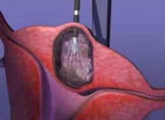
Fig. 10.4
Laparoscopically, the Handpiece tip is advanced into the fibroid with ultrasound guidance (Source: www.haltmedical.com)
Fig. 10.5
(a) Placement of bipolar needles in fibroid in order to do trans-cervical ablation. The ultrasound probe is directed downwards right before the surface of the fibroid (Source: Gynesonics Inc.). (b) MRI with contrast of uterus with single fibroid. On the left image the situation prior to ablation is visible. On the right side the MRI is repeated demonstrating no flow in the fibroid anymore (Source: Gynesonics Inc)
Laparoscopic RFA
Recently one RCT has been published comparing laparoscopic RFA with laparoscopic myomectomy [52]. Although a 5 year follow up period was planned with outcomes such as pregnancy outcome after RFA, symptom improvement, re-intervention rates and recurrence of fibroids, this first publication describes short term outcomes only. In total 51 patients were randomized between RFA and laparoscopic myomectomy. RFA was significantly better in terms of mean hospitalization time (10 versus 30 h), operative blood loss (16 versus 51 ml). Not significantly different were mean number of excised fibroids per patient (2.8 versus 2.0), operation time (1.1 h versus 1.3 h). No major complications or conversions occurred.
Several prospective studies have been published. Garza Leal et al. published a prospective study with 31 patients that underwent laparoscopic RFA for symptomatic fibroids, where fibroid symptoms and volumes were successfully reduced. At 3, 6, and 12 months, mean symptom severity score improved significantly compared with baseline: by 59.7, 71.7, and 82.0 % respectively. The increase in mean health related quality of life scores over time was statistically significant (p < 0.001): 60 at baseline and 98 at 12 months. Mean uterine volume decreased from 194.4 (standard deviation: 105.9 cm3) at baseline to 113.2 (standard deviation 53.5 cm3) at 12 months (p = 0.006) [53].
Stay updated, free articles. Join our Telegram channel

Full access? Get Clinical Tree


