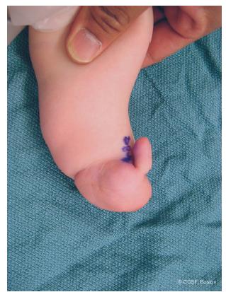
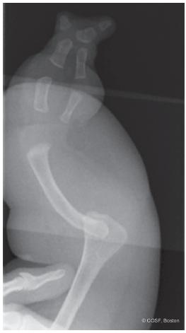
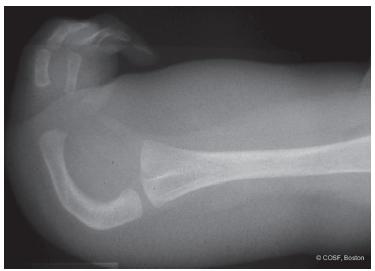
FIGURE 14-1 A: Ulnar longitudinal deficiency with elbow synostosis, foreshortened forearm, and three-digit hand. The thumb is in the same plane as the other two digits, which limits pinch. B: Radiograph of same patient. Note carpal coalition.
There have been many classification systems since Kummel in 1895. The classification systems have variably focused on the forearm, hand, thumb, or some combination thereof.2,3,6–10 Bayne’s classification indentllels the radial longitudinal classification and makes it easy for most to remember. Type I is a hypoplastic ulna, type II is an absent distal ulna, type III is complete absence of the ulna, and type IV is complete absence of the ulna with radial-humeral fusion. Manske emphasized the status of the thumb in surgical decision making, similar to his classification for cleft hands.
Clinical Evaluation
As with any congenital difference, there is parental and primary care concern about malformations of other organ systems. However, these cases occur almost always in isolation or less commonly, with associated musculoskeletal malformations. Evaluation of the upper limb deficiency is by serial clinical exams and plain radiographs. Deformity is monitored over time to see if there is progressive wrist ulnar deviation, forearm instability, limb foreshortening, or functional deficits. The presence or absence of pain is recorded in older children. Their emotional well-being and social status is followed. Anyone who cares for children with congenital differences needs to be a highranking amateur psychologist; but, we also need to know the boundaries of our skills and knowledge.
The spectrum of ulnar longitudinal deficiency is broad. The simplest is absence of ulnar digital ray(s). These children are normal functionally. Since most people do not count fingers, cosmetically their deficiency is often not noted socially. Reassurance and time for the parents to be convinced that all is well is the major form of care for these children.
When the deficiency extends into the forearm, there is more extensive involvement of the hand, wrist, and elbow. Besides absent rays, hand and carpal involvement can include: digital hypoplasia, limited joint development, extrinsic and/or intrinsic weakness or absence, carpal absence or coalitions,11 and thumb out of anatomic plane. Syndactyly can be present. The extreme form of hand malformation with ulnar longitudinal deficiency is monodactyly (Figure 14-2). Care for the more involved hand is geared to improving alignment and maximizing motion and strength. Passive stretching and progressive nighttime splinting programs are commonplace. Surgery to improve independent digital and combined hand function is indicated.
Wrist involvement can extend from a mild negative ulnar variance all the way to an unstable forearm. The wrist ulnar deviation can be static or progressive. Historically the ulnar anlage, a fibrocartilaginous connection between the residual ulna and the wrist, was thought to be responsible for progressive ulnar deviation deformity. Surgical excision in infancy was recommended for nearly every infant with ulnar longitudinal deficiency.9,12 Now, ulnar anlage excision is rarely performed in our institution.8,13,14 If the deformity is extreme, limits function, and/ or is progressive, then a radial osteotomy and/or ulnar soft tissue release is considered.

FIGURE 14-2 Ulnar deficiency with single floating digit.
The unstable forearm and elbow are also less clear in terms of treatment12,13 (Figure 14-3). In the absence of pain, many of these children can be more functional with an unstable, mobile forearm and elbow than a surgical single bone forearm. It can be alarming to the parents that we endorse no treatment. In some respects, they need to be conditioned to the same limits of care available that we have over time. The best treatment here is often to get out of the way of the child and let them maximize their native intelligence, whole body capabilities, and adaptive abilities. Occupational and physical therapy can help with specific tasks.
The fused elbow is even more of an enigma to the parents (Figure 14-4). They cannot understand why in this age of artificial joints and biologic wonders, we cannot provide their children with a mobile, stable elbow that grows. Our job is to (1) provide them with reality-based evaluations and (2) continue research that hopefully one day achieves its goal.
Surgical Indications
Above all, keep it simple .
—Auguste Escoffier

FIGURE 14-3 Complete absence of ulna with proximal radius dislocation (type III) and complete, complex syndactylized digits.
Children with ulnar longitudinal deficiency can have a combination of problems with limb shortening and malalignment; unstable, malaligned, and/or fused joints; missing digital rays; limitations in grasp and pinch; and syndactyly. We have established operations for all these conditions. However, multiple extensive surgeries have not necessarily made these children have more function. Surgery for the hand has a clear benefit. Syndactyly reconstruction gives more independent digital use. Realignment of the thumb and deepening of the first web space provide better pinch and grasp. The indications for derotational and/or angular corrective osteotomy, ulnar anlage resection, forearm and elbow stabilization, and limb lengthening are progressively less clear. We usually let the functional status of the child and our knowledge of surgical outcomes, rather than parental pressure to normalize the situation, be the main drivers in surgical decision making. Aside from thumb and digital surgery, changing one element of the limb in these children can affect functional use in another part. This makes surgical intervention complex in the forearm, elbow, and humerus. At present, collectively we are not as smart and talented as patients and families want us to be.

FIGURE 14-4 Complete absence of ulna with radiohumeral synostosis. Forearm is markedly bowed and foreshortened with a two-digit hand.
Stay updated, free articles. Join our Telegram channel

Full access? Get Clinical Tree


