Tympanocentesis
Marla J. Friedman
Introduction
Tympanocentesis involves needle aspiration of middle ear contents to obtain fluid for culture or to provide symptomatic relief in cases of especially painful otitis media. It can be safely performed by pediatricians, emergency physicians, and other qualified clinicians in the outpatient setting (1,2,3,4,5).
Otitis media is one of the most common childhood infections, with a peak incidence during the first 2 years of life. The incidence of otitis media is on the rise, with approximately 70% of children having at least one episode in the first year of life (6,7). The growing number of cases may be due to the increased use of day-care facilities in this country (7,8,9). Otitis media is a general term used to describe acute or chronic inflammation of the middle ear. Acute otitis media is defined as the presence of middle ear effusion with symptoms (pain, fever, irritability) and signs (redness, fullness, bulging of the tympanic membrane) of acute suppurative inflammation. Otitis media with effusion describes the presence of fluid in the middle ear without the signs or symptoms of acute suppurative inflammation (8,10,11). Otitis media with effusion frequently follows an episode of acute otitis media and usually resolves within about 3 months (12).
Middle ear infections must be effectively treated to prevent complications and minimize the morbidity that can be associated with otitis media. Conductive hearing loss due to middle ear fluid is the most common complication of otitis media. Even a mild unilateral hearing impairment can lead to speech and language problems and affect both the school performance and the social interactions of a young child. Other less common complications include chronic suppurative otitis media, tympanic membrane perforation and scarring, cholesteatoma, facial paralysis, and extension of the infection into the mastoids or intracranial structures (7,9).
Antibiotic therapy is generally effective in treating acute otitis media, and it remains the initial recommended approach. Tympanocentesis is recommended for patients who fail antimicrobial therapy (13,14). With the increasing prevalence of antimicrobial resistance, tympanocentesis is likely to have an increasingly important role in the management of otitis media.
Anatomy and Physiology
The anatomy of the ear is illustrated in Figure 52.2. The tympanic membrane separates the external auditory canal from the middle ear. The middle ear, or tympanic cavity, consists of the auditory ossicles (malleus, incus, and stapes), muscles, nerves, blood vessels, and the eustachian tube. The eustachian tube, which opens and closes during swallowing, connects the nasopharynx with the middle ear cavity and functions to equalize the pressure in the middle ear with atmospheric pressure. It protects the middle ear from reflux of nasopharynx secretions and drains secretions from the middle ear into the nasopharynx. Most cases of otitis media occur as a result of an abnormally functioning eustachian tube (15,16).
The structure of the middle ear is illustrated in Figure 56.1A. Landmarks that must be clearly identified before performing tympanocentesis include (a) the lateral (short) process of the malleus, a protrusion at the superior aspect of the malleus that points toward the anterior quadrant; (b) the handle (long) process of the malleus, the thin structure that divides the membrane into anterior and posterior segments; and (c) the umbo, which is the inferior tip of the malleus that separates the superior and inferior quadrants of the tympanic membrane. The four quadrants of the tympanic membrane are roughly delineated by drawing one line through the long handle of the malleus and a second perpendicular line through the umbo (16,17). The umbo is perhaps the most important
bony landmark to identify. It points toward the posteroinferior quadrant of the tympanic membrane, which is where tympanocentesis should be performed. The ossicles and nerves, including a branch of the facial nerve, lie within the superior quadrants of the tympanic membrane. Consequently, this area should be avoided during tympanocentesis.
bony landmark to identify. It points toward the posteroinferior quadrant of the tympanic membrane, which is where tympanocentesis should be performed. The ossicles and nerves, including a branch of the facial nerve, lie within the superior quadrants of the tympanic membrane. Consequently, this area should be avoided during tympanocentesis.
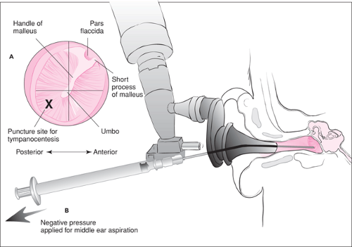 Figure 56.1 A. Landmarks of the middle ear. B. Technique for tympanocentesis.
Stay updated, free articles. Join our Telegram channel
Full access? Get Clinical Tree
 Get Clinical Tree app for offline access

|