Fig. 22.1
Multiple angiomatous papular lesions of verruga peruana
Complications are not uncommon in the acute phase and include super-infections, most commonly with salmonella species but also with toxoplasma, histoplasma, and others [8]. Hematological, gastrointestinal, cardiovascular, and neurological complications also occur during pregnancy and infection can lead to miscarriage, premature labor, and maternal death. Young children are the most affected in endemic communities, partly because of predominantly younger population but also due to the presumed protective immunity that develops with repeated infections [2].
Differential Diagnosis
The cutaneous lesions of verruga peruana have a wide differential that includes pyogenic granuloma, hemangioma, bacillary angiomatosis, Kaposi’s sarcoma, fibrosarcoma, leprosy (cystoid form), malignant lymphoma, reticuloendotheliosis, yaws, lymphomatoid papillomatosis, angiolymphoid hyperplasia with eosinophilia, varicella, molluscum contagiosum, Spitz nevus, and sarcoma [9].
Treatment
Only studies looking at quinolones and chloramphenicol were found in the literature but none comparing the two. Rifampicin is now the most widely used antibiotic in the chronic phase and most patients respond well: 80 % and 93.1 % respectively in two studies one of which has level 3a evidence [10].
Ongoing Research
Development of effective surveillance tools to increase understanding of the epidemiology of disease, harmonized case and outcome definitions, readiness for outbreak investigation including evaluation of vector control strategies, contact investigations, and environmental and reservoir host studies will all be important [2]. Understanding of immunity and strain diversity is vestigial and advances might yield important insights into pathophysiology.
Free-Living Amoebiasis
Introduction
Free-living amoebiasis is a severe protozoan disease.
Caused by free-living amoebas.
Usually causes neurological disease, and sometimes cutaneous disease.
Free-living amoebiasis (FLA) is a protozoan disease caused by free-living amoebae belonging to genera Naegleria, Acanthamoeba, and Balamuthia, which usually produces severe central nervous system (CNS) involvement, and sometimes is associated with cutaneous disease.
Initially cases of cerebral FLA were ascribed to genus Acanthamoeba, but later were thought to be produced by order Leptomyxida [11]. However, now the organism is reclassified into the genus Balamuthia, to honor William Balamuth. Balamuthia mandrillaris denotes the origin of the species isolated from a pregnant baboon that died at the San Diego Zoo [12].
At present FLA is considered an emerging disease causing skin lesions as well as CNS involvement with a fatal outcome if untreated. The infection is more commonly described in inmunocompetent individuals, mostly males, and children. Commonly patients debut with neurological symptoms, whereas in our country, Peru, the skin lesion precedes other manifestations [13].
Epidemiology
Cases of FLA have been reported worldwide, mostly in the American continent, but also in Asia, Australia, and Europe [14]. In South America many cases occur in Mexico, Venezuela, Argentina, Brasil, Chile, and specially in Peru At present, more than 200 cases of primary amoebic meningoencephalitis (PAM) produced by N. fowleri have been reported; around 100 cases of granulomatous amoebic encephalitis (GAE) are related to Acanthamoeba; and approximately 66 human Balamuthia cases have been recognized worldwide [13].
Amoebae are usually found in freshwater lakes, thermal discharge of power plants, hot springs, ponds, streams with still water, sewage, and even in the nasal cavity of healthy persons. Immersion in swimming pools, freshwater lakes, or ponds and any form of immunodeficiency are predisposing factors for Acanthamoeba species [15]. There are no identified predisposing factors for Balamuthia infections.
Although the disease may occur at any age, most reported cases are in children and adolescents, especially those produced by B. mandrillaris [16].
In Peru a number of cases, 55, have been identified since 1975, all of them with a confirmed diagnosis of amoebic infection of the skin and CNS. Half of the cases occurred in pediatric patients 15 years old or younger; and at least 22 were confirmed cases of Balamuthia mandrillaris infection [17].
Clinical Features
Skin lesion may precede other manifestations.
Centrofacial plaque with granulomatous appearance and elevated and serpiginous borders.
Neurological involvement has fatal course.
FLA produces mainly CNS disease, but may also involve other organs such as eyes, nasal mucosa, and especially skin. In countries like Peru, skin lesion precedes other symptoms. This primary cutaneous lesion can be present for weeks or even months. However, the appearance of neurological disease predicts a poor prognosis [18]. Certainly, the diagnosis is very difficult and its prognosis is very ominous if the patient debuts with CNS involvement; on the other hand, if the patient debuts with skin lesions, there is the possibility of an earlier diagnosis and treatment; therefore it is very important to recognize the cutaneous manifestation of the disease.
Cutaneous FLA
Skin lesions in otherwise healthy children are the most common presentation in Peruvian cases, but in general the disease starts with CNS symptoms. The primary cutaneous lesion occurs in the nasal pyramid and rarely in other areas of the skin. The centrofacial plaque has a granulomatous appearance with typical elevated and serpiginous borders that slowly spread peripherally (Fig. 22.2); ulceration seems to be the exception rather than the rule at least in our cases. Other locations are seen, most frequently the knees, the elbows, and the chest. Many patients present a single lesion but satellite lesions near the larger lesion are not uncommon. Some patients may develop simultaneous lesions on the face and extremities. A few cases have oral involvement, especially of the hard palate. Central facial lesions in untreated patients may progress to a more diffuse infiltration and tumor stage, and CNS symptoms may develop months later, with progressive deterioration and death.
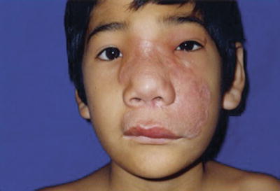
Fig. 22.2
This picture shows centrofacial plaque with a granulomatous appearance and the typical elevated and serpiginous borders
Neurological FLA
The neurological symptoms are classically those of an encephalitis, including headache, fever, and photophobia. As the disease progresses, and some times quite rapidly, other signs, such as seizures, lethargy, and anisocoria, related to increased intracranial pressure, become evident. The patient may present focal sensorial and motor deficits. The level of consciousness deteriorates, going from lethargy to coma and death.
N. fowleri produces an acute and fulminating primary amoebic meningoencephalitis (PAM), which is characterized by an abrupt onset and fulminant course, that usually affects children and young adults. Patients with PAM present signs and symptoms of meningeal irritation and encephalitis; sudden onset of headache, fever, nausea, vomiting, and stiff neck may progress rapidly to lethargy, confusion, coma, and death in 48–72 h [19].
Acanthamoeba or Balamuthia species in debilitated or immunosuppressed patients, produce the so-called granulomatous amoebic encephalitis (GAE), which is a chronic and fatal disease characterized by neurological signs depending on multifocal hemorrhagic and necrotic areas of the brain [20]. Symptoms resemble those of single or multiple space-occupying lesions; hemiparesis, drowsiness, personality changes, and seizures appear early; insidious headache, low-grade fever, stiff neck, nausea, vomiting, and lethargy may be present. Death occurs from 1 week to several months after the onset.
Disseminated FLA occurs especially in the immunosuppressed. Granulomas and amoebae have been found in the adrenal glands, kidneys, liver, lungs, pancreas, spleen, thyroid gland, and uterus [21].
Diagnosis
Direct examination of cerebrospinal fluid in PAM and GAE using a phase-contrast microscope discloses pear-shaped biflagellated motile amoebae. Smears stained with Giemsa, Wright, or hematoxylin and eosin (H&E) disclose the morphology of the amoebae.
N. fowleri and Acanthamoeba species can be cultured easily in agar seeded with bacteria like E. coli at 37 °C. Cultures of B. mandrillaris require monkey kidney cells but are often unsuccessful; a new axenic (cell-free) medium may be more suitable [22].
The skin biopsy may serve as the first clue for the diagnosis of FLA; histopathology of the skin lesions discloses a granulomatous reaction, with multinucleated giant cells, and a mixed inflammatory infiltrate. The amoebae can be seen using routine H&E stains [17].
Differential diagnosis of cutaneous FLA includes mucocutaneous leishmaniasis, rhinosporidiosis, rhinoescleroma, conidiobolomycosis, Wegener’s granulomatosis, sarcoidosis, Melkersson Rosenthal syndrome, mid-line granuloma, and cutaneous T-cell lymphoma.
Pathogenesis
The organisms enter the nasal cavity by inhalation of dust, through contaminated water, or in air that contains trophozoites or cysts. After the parasite enters through nasal mucosa, initially involving the olfactory neuroepithelium, it subsequently reaches the CNS by active phagocytosis of sustentacular cells. Due to the contiguity with subarachnoid membranes, amoebae may easily disseminate to other areas of the CNS, producing an acute and fulminating PAM [19].
Therapeutics and Prognosis
Successful treatment of Naegleria encephalitis with intravenous and intrathecal amphotericin B has been reported [23]. There is no known effective therapy for Balamuthia infection. Miconazole, 5-fluorocytosine, pentamidine, sulfadiazine, fluconazole, itraconazole, amphotericin b, azithromycin, and clarithromycin have been used with variable results.
A report of successful treatment of two cases with CNS involvement included flucytosine, pentamidine, fluconazole, sulfadiazine, and a macrolide; phenothiazines were also added in one case [24]. Both patients received long-term therapy for more than 5 years.
Long-term albendazole 20 mg/kg/day and itraconazole 10 mg/kg/day have been used as therapeutic alternatives in some of our patients with promising results.
Ongoing Researches
In the past years, advances have been made regarding knowledge about the ubiquity of the amoeba in the environment, its worldwide distribution especially in South America, the patients at risk particularly those of Hispanic origin, the diagnostic methods including those based on molecular biology, and the different therapeutic strategies that have resulted in survival of patients. A recent report dealing with solid organ transplant transmission of this infection has made it a subject of interest in transplant medicine [25].
Conclusion
As more and more cases are reported, this entity should be called to the attention of dermatologists, infectious disease specialists, neurologists, pediatricians, and internists. Cases do occur all over the world, and many diagnoses will be missed, unless we have a high index of suspicion. It is very likely that this condition may explain some of the cases previously described as lethal mid-line granuloma.
Myiasis
Introduction
Myiasis is an infestation with maggots.
Occurs in poor socioeconomic regions and forests.
Treatment consists of many local remedies.
It is an infestation of live human and vertebrate animals with dipterous (two-winged) larvae (maggots) which, at least for a certain period, feed on the host’s dead or living tissue, liquid body substance, or ingested food [26]. The distribution of human myiasis is worldwide, with more species and greater abundance in poor socioeconomic regions of tropical and subtropical countries [27].
The anatomical classification system is based on the one proposed by Bishopp and later modified by James and by Zumpt [28] (Table 22.1).
Sanguinivorous or bloodsucking
Cutaneous myiasis, furuncular and migratory
Wound myiasis
Cavitary myiasis, where the infestation receives the name of the affected organ, e.g., cerebral myiasis, aural myiasis, nasal myiasis, and ophthalmo-myiasis.
Ecological classification
Description
Specific/obligatory
Parasite dependent on host for part of its life cycle
Semispecific/facultative
Primary
Free living and may initiate myiasis
Secondary
Free living and unable to initiate myiasis; may be involved once animal is infested by other species
Tertiary
Free living and unable to initiate myiasis; may be involved when host is near death
Accidental/pseudomyiasis
Free-living larva and not able to complete its life cycle; causes pathological reaction when accidentally in contact with the host
Epidemiology
Myiasis is worldwide, the most common flies that cause the human infestation are Dermatobia hominis (human botfly) (Fig. 22.3) and Cordylobia anthropophaga (tumbu fly).
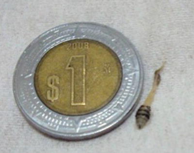
Fig. 22.3
Dipteran larva obtained from a furuncle-like nodule. The patient is from the south of Mexico. Courtesy Dr. Natalia Deveaux. Med Vet
Dermatobia hominis is the most important cuterebra fly and the most common cause of myiasis in the Americas. American warble fly, tropical both fly, colmoyote, moyocuil (both in Mexico, meaning “mosquito worm”), suglacuru (from “between breasts” in French Guyana), and berne (in Brazil).
Stay updated, free articles. Join our Telegram channel

Full access? Get Clinical Tree


