Fig. 15.1
Urethral erosion as demonstrated by a wide, patulous urethra
Management options for these patients are limited. Unfortunately many of these patients are debilitated secondary to their medical comorbidities and poor nutritional status. The use of pads or diapers can be problematic for management of pressure ulcers and wounds, which are commonplace in this subset of patients. Suprapubic catheters have been successfully used in some patients; however, many patients will still have significant leakage per urethra due to the damage caused by the initial indwelling urethral catheter. Transvaginally placed slings, although theoretically are useful as they allow for continued access to the bladder through the native urethra, do not usually give enough support to achieve continence. In addition, there may not be an adequate amount of residual urethral length to allow for sling placement if the urethral damage is severe enough.
Reconstruction of the lower urinary with various methods have been described but many patients are not willing or medically appropriate to undergo such procedures. In patients who are willing and able to undergo urinary tract reconstruction, closure of the bladder neck is usually achieved transabdominally at the same time as their reconstruction. This type of closure is more invasive but has been reported to have lower rates of fistula formation postoperatively compared to transvaginal repair.
Transvaginal bladder neck closure (BNC) with SPT placement is reserved for those patients whom are not candidates for more invasive reconstruction. The primary concern with this procedure is fistula formation between the closed bladder neck and the vagina and may be more technically challenging for inexperienced pelvic surgeons.
Preoperative Evaluation
Preoperatively the most important decision is which approach to take in managing the patient’s incontinence. Andrews et al. described a series of 57 patients with long-term indwelling catheters of which 39 were managed successfully with SPT alone [1]. Similarly, Eckford et al. reported that 11 of 50 women with multiple sclerosis managed with indwelling catheters were happy with an SPT alone even with a small degree of intermittent leakage from the urethra [3]. It is important to recognize that some patients, depending on the degree of urethral destruction, may have enough improvement in their incontinence with an SPT alone that they may not need further surgical intervention. If this fails due to a poor outlet, then treatment will likely focus on an obstructing sling vs. bladder neck closure. Wantanabe et al. reported that candidates for pubovaginal sling must have an intact bladder neck with 1 cm of proximal urethral tissue in order to obtain effective compression of the urethra, which may or may not be the case in patients with a chronic indwelling catheter [4]. If bladder neck closure is to be done, then the decision between transvaginal versus transabdominal approach needs to be resolved.
When considering abdominal versus vaginal approaches, various factors must be considered: morbidity of the procedure, planned concomitant procedures, prior surgeries, surgeon experience [1, 5]. Certainly avoiding an abdominal incision allows for decreased morbidity, but the transvaginal approach is associated with higher rates of fistula and/or failure [1, 2, 5]. A discussion with the patient regarding the lower morbidity but higher risk of failure and potential need for reoperation must take place to set appropriate expectations. Surgeon experience should also be taken into account, as less experienced vaginal surgeons may not fare as well with this approach. Levy et al. reported on a series of 12 patients, 4 of whom underwent transvaginal closure of the bladder neck alone with a 50 % success rate [5]. The two patients who failed and the subsequent 8 patients underwent combined abdominal and transvaginal approach with 100 % success. Levy suggests that surgeons without significant experience operating vaginally should consider an abdominal approach to achieve higher success. Ginger et al. also reported an 11 % leakage rate in 26 patients undergoing abdominal BNC as opposed to a 100 % leakage rate in 2 patients who underwent transvaginal approach [2].
As already discussed, patients with chronic indwelling catheters are often debilitated and malnourished. Poor preoperative nutrition status is associated with poor wound healing, increased infection rate, higher pulmonary complication rate, prolonged hospitalization, and higher mortality rates [6]. Hebbar et al. reviewed studies looking at the use of total parenteral nutrition (TPN) or enteral feeds preoperatively and the rates of complications. From the VA TPN Cooperative Study which used 7–15 days of preoperative TPN, patients with severe malnourishment were found to have a dramatic drop in complication rate from 42.9 to 5.3 % with the use of TPN; however, pooled data of all patients did not show any significant difference. There was no difference between the use of TPN vs. enteral feeds. Therefore, one could consider using preoperative nutrition in the severely malnourished patient.
Procedure
Various techniques have been described to perform a transvaginal closure of the bladder neck [1–3, 7–10]. The patient is placed in dorsal lithotomy position with adequate exposure to the anterior vaginal wall using labial retraction sutures and/or a self-retaining retractor, with or without a posterior weighted speculum. If an SPT is not already present, one can be placed with various techniques. Eckford et al. describe a two stage technique in which an SPT is placed during the first procedure (percutaneously or open) followed by a second procedure for transvaginal BNC if the patient continues to leak [3]. A Lowsley suprapubic tractor may be used to aid in SPT placement if desired [7].
The anterior vaginal wall is infiltrated with injectable saline to aid in the dissection of the vaginal wall from the urethra and bladder. Two incisions are made along the anterior vaginal wall: one circumferentially around the urethral meatus and the second as an inverted wide-based anterior vaginal wall flap beginning from the urethral meatus extending past the bladder neck (Fig. 15.2a–f) [9]. The urethra is dissected laterally over the periurethral fascia to the retropubic space and off of the urethropelvic ligaments followed by transection of the urethra off of the urethropelvic ligament dorsally to the inferior margin of the pubic symphysis. This allows for complete mobilization of the remaining urethra and bladder neck (Fig. 15.2b). The necrotic urethral tissue is then removed, which may in fact be the entire urethra, thus making bladder neck mobility extremely important. If there is viable urethral tissue, one can utilize a technique described by Rovner et al. in which the anterior urethra is divided toward the bladder neck and the bivalved urethra is rotated in an anterior and cephalad direction and secured to the anterior bladder wall with two layers of absorbable suture (Fig. 15.2d). This rotates the suture line anteriorly, toward the retropubic space and underneath the pubic symphysis, minimizing overlying suture lines during closure of the vaginal wall flap (Fig. 15.2e, f). In addition, if possible, we also try to then secure the sutures used to close the bladder neck to the undersurface of the pubic symphysis, further placing the bladder neck closure anteriorly. Mobilization of the closure upward should minimize the risk of postoperative fistula formation. The vaginal wall flap is closed with absorbable suture and packing is placed.
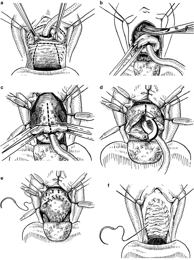
Fig. 15.2
(a–f) TV BNC with posterior urethral flap. (a) Vaginal wall flap developed and dorsal semilunar incision is made above the urethra. (b) Dissection continued above urethra into retropubic space. Pubourethral and urethropelvic ligaments taken down, mobilizing urethra and anterior bladder neck. (c) Dorsal urethra bivalved and incision carried onto anterior bladder neck for 2–3 cm. (d) Dorsally bivalved urethra then rotated cephalad toward anterior bladder wall incision. (e) Ventral urethral flap affixed high on anterior bladder wall, such that when bladder rotates into anatomic position, suture line rotates under symphysis pubis. (f) Vaginal wall closed as second layer with no overlapping suture lines (All: Used with permission from Rovner ES, Goudelocke CM, Gilchrist A, Lebed B. Transvaginal bladder neck closure with posterior urethral flap for devastated urethra. Urology 2011;78(1):208-212)
Nielson et al. describe a technique in which two chromic sutures are passed through the SPT site via the Lowsley tractor and used to tag the edges of the urethral closure [7]. These sutures are then later used to invaginate the urethral mucosa and pull the urethra away from the vaginal closure.
Flaps or graft placements are generally not necessary in primary repair. In cases where the perivesical tissues may be compromised or in patients with history of prior pelvic radiation, one can consider using a Martius flap for interposition [8]. In patients who have failed prior attempts, a combined abdominal and vaginal approach with omental, peritoneal, or Martius flaps have been described [3, 9].
Stay updated, free articles. Join our Telegram channel

Full access? Get Clinical Tree


