The Breast


The Breast


image

The form, function, and pathology of the human female breast are major concerns of medicine and society. As mammals, we define our biologic class by the function of the breast in nourishing our young. Breast contours occupy our attention. As obstetricians, we seek to enhance or diminish function, and as gynecologists, the appearance of inappropriate lactation (galactorrhea) may signify serious disease. Cancer of the breast is the most prevalent cancer in women.

This chapter reviews the factors involved in normal growth and development of the breast, including the physiology of normal lactation, describes the numerous factors leading to inappropriate lactation, and, finally, discusses the endocrine aspects of breast cancer.


Growth and Development

The basic component of the breast lobule is the hollow alveolus or milk gland lined by a single layer of milk-secreting epithelial cells, derived from an ingrowth of epidermis into the underlying mesenchyme at 10-12 weeks of gestation. Each alveolus is encased in a crisscrossing mantle of contractile myoepithelial strands. Also surrounding the milk gland is a rich capillary network.


The lumen of the alveolus connects to a collecting intralobular duct by means of a thin nonmuscular duct. Contractile muscle cells line the intralobular ducts that eventually reach the exterior via 15-20 collecting ducts in a radial arrangement, corresponding to the 15-20 distinct mammary lobules in the breast, each of which contains many alveoli.

Growth of this milk-producing system is dependent on numerous hormonal factors that occur in two sequences, first at puberty and then in pregnancy. Although there is considerable overlapping of hormonal influences, the differences in quantities of the stimuli in each circumstance and the availability of entirely unique inciting factors (human placental lactogen and prolactin) during pregnancy permit this chronologic distinction. The strength of the hormonal stimulus to breast tissue during pregnancy is responsible for the fact that nearly half of male and female newborns have breast secretions.


image

The major influence on breast growth at puberty is estrogen. In most girls, the first response to the increasing levels of estrogen is an increase in size and pigmentation of the areola and the formation of a mass of breast tissue just underneath the areola. Breast tissue binds estrogen in a manner similar to the uterus and vagina. The human breast expresses both estrogen receptors, ER-a and ER-b.1 The development of estrogen receptors in the breast does not occur in the absence of prolactin. The primary effect of estrogen in subprimate mammals is to stimulate growth of the ductal portion of the gland system. Progesterone in these animals, in the presence of estrogen, influences growth of the alveolar components
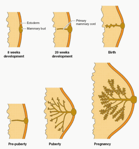
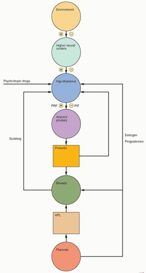
of the lobule that later become the milk-producing structures.2 However, neither hormone alone, or in combination, is capable of yielding optimal breast growth and development. Full differentiation of the gland requires insulin, cortisol, thyroxine, prolactin, and especially, growth hormone-induced insulin-like growth factor-I.3,4 Experimental evidence in mice knock-out models supports the combined actions of estrogen and progesterone, mediated primarily by estrogen receptor-a and progesterone receptor-B, but dependent on epidermal growth factor and IGF-I.5,6 and 7 Estrogen and progesterone receptors in normal breast tissue are located in non-dividing epithelial cells and in stromal cells adjacent to proliferating epithelial cells, indicating the importance of paracrine communication using growth factors. Growth hormone-induced IGF-I is essential in both mammary development and function.4

The pubertal response is a manifestation of closely synchronized central (hypothalamuspituitary) and peripheral (ovary-breast) events. For example, gonadotropin-releasing hormone (GnRH) is known to stimulate prolactin release, and this action is potentiated by estrogen.8 This suggests a paracrine interaction between gonadotrophs and lactotrophs, linked by estrogen, ultimately with an impact on the breast.

Changes occur routinely in response to the estrogen-progesterone sequence of a normal menstrual cycle. Maximal size of the breast occurs late in the luteal phase. Fluid secretion,
mitotic activity, and DNA production of nonglandular tissue and glandular epithelium peak during the luteal phase.9,10 and 11 This accounts for cystic and tender premenstrual changes.


image

During the normal menstrual cycle, estrogen receptors in mammary gland epithelium decrease in number during the luteal phase, whereas progesterone receptors remain at a high level throughout the cycle.12 Studies using tissue from reduction mammoplasties or from breast tissue near a benign or malignant lesion have demonstrated a peak in mitotic activity during the luteal phase.10,13,14 Using fine-needle biopsy tissue, an immunocytochemical marker of proliferation was higher in the luteal phase than in the proliferative phase.12 And in this study there was a direct correlation with serum progesterone levels. However, important studies indicate that with increasing duration of exposure, progesterone imposes a limitation on breast cell proliferation.15,16 and 17 Therefore, breast and endometrium epithelial cells may be more similar than conventionally proposed.

Final differentiation of the alveolar epithelial cell into a mature milk cell is accomplished by the gestational increase in estrogen and progesterone, combined with the presence of prolactin, but only after prior exposure to cortisol and insulin. The complete reaction depends on the availability of minimal quantities of thyroid hormone. Thus, the endocrinologically intact individual in whom estrogen, progesterone, thyroxine, cortisol, insulin, prolactin, human placental lactogen, and growth hormone are available can have appropriate breast growth and function. During the first trimester of pregnancy, growth and proliferation are maximal, changing to differentiation and secretory activity as pregnancy progresses.

Breast tissue changes with aging. During teenage years the breasts are dense and predominantly glandular. As the years go by, the breasts contain progressively more fat, but after menopause, this process accelerates so that soon into the postmenopausal years, the breast glandular tissue is mostly replaced by fat.


Abnormal Shapes and Sizes

Early differentiation of the mammary gland anlage is under fetal hormonal control. Abnormalities in adult size or shape may reflect the impact of hormones (especially the presence or absence of testosterone) during this early period of development. This prenatal hormonal influence programs the breast development that will occur in response to the increase in hormones at puberty. Occasionally, the breast bud begins to develop on one side first. Similarly, one breast may grow faster than the other. These inequalities usually disappear by the time development is complete. However, exact equivalence in size may not be attained. Significant asymmetry is correctable only by a plastic surgeon. Likewise hypoplasia and hypertrophy can be treated only by corrective surgery. Hormone therapy is totally ineffective in producing a permanent change in breast shape or size, with one exception, in patients with primary amenorrhea due to deficient ovarian function, estrogen treatment induces significant and gratifying breast growth. Breast size can be increased in current users of oral contraceptives, but there is no lasting effect associated with past use.18

Accessory nipples (almost always without underlying breast tissue) can be found anywhere from the groin to the neck, remnants of the mammary line that extends early in embryonic life (sixth week) along the ventral, lateral body wall. They occur in approximately 1% of women (sporadic or familial) and require no therapy. The presence of polythelia has been reported to be associated with a variety of renal and urinary tract malformations.19,20 However, in three series, each with a large number of children, the presence of supernumerary nipples was not associated with a higher prevalence of kidney and urinary tract malformations.21,22 and 23 Nevertheless, it is prudent to investigate the renal-urinary tract in the presence of polythelia.24


Accessory breast tissue occurs because of incomplete embryologic regression of the mammary ridges, and for this reason, the location is along the mammary line that extends from the axilla to the pubic area. Ectopic breast tissue is usually detected during puberty, pregnancy, or lactation, a consequence of hormonally-induced enlargement. Accessory breasts are commonly bilateral, and occasionally are found in unusual locations such as the axilla, scapula, thigh, or labia majora, and when nipple and areola are absent, the mass can be a diagnostic dilemma.25 Even when the diagnosis is obvious, surgical excision is indicated for cosmetic and comfort reasons.26 Accessory breast tissue is subject to the same risk of cancer as normal breasts.


Pregnancy and Lactation


Prolactin Secretion

In most mammalian species, prolactin is a single-chain polypeptide of 199 amino acids, 40% similar in structure to growth hormone and placental lactogen. All three hormones are believed to have originated from a common ancestral protein about 400 million years ago.

Prolactin is encoded by a single gene on chromosome 6, producing a molecule that in its major form is maintained in three loops by disulfide bonds.27 Most, if not all, variants of prolactin are the result of posttranslational modifications. Little prolactin represents a splicing variant resulting from the proteolytic deletion of amino acids. Big prolactin can result from the failure to remove introns; it has little biologic activity and does not cross-react with antibodies to the major form of prolactin. The so-called big big variants of prolactin are due to separate molecules of prolactin binding to each other, either noncovalently or by interchain disulfide bonding. Some of the apparently larger forms of prolactin are prolactin molecules complexed to binding proteins. High levels of relatively inactive prolactin in the absence of a tumor can be due to the creation of macromolecules of prolactin by antiprolactin autoantibodies.28,29 Overall, big prolactins account for somewhere between 10% and 25% of the hyperprolactinemia reported by commercial assays.30

Other variations exist. Enzymatic cleavage of the prolactin molecule yields fragments that may be capable of biologic activity, and prolactin that has been glycosylated continues to exert activity. Differences in the carbohydrate moieties can produce differences in biologic activity and immunoreactivity. However, the nonglycosylated form of prolactin is the predominant form of prolactin secreted into the circulation.31 Modification of prolactin also includes phosphorylation, deamidation, and sulfation.

At any one point of time, the bioactivity (e.g., galactorrhea) and the immunoreactivity (circulating levels by immunoassay) of prolactin represent the cumulative effect of the family of structural variants. Remember, immunoassays do not always reflect the biologic situation (e.g., a normal prolactin level in a woman with galactorrhea). Nevertheless, the routine radioimmunoassay of prolactin is generally clinically reliable, especially at extremely high levels associated with prolactin-secreting pituitary tumors.

The anterior pituitary cells that produce prolactin, growth hormone, and thyroid-stimulating hormone (lactotrophs, somatotrophs, and thyrotrophs) require the presence of Pit-1, a transcription factor, for transactivation. Pit-1 binds to the prolactin gene in multiple sites in both the promoter region and in an adjacent region, designated as a distal enhancer; Pit-1 binding is a requirement for prolactin promoter activity and gene transcription. Many hormones, neurotransmitters, and growth factors influence the prolactin gene, involved in a level of function beyond that allowed by Pit-1. Fundamental modulation of prolactin
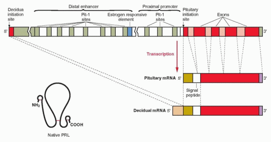
secretion is exerted by estrogen, producing both differentiation of lactotrophs and direct stimulation of prolactin production.32,33 An estrogen response element is adjacent to one of the Pit-1 binding sites in the distal enhancer region, and estrogen stimulation of the prolactin gene involves interaction with this Pit-1 binding site. Estrogen additionally influences prolactin production by suppressing dopamine secretion.34 Prolactin is also synthesized in extrapituitary tissues, including breast tissue and endometrial decidua.35 In extrapituitary sites, the active promoter site is upstream of the pituitary initiation site, and is not regulated by Pit-1, estrogens, or dopamine. Progesterone increases prolactin secretion in the decidua, but has no effect in the pituitary.


image

Prolactin is involved in many biochemical events during pregnancy. Surfactant synthesis in the fetal lung is influenced by prolactin, and decidual prolactin modulates prostaglandinmediated uterine muscle contractility.36,37 Prolactin also contributes to the prevention of the immunologic rejection of the conceptus by suppressing the maternal immune response. Prolactin is both produced and processed in breast cells. The mechanisms and purpose for mammary production of prolactin remain to be determined, but prolactin in milk is believed to be derived from local synthesis. Transmission of this prolactin to the newborn may be important for immune functions.


Prolactin-Inhibiting Factor

The hypothalamus maintains suppression of pituitary prolactin secretion by delivering a prolactin-inhibiting factor (PIF) to the pituitary via the portal circulation. Suckling suppresses the formation of this hypothalamic substance, which is believed to be dopamine (as discussed in Chapter 5).38 Dopamine is secreted by the basal hypothalamus into the portal system and conducted to the anterior pituitary. Dopamine binds specifically to lactotroph cells and suppresses the secretion of prolactin into the general circulation; in its absence, prolactin is secreted. Dopamine binds to a G-protein-coupled receptor (Chapter 2) that
exists in a long form and a short form, but only the D2 (long form) is present on lactotrophs. The molecular mechanism for dopamine’s inhibitory action is still not known. There are several other PIFs, but a specific role has been established only for dopamine.


Prolactin Releasing Factor

Prolactin secretion may also be influenced by a positive hypothalamic factor, prolactinreleasing factor (PRF). PRF does exist in various fowl (e.g., pigeon, chicken, duck, turkey, and the tricolored blackbird). While the identity of this material has not been elucidated, or its function substantiated in normal human physiology, it is possible that thyrotropinreleasing hormone (TRH) is a potent stimulant of prolactin secretion in humans. The smallest doses of TRH that are capable of producing an increase in TSH also increase prolactin levels, a finding that supports a physiologic role for TRH in the control of prolactin secretion, at least in response to suckling.39 TRH stimulation of prolactin release involves calcium mechanisms (both internal release and influx via calcium channels) in response to the TRH receptor, also a member of the G-protein family. However, except in hypothyroidism, normal physiologic changes as well as abnormal prolactin secretion are easily explained and understood in terms of variations in the prolactin-inhibiting factor, dopamine. A large collection of peptides has been reported to stimulate the release of prolactin in vitro. These include growth factors, angiotensin II, GnRH, vasopressin, and others. But it is unknown whether these peptides participate in the normal physiologic regulation of prolactin secretion.


The Prolactin Receptor

The prolactin receptor is encoded by a gene on chromosome 5p13-14 that is near the gene for the growth hormone receptor. The prolactin receptor belongs to a receptor family that includes many cytokines and some growth factors, supporting a dual role for prolactin as a classic hormone and as a cytokine.27

Prolactin receptors exist in more than one form, all containing an extracellular region, a single transmembrane region, and a relatively long cytoplasmic domain. There is evidence for more than one receptor, depending on the site of action (e.g., decidua and placenta).40 The similar amino acid identity between prolactin and growth hormone receptors is approximately 30%, with certain regions having up to 70% homology.41 Prolactin receptors are expressed in many tissues throughout the body. Because of the various forms and functions of prolactin, it is likely that multiple signal mechanisms are involved, and for that reason, no single second messenger for prolactin’s intracellular action has been identified. A protein also exists that functions as a receptor/transporter, translocating prolactin from the blood into the cerebrospinal fluid, the amniotic fluid, and milk.


Amniotic Fluid Prolactin

Amniotic fluid concentrations of prolactin parallel maternal serum concentrations until the 10th week of pregnancy, rise markedly until the 20th week, and then decrease.
Maternal prolactin does not pass to the fetus in significant amounts. Indeed, the source of amniotic fluid prolactin is neither the maternal pituitary nor the fetal pituitary. The failure of dopamine agonist treatment to suppress amniotic fluid prolactin levels, and studies with in vitro culture systems indicate a primary decidual source with transfer via amnion receptors to the amniotic fluid, requiring the intactness of amnion, chorion, and adherent decidua. This decidual synthesis of prolactin is initiated by progesterone, but once decidualization is established, prolactin secretion continues in the absence of both progesterone and estradiol.42 Various decidual factors regulate prolactin synthesis and release, including relaxin, insulin, and insulin-like growth factor-I. Prolactin produced in extrapituitary sites involves an alternative exon upstream of the pituitary start site, generating a slightly larger RNA transcript compared with the pituitary product. However, the amino acid sequence and the chemical and biologic properties of decidual prolactin are identical to those of pituitary prolactin. It is hypothesized that amniotic fluid prolactin plays a role in modulating electrolyte economy not unlike its ability to regulate sodium transport and water movement across the gills in fish (allowing the ocean-dwelling salmon and steelhead to return to freshwater streams for reproduction). Thus prolactin would protect the human fetus from dehydration by control of salt and water transport across the amnion. Prolactin reduces the permeability of the human amnion in the fetal to maternal direction by a receptor-mediated action on the epithelium lining the fetal surface.43 Decidual and amniotic fluid prolactin levels are lower in hypertensive pregnancies and in patients with polyhydramnios.44,45 Prolactin receptors are present in the chorion laeve, and their concentration is lower in patients with polyhydramnios.46 Thus, idiopathic polyhydramnios may be a consequence of impaired prolactin regulation of amniotic fluid.


Lactation

During pregnancy, prolactin levels rise from the normal level of 10-25 ng/mL to high concentrations, beginning about 8 weeks and reaching a peak of 200-400 ng/mL at term.47,48 The increase in prolactin parallels the increase in estrogen beginning at 7-8 weeks’ gestation, and the mechanism for increasing prolactin secretion (discussed in Chapter 5) is believed to be estrogen suppression of the hypothalamic prolactin-inhibiting factor, dopamine, and direct stimulation of prolactin gene transcription in the pituitary.49,50 There is marked variability in maternal prolactin levels in pregnancy, with pulsatile secretion and a diurnal variation similar to that found in nonpregnant subjects. The peak level occurs 4-5 hours after the onset of sleep.51

Made by the placenta and actively secreted into the maternal circulation from the sixth week of pregnancy, human placental lactogen (hPL) rises progressively, reaching a level of approximately 6,000 ng/mL at term. hPL, though displaying less activity than prolactin, is produced in such large amounts that it may exert a lactogenic effect.

Although prolactin stimulates significant breast growth, and is available for lactation, only colostrum (composed of desquamated epithelial cells and transudate) is produced during gestation. Full lactation is inhibited by progesterone, which interferes with prolactin action at the alveolar cell prolactin receptor level. Both estrogen and progesterone are necessary for the expression of the lactogenic receptor, but progesterone antagonizes the positive action of prolactin on its own receptor while progesterone and pharmacologic amounts of androgens reduce prolactin binding.41,52,53 In the mouse, inhibition of milk protein production is due to progesterone suppression of prolactin receptor expression.54 The effective use of high doses of estrogen to suppress postpartum lactation indicates that pharmacologic amounts of estrogen also block prolactin action.


image

Progesterone can directly suppress milk production. A nuclear peptide (a corepressor) has been identified that binds to specific sites in the promoter region of the casein gene, thus inhibiting transcription.55 Progesterone stimulates the generation of this corepressor. After delivery, the loss of progesterone leads to a decrease in this inhibitory peptide.

The principal hormone involved in milk biosynthesis is prolactin. Without prolactin, synthesis of lactose, lipids and the primary protein, casein, will not occur, and true milk secretion will be impossible. The hormonal trigger for initiation of milk production within the alveolar cell and its secretion into the lumen of the gland is the rapid disappearance of estrogen and progesterone from the circulation after delivery. The clearance of prolactin is much slower, requiring 7 days to reach nonpregnant levels in a nonbreastfeeding woman. These discordant hormonal events result in removal of the estrogen and progesterone inhibition of prolactin action on the breast. Breast engorgement and milk secretion begin 3-4 days postpartum when the sex steroids have been sufficiently cleared. Maintenance of steroidal inhibition or rapid reduction of prolactin secretion (with a dopamine agonist) are effective in preventing postpartum milk synthesis and secretion. Augmentation of prolactin (by TRH or sulpiride, a dopamine receptor blocker) results in increased milk yield.

In the first postpartum week, prolactin levels in breastfeeding women decline approximately 50% (to about 100 ng/mL). Suckling elicits increases in prolactin, which are important in initiating milk production. Until 2-3 months postpartum, basal levels are approximately 40-50 ng/mL, and there are large (about 10-20-fold) increases after suckling. Throughout breastfeeding, baseline prolactin levels remain elevated, and suckling produces a 2-fold increase that is essential for continuing milk production.56,57 The pattern or values of prolactin levels do not predict the postpartum duration of amenorrhea or infertility.58 The failure to lactate within the first 7 days postpartum may be the first sign of Sheehan’s syndrome (hypopituitarism following intrapartum infarction of the pituitary gland).


Maintenance of milk production at high levels is dependent on the joint action of both anterior and posterior pituitary factors. By mechanisms to be described in detail shortly, suckling causes the release of both prolactin and oxytocin as well as thyroid-stimulating hormone (TSH).59,60 Prolactin sustains the secretion of casein, fatty acids, lactose, and the volume of secretion, while oxytocin contracts myoepithelial cells and empties the alveolar lumen, thus enhancing further milk secretion and alveolar refilling. The increase in TSH with suckling suggests that thyrotropin-releasing hormone (TRH) may play a role in the prolactin response to suckling. The optimal quantity and quality of milk are dependent upon the availability of thyroid, insulin and the insulin-like growth factors, cortisol, and the dietary intake of nutrients and fluids.

Secretion of calcium into the milk of lactating women approximately doubles the daily loss of calcium.61,62 In women who breastfeed for 6 months or more, this is accompanied by significant bone loss even in the presence of a high calcium intake.63 However, bone density rapidly returns to baseline levels in the 6 months after weaning.64,65 The bone loss is due to increased bone resorption, probably secondary to the relatively low estrogen levels associated with lactation. It is possible that recovery is impaired in women with inadequate calcium intake; total calcium intake during lactation should be at least 1,500 mg per day. Nevertheless, calcium supplementation has no effect on the calcium content of breast milk or on bone loss in lactating women who have normal diets.66 In addition, fetuses and lactating mothers, except in unusual circumstances, do not suffer from a significant deficiency in vitamin D.67 Furthermore, studies indicate that any loss of calcium and bone associated with lactation is rapidly restored, and, therefore, there is no impact on the risk of postmenopausal osteoporosis.68,69,70,71 and 72 Rarely, a pregnant woman can present with osteoporosis and vertebral fractures, probably a consequence of very inadequate calcium intake and severe vitamin D deficiency.73 Case reports of pregnancy-associated osteoporosis indicate that this acute condition can be successfully treated with either bisphosphonates or teriparatide, the parathyroid hormone fragment.74,75

Antibodies are present in breast milk and contribute to the health of an infant. Besides the proteins, carbohydrates, and fats that provide a complete and balanced diet, human milk prevents infections in infants both by transmission of immunoglobulins and by modifying the bacterial flora of the infant’s gastrointestinal tract. Viruses are transmitted in breast milk, and although the actual risks are unknown, women infected with cytomegalovirus, hepatitis B, or human immunodeficiency viruses are advised not to breastfeed. Vitamin A, vitamin B12, and folic acid are significantly reduced in the breast milk of women with poor dietary intake. As a general rule approximately 1% of any drug ingested by the mother appears in breast milk. In a study of Pima Indians, exclusive breastfeeding for at least 2 months was associated with a lower rate of adult-onset noninsulin-dependent diabetes mellitus, probably because overfeeding and excess weight gain are more common with bottle-feeding.76

Frequent emptying of the lumen is important for maintaining an adequate level of secretion. Indeed, after the fourth postpartum month, suckling appears to be the only stimulant required; however, environmental and emotional states also are important for continued alveolar activity. Vigorous aerobic exercise does not affect the volume or composition of breast milk, and therefore infant weight gain is normal.77 Maternal diet and hydration have little impact on lactation; the primary control of milk output is under the control of the infant’s suckling.78

Suckling studied with ultrasonography indicates that the infant’s instinctive attachment to a nipple immediately establishes a vacuum seal.79 The tongue moves up and down, increasing the vacuum and producing milk flow during the downward motion. However, the ejection of milk from the breast does not occur only as the result of a mechanically induced negative pressure produced by suckling. Tactile sensors concentrated in the areola activate, via thoracic sensory nerve roots 4, 5, and 6, an afferent sensory neural arc that stimulates
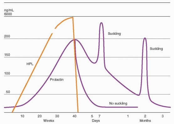
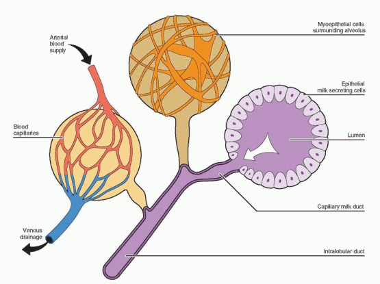
the paraventricular and supraoptic nuclei of the hypothalamus to synthesize and transport oxytocin to the posterior pituitary. The efferent arc (oxytocin) is blood-borne to the breast alveolus-ductal systems to contract myoepithelial cells and empty the alveolar lumen. Milk contained in major ductal repositories is ejected from 15 to 20 openings in the nipple. This rapid release of milk is called “let-down.” This important role for oxytocin is evident in knockout mice lacking oxytocin who undergo normal parturition, but fail to nurse their offspring.80 The milk ejection reflex involving oxytocin is present in all species of mammals. Oxytocin-like peptides exist in fish, reptiles, and birds, and a role for oxytocin in maternal behavior may have existed before lactation evolved.78

In many instances, the activation of oxytocin release leading to let-down does not require initiation by tactile stimuli. The central nervous system can be conditioned to respond to the presence of the infant, or to the sound of the infant’s cry, by inducing activation of the efferent arc. These messages are the result of many stimulating and inhibiting neurotransmitters. Suckling, therefore, acts to refill the breast by activating both portions of the pituitary (anterior and posterior) causing the breast to produce new milk and to eject milk. The release of oxytocin is also important for uterine contractions that contribute to involution of the uterus.

The oxytocin effect is a release phenomenon acting on secreted and stored milk. Prolactin must be available in sufficient quantities for continued secretory replacement of ejected milk. This requires the transient increase in prolactin associated with suckling. The amount of milk produced correlates with the amount removed by suckling. The breast can store milk for a maximum of 48 hours before production diminishes.


Breastfeeding by Adopting Mothers

Adopting mothers occasionally request assistance in initiating lactation.81 Successful breastfeeding can be achieved by ingestion of 25 mg chlorpromazine t.i.d. together with vigorous nipple stimulation every 3-4 hours. Milk production will not appear for several weeks. This preparation ideally should be begin about a month before the expected baby is due. An electric breast pump should be used, again preferably beginning about a month before the expected baby is due to deliver. Stasis of milk within the breast, without stimulation, will lead to cessation of lactation. Metoclopramide, 10 mg t.i.d., is another drug that has produced success in increasing prolactin levels and inducing lactation.82 Metoclopramide can also be used when nursing mothers have an inadequate milk supply. Once adequate lactation is established (usually in 7 to 10 days), drug treatment should be discontinued, tapering the dose over 3 weeks.


Cessation of Lactation

Lactation can be terminated by discontinuing suckling. The primary effect of this cessation is loss of milk let-down via the neural evocation of oxytocin. With passage of a few days, the swollen alveoli depress milk formation probably via a local pressure effect (although milk itself may contain inhibitory factors). With resorption of fluid and solute, the swollen engorged breast diminishes in size in a few days. In addition to the loss of milk let-down the absence of suckling reactivates dopamine (PIF) production so that there is less prolactin stimulation of milk secretion. Routine use of a dopamine agonist for suppression of lactation is not recommended because of reports of hypertension, seizures, myocardial infarctions, and strokes associated with its postpartum use.


Contraceptive Effect of Lactation

A moderate contraceptive effect accompanies lactation and produces child-spacing, which is very important in the developing world as a means of limiting family size. The contraceptive effectiveness of lactation, i.e., the length of the interval between births, depends on the intensity of suckling, the extent to which supplemental food is added to the infant diet, and the level of nutrition of the mother (if low, the longer the contraceptive interval; however well-nourished and undernourished women resume ovulating at the same time postpartum.83) If suckling intensity and/or frequency is diminished, contraceptive effect is reduced. Only amenorrheic women who exclusively breastfeed (full breastfeeding) at regular intervals, including nighttime, during the first 6 months have the contraceptive protection equivalent to that provided by oral contraception (98% efficacy); with menstruation or after 6 months, the chance of ovulation increases.84,85 With full or nearly full breastfeeding, approximately 70% of women remain amenorrheic through 6 months and only 37% through 1 year; nevertheless with exclusive breastfeeding, the contraceptive efficacy at 1 year is high, at 92%.85 Fully breastfeeding women commonly have some vaginal bleeding or spotting in the first 8 postpartum weeks, but this bleeding is not due to ovulation.86

Supplemental feeding increases the chance of ovulation (and pregnancy) even in amenorrheic women.87 Total protection is achieved by the exclusively breastfeeding woman for a duration of only 10 weeks.86 Half of women studied who are not fully breastfeeding ovulate before the sixth week, the time of the traditional postpartum visit; a visit during the third postpartum week is strongly recommended for contraceptive counseling.


Rule of 3’S for Postpartum Initiation of Contraception


Full breastfeeding;


Begin in 3rd postpartum month.


Partial or no breastfeeding:


Begin in 3rd postpartum week.


In non-breastfeeding women, gonadotropin levels remain low during the early puerperium and return to normal concentrations during the third to fifth week when prolactin levels have returned to normal. In an assessment of this important physiologic event (in terms of the need for contraception), the mean delay before first ovulation was found to be approximately 45 days, while no woman ovulated before 25 days after delivery.84 Of the 22 women, however, 11 ovulated before the sixth postpartum week, underscoring the need to move the traditional postpartum medical visit to the third week after delivery. In women who do receive dopamine agonist treatment at or immediately after delivery, return of ovulation is slightly accelerated, and contraception is required a week earlier, in the second week postpartum.88,89

Prolactin concentrations are increased in response to the repeated suckling stimulus of breastfeeding. Given sufficient intensity and frequency, prolactin levels remain elevated. Under these conditions, follicle-stimulating hormone (FSH) concentrations are in the low normal range (having risen from extremely low concentrations at delivery to follicular range in the 3 weeks postpartum) and luteinizing hormone (LH) values are also in the low normal range. These low levels of gonadotropins do not allow the ovary, during lactational hyperprolactinemia, to display follicular development and secrete estrogen. Therefore, vaginal dryness and dyspareunia are commonly reported by breastfeeding women. The use of vaginal estrogen preparations is discouraged because absorption of the estrogen can lead to inhibition of milk production. Vaginal lubricants should be used until ovarian function and estrogen production return.


The mechanism of the contraceptive effect is of interest because a similar interference with normal pituitary-gonadal function is seen with elevated prolactin levels in nonpregnant women, the syndrome of galactorrhea and amenorrhea. Earlier experimental evidence suggested that the ovaries might be refractory to gonadotropin stimulation during lactation, and, in addition, the anterior pituitary might be less responsive to GnRH stimulation. Other studies, done later in the course of lactation, indicated, however, that the ovaries as well as the pituitary were responsive to adequate tropic hormone stimulation.90

These observations suggest that high concentrations of prolactin can work at both central and ovarian sites to produce lactational amenorrhea and anovulation. Prolactin appears to affect granulosa cell function in vitro by inhibiting the synthesis of progesterone. It also may change the testosterone/dihydrotestosterone ratio, thereby reducing aromatizable substrate and increasing local antiestrogen concentrations. Nevertheless, a direct effect of prolactin on ovarian follicular development does not appear to be a major factor. The central action predominates.

Elevated levels of prolactin inhibit the pulsatile secretion of GnRH.91,92 Prolactin excess has short-loop positive feedback effects on dopamine. Increased dopamine reduces GnRH by suppressing arcuate nucleus function, perhaps in a mechanism mediated by endogenous opioid activity.93,94 However, blockade of dopamine receptors with a dopamine antagonist or the administration of an opioid antagonist in breastfeeding women does not always affect gonadotropin secretion.95 The exact mechanism for the suppression of GnRH secretion remains to be unraveled. The principle of GnRH suppression by prolactin is reinforced by the demonstration that treatment of amenorrheic, lactating women with pulsatile GnRH fully restores pituitary secretion and normal ovarian cyclic activity.96

At weaning, as prolactin blood concentrations fall to normal, gonadotropin levels increase, and estradiol secretion rises. This prompt resumption of ovarian function is followed by the occurrence of ovulation within 14-30 days of weaning.


Inappropriate Lactation—Galactorrheic Syndromes

Galactorrhea refers to the mammary secretion of a milky fluid, which is nonphysiologic in that it is inappropriate (not immediately related to pregnancy or the needs of a child), persistent, and sometimes excessive. Although usually white or clear, the color may be yellow or even green. In the latter circumstance, local breast disease should be considered. To elicit breast secretion, pressure should be applied to all sections of the breast beginning at the base of the breast and working up toward the nipple. Hormonally-induced secretions usually come from multiple duct openings in contrast to pathologic discharge that usually comes from a single duct. A bloody discharge is more typical of cancer. The quantity of secretion is not an important criterion. Amenorrhea does not necessarily accompany galactorrhea, even in the most serious provocative disorders. Any galactorrhea demands evaluation in a nulliparous woman and if at least 12 months have elapsed since the last pregnancy or weaning in a parous woman. Galactorrhea can involve both breasts or just 1 breast. This recommendation has evolved empirically, knowing that many women have the persistence of galactorrhea for many months after breastfeeding, and therefore the rule is a soft one. The exact numbers have never been established by appropriate studies. Thus, there is room for clinical judgment with this clinical problem.


Differential Diagnosis of Galactorrhea

The differential diagnosis of galactorrhea is a difficult and complex clinical challenge. The difficulty arises from the multiple factors involved in the control of prolactin release. In most pathophysiologic states the final common pathway leading to galactorrhea is an inappropriate augmentation of prolactin release. The following considerations are important:


  • Increased prolactin release can be a consequence of prolactin elaboration and secretion from pituitary tumors (discussed in Chapter 11), which function independently of the otherwise appropriate restraints exerted by PIF from a normally functioning hypothalamus. This infrequent but potentially dangerous tumor, which has endocrine, neurologic, and ophthalmologic liabilities that can be disabling, makes the differential diagnosis of persistent galactorrhea a major clinical challenge. Beyond producing prolactin, the tumor may also suppress pituitary parenchyma by expansion and compression, interfering with the secretion of other tropic hormones. Other pituitary tumors may be associated with lactotroph hyperplasia and present with the characteristic syndrome of hyperprolactinemia and amenorrhea.


  • A variety of drugs can inhibit hypothalamic dopamine.97 There are nearly 100 phenothiazine derivatives with indirect mammotropic activity. In addition, there are many phenothiazine-like compounds, reserpine derivatives, amphetamines, and an unknown variety of other drugs (opiates, diazepams, butyrophenones, verapamil, a-methyldopa, and tricyclic antidepressants) that can initiate galactorrhea via hypothalamic suppression. The final action of these compounds is either to deplete dopamine levels or to block dopamine receptors. Chemical features common to many of these drugs are an aromatic ring with a polar substituent as in estrogen and at least two additional rings or structural attributes making spatial arrangements similar to estrogen. Thus, these compounds may act in a manner similar to estrogens to decrease PIF or to act directly on the pituitary. In support of this conclusion, it has been demonstrated that estrogen and phenothiazine derivatives compete for the same receptors in the median eminence. Prolactin is uniformly elevated in patients on therapeutic amounts of these drugs, but essentially never as high as 100 ng/mL. Approximately 30-50% exhibit galactorrhea that should not persist beyond 3-6 months after drug treatment is discontinued.


  • Hypothyroidism (juvenile or adult) can be associated with galactorrhea. With diminished circulating levels of thyroid hormone, hypothalamic TRH is produced in excess and acts as a PRF to release prolactin from the pituitary. Reversal with thyroid hormone is strong circumstantial evidence to support the conclusion that TRH stimulates prolactin.


  • Excessive estrogen (e.g., oral contraceptives) can lead to milk secretion via hypothalamic suppression, causing reduction of dopamine and release of pituitary prolactin, and direct stimulation of the pituitary lactotrophs. Galactorrhea developing during oral contraceptive administration may be most noticeable in the traditional dosing regimen during the 7 days free of medication (when the steroids are cleared from the body and the prolactin interfering action of the estrogen and progestin on the breast wanes). Galactorrhea caused by excessive estrogen disappears within 3-6 months after discontinuing medication. This is now a rare occurrence with the lower-dose pills.98 A longitudinal study of 126 women did demonstrate a 22% increase in prolactin values over mean control levels, but the response to low-dose oral contraceptives was not out of the normal range.99


  • Prolonged intensive suckling can also release prolactin, via hypothalamic reduction of dopamine. Similarly, thoracotomy scars, cervical spinal lesions, and herpes

    zoster can induce prolactin release by activating the afferent sensory neural arc, thereby simulating suckling. Galactorrhea and elevated prolactin levels have been observed secondary to nipple piercing.100


  • Stresses can inhibit hypothalamic dopamine, thereby inducing prolactin secretion and galactorrhea. Trauma, surgical procedures, and anesthesia can be seen in temporal relation to the onset of galactorrhea.


  • Hypothalamic lesions, stalk lesions, or stalk compression (events that physically reduce production or delivery of dopamine to the pituitary) allow release of excess prolactin leading to galactorrhea.


  • Increased prolactin concentrations can result from nonpituitary sources such as lung, ovarian, and renal tumors and even a uterine leiomyoma. Severe renal disease requiring hemodialysis is associated with elevated prolactin levels due to the decreased glomerular filtration rate.


image


The Clinical Problem of Galactorrhea

A variety of eponymic designations were applied in the past to variants of the lactation syndromes. These were based on the association of galactorrhea with intrasellar tumor (Forbes, et al. 1951), antecedent pregnancy with inappropriate persistence of galactorrhea (Chiari and Frommel 1852), and in the absence of previous pregnancy (Argonz and del Castillo 1953). In all, the association of galactorrhea with eventual amenorrhea was noted. On the basis of currently available information, categorization of individual cases according to these eponymic guidelines neither is helpful nor does it permit discrimination of patients who have serious intrasellar or suprasellar pathology.

Hyperprolactinemia may be associated with a variety of menstrual cycle disturbances: oligo-ovulation, corpus luteum insufficiency, as well as amenorrhea. About one-third of women with secondary amenorrhea have elevated prolactin concentrations. Pathologic hyperprolactinemia inhibits the pulsatile secretion of GnRH, and the reduction of circulating prolactin levels restores menstrual function.

Mild hirsutism may accompany ovulatory dysfunction caused by hyperprolactinemia. Whether excess androgen is stimulated by a direct prolactin effect on adrenal cortex synthesis of dehydroepiandrosterone (DHEA) and its sulfate (DHEAS) or is primarily related to the chronic anovulation of these patients (and hence ovarian androgen secretion) is not settled. Another possibility is hyperinsulinemia. Women with elevated prolactin levels have been reported to have an association with hyperinsulinemia because of an increase in peripheral insulin resistance.101,102,103,104,105,106,107 and 108 This association is independent of obesity; however, there is considerable variation and the mechanism is uncertain. We recommend that in patients with hyperprolactinemia who have a family history of early coronary heart disease or who have an abnormal lipid profile, consideration should be given to the evaluation and management of hyperinsulinemia as described in Chapter 12.

Not all patients with hyperprolactinemia display galactorrhea. The reported incidence is about 33% (Chapter 11). The disparity may not be due entirely to the variable zeal with which the presence of nipple milk secretion is sought during physical examination. The absence of galactorrhea may be due to the usually accompanying hypoestrogenic state. A more attractive explanation focuses on the concept of heterogeneity of tropic hormones (Chapter 2). The immunoassay for prolactin may not discriminate among heterogeneous molecules of prolactin. A high circulating level of prolactin may not represent material
capable of interacting with breast prolactin receptors. On the other hand, galactorrhea can be seen in women with normal prolactin serum concentrations. Episodic fluctuations and sleep increments may account for this clinical discordance, or, in this case, bioactive prolactin may be present that is immunoreactively not detectable. Remember that at any one point in time, the bioactivity (galactorrhea) and the immunoreactivity (immunoassay result) of prolactin represent the cumulative effect of the family of structural and molecular prolactin variants present in the circulation.

In the pathophysiology of male hypogonadism, hyperprolactinemia is much less common, and the incidence of actual galactorrhea quite rare. Hyperprolactinemia in men usually presents with decreased libido and potency.

If galactorrhea has been present for 6 months to 1 year, or hyperprolactinemia is noted in the process of working up menstrual disturbances, infertility, or hirsutism, the probability of a pituitary tumor must be recognized. The evaluation and management of hyperprolactinemia are presented in detail in Chapter 11.

Galactorrhea as an isolated symptom of hypothalamic dysfunction existing in an otherwise healthy woman does not require treatment. Periodic prolactin levels, if within normal range, confirm the stability of the underlying process. However, some patients find the presence or amount of galactorrhea sexually, cosmetically, and emotionally burdensome. Treatment with combined oral contraceptives, androgens, danazol, and progestins has met with minimal success. Dopamine agonist treatment, as described in Chapter 11, therefore, is the therapy of choice. Even with normal prolactin concentrations and normal imaging, treatment with a dopamine agonist can eliminate galactorrhea.


The Management of Mastalgia

The cyclic premenstrual occurrence of breast discomfort is a common problem and is occasionally associated with dysplastic, benign histologic changes in the breast. Neither a specific etiology (although the response is probably secondary to the hormonal stimulation of the luteal phase) nor an adverse consequence (such as an increased risk of breast cancer) has been established.109 Approximately 70% of women report premenstrual breast discomfort in surveys, and interference with activities is recorded in 10-30%.109

Medical treatment of mastalgia has historically included a bewildering array of options. Several are of questionable value. Diuretics have little impact, and thyroid hormone treatment is indicated only when hypothyroidism is documented. Steroid hormone treatment has been tried in many combinations, mostly unsupported by controlled studies. An old favorite, with many years of clinical experience testifying to its effectiveness, is testosterone. One must be careful, however, to avoid virilizing doses. In recent years, these methods have been supplanted by several new approaches.

Danazol in a dose of 100-200 mg/day is effective in relieving discomfort as well as decreasing nodularity of the breast.110,111 A daily dose is recommended for a period of 6 months. This treatment may achieve long-term resolution of histologic changes in addition to the clinical improvement. Doses below 400 mg daily do not assure inhibition of ovulation, and a method of effective contraception is necessary because of possible teratogenic effects of the drug. Significant improvement has been noted with vitamin E, 600 units/day of the synthetic tocopherol acetate. No side effects have been noted, and the mechanism of action is unknown. Bromocriptine (2.5 mg/day, which can be administered vaginally if side effects are a problem) and antiestrogens such as tamoxifen (10 or 20 mg daily) are also
effective for treating mammary discomfort and benign disease.111,112 and 113 In a comparison study, tamoxifen was more effective than danazol.111

Clinical observations suggested that abstinence from methylxanthines leads to resolution of symptoms. Methylxanthines (caffeine, theophylline, and theobromine) are present in coffee, tea, chocolate, and cola drinks. In controlled studies, however, a significant placebo response rate (30-40%) has been observed. Careful assessments of this relationship in controlled studies failed to demonstrate a link between methylxanthine use and mastalgia, mammographic changes, or atypia (premalignant tissue changes).114,115 In addition, studies have consistently failed to detect a convincing link between methylxanthine-containing beverages and the risk of breast cancer.116,117,118,119 and 120


image


Cancer of the Breast


Scope of the Problem

Currently, female American newborns have a lifetime probability of developing breast cancer of 12%, about one in eight, double the risk in 1940.121,122 There are about 182,000 new cases of invasive breast cancer and 68,000 new cases of in situ breast cancer per year in the U.S. Since 1990, breast cancer incidence has decreased, by about 3% per year.123
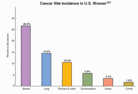
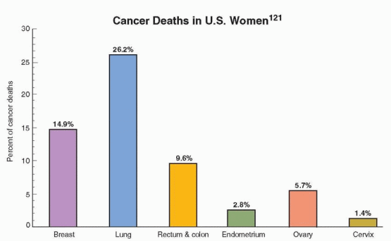
This decrease is believed to reflect a reduction in the use of postmenopausal hormone therapy following the publicized results of the Women’s Health Initiative and a decrease in the utilization of mammography; discussed in Chapter 18. About 87% of all breast cancers in the U.S. occur in women over age 44; only 1.9% of all cases occur under age 35, 12.5% under age 45, and 97% of breast cancer deaths in the U.S. occur in women over age 40.122

Mortality rates remained disappointingly constant until a decline began in the 1990s. The 5-year survival rate for localized breast cancer (about 61% of breast cancers) has risen from 72% in the 1940s to 98%.121 This is attributed to better therapy and earlier diagnosis because of the greater utilization of screening mammography. With regional spread, the 5-year survival rate for breast cancer is 84%; with distant metastases, the rate is 27%. The breast is the leading site of cancer in U.S. women and is now, unfortunately (because smoking is obviously the reason), exceeded by lung and bronchus cancer as the leading cause of death from cancer in women.121


The Chances of Developing Breast Cancer in the U.S. according to Age124


Birth to age 39


1 in 228


Age 40 to 59


1 in 24


Age 60 to 79


1 in 14


Birth to death


1 in 8


image


image

Over the years, breast cancer continued to have a deadly impact despite advances in surgical and diagnostic techniques. Classically, the single most useful prognostic information in women with operable breast cancer has been the histologic status of the axillary lymph nodes.127,128 The survival rate is higher with axillary lymph nodes negative for disease compared with positive nodes. Because of this recognition of the importance of the axillary nodes, the traditional surgical approach to breast cancer was based on the concept that breast cancer is a disease of stepwise progression. There is an important change in concept. Breast cancer is now viewed as a systemic disease, with spread to local and distant sites at the same time. Breast cancer is best viewed as occultly metastatic at the time of presentation. Therefore, dissemination of tumor cells has occurred by the time of surgery in many patients. However, this is not the story for all patients. Surely, some (if not many) cancers prior to invasion (and perhaps even some small invasive cancers) are not systemic at the time of diagnosis. For this reason, surgery is curative for many early cases of breast cancer.

Because we have been dealing with a disease that has already reached the point of dissemination in many patients, we must move the diagnosis forward several years in order to have an impact on breast cancer mortality. Earlier diagnosis requires that we be aware of what it is that makes a high-risk patient. However, keep in mind that the great majority of women (85%) who develop breast cancer do not have an identifiable risk factor other than age, and, therefore, every woman must be considered at risk.


Risk Factors

A constellation of factors influences the risk for breast cancer. These include reproductive experience, ovarian activity, benign breast disease, familial tendency, genetic differences, dietary considerations, and specific endocrine factors. Clinicians can calculate the risk for an individual patient at the National Cancer Institute Internet site: http://www.cancer.gov/bcrisktool/.


Risk Factors for Breast Cancer121


Relative risk greater than 4.0:


Over age 65


Inherited mutations


Two or more first-degree relatives with early disease


Postmenopausal breasts that are at least 75% dense on mammography


Relative risk 2.1-4.0:


One first-degree relative with breast cancer


Atypical hyperplasia on breast biopsy


High-dose radiation to the chest


High postmenopausal bone density


Relative risk 1.1-2.0:


First full-term pregnancy after age 30


Menarche before age 12


Menopause after age 55


Nulliparity


Never breastfed


Postmenopausal obesity


Previous cancer of endometrium, ovary, or colon


Alcohol consumption, 2 to 5 drinks daily


Reproductive Experience

The risk of breast cancer increases with the increase in age at which a woman bears her first full-term child. A woman pregnant before the age of 18 has about one-third the risk of one who first delivers after the age of 35. To be protective, pregnancy must occur before the age of 30. Age at first birth and multiparity in women who experience their first birth before age 25 reduce the risk of breast cancer that is positive for estrogen and progesterone receptors.127,128 Women over the age of 30 years at the time of their first birth have a greater risk than women who never become pregnant.129 Indeed, there is reason to believe that the age at the time of birth of the last child is the most important influence (an increasing risk with increasing age).130 There is, however, a significant protective effect with increasing parity, present even when adjusted for age at first birth and other risk factors.131,132 Delayed childbearing and fewer children in modern times are believed to have contributed significantly to the increased incidence of breast cancer observed over the last decades.

Although pregnancy at an early age produces an overall lifetime reduction in risk, there is evidence that the first few years after delivery are associated with a transient increase in risk.133 This increase probably reflects accelerated growth of an already present malignancy by the hormones of pregnancy. A very large case-control study concluded that pregnancy transiently increases the risk (perhaps for up to 3 years) after a woman’s first childbirth, and this is followed by a lifetime reduction in risk.134 And some have found that a concurrent or recent pregnancy (3-4 years previously) adversely affects survival (even after adjustment for size of tumor and number of nodes).135,136 It is argued that breast cells that have already begun malignant transformation are adversely affected by the hormones of pregnancy, while normal stem cells become more differentiated and resistant, reducing the number of stem cells capable of malignant change. The number of breast stem cells available for this beneficial response diminishes with age and succeeding pregnancies.137 Although it is likely this effect is mediated by estrogen and progesterone, experimental evidence indicates the presence of LH receptors in breast tissue, and it is possible that human chorionic gonadotropin (hCG) contributes to the protective differentiation of breast cells.138,139 and 140 Another possibility is an antiproliferative action of alpha-fetoprotein, a peptide that is secreted in the fetal liver and stimulated by

the hormones of pregnancy.141


Initially, conflicting results were reported in over 20 studies examining the risk of breast cancer associated with the number of abortions (both spontaneous and induced abortions) experienced by individual patients.142,143 Concern for an adverse effect was based on the theoretical suggestion that a full-term pregnancy protects against breast cancer by invoking complete differentiation of breast cells, but abortion increases the risk by allowing breast cell proliferation in the first trimester of pregnancy, but not allowing the full differentiation that occurs in later pregnancy. In these studies there was a major problem of recall bias; women who develop breast cancer are more likely to truthfully reveal their history of induced abortion than healthy women. In studies that avoided recall bias (e.g., by deriving data from national registries instead of personal interviews), the risk of breast cancer was identical in women with and without induced abortions.144,145 More careful case-control studies failed to link a risk of breast cancer with either induced or spontaneous abortions.146,147 Similarly, newer prospective cohort studies, including the Nurses’ Health Study, also reported no association between the incidence of breast cancer and induced or spontaneous abortions.148,149 and 150

The fact that pregnancy early in life is associated with a reduction in the risk of breast cancer implies that etiologic factors are operating during that period of life. The protection afforded only by the first pregnancy suggests that the first full-term pregnancy has a trigger effect that either produces a permanent change in the factors responsible for breast cancer or changes the breast tissue and makes it less susceptible to malignant transformation. There is evidence for a lasting impact of a first pregnancy on a woman’s hormonal milieu. A small but significant elevation of estriol, a decrease in dehydroepiandrosterone and dehydroepiandrosterone sulfate, and lower prolactin levels all persist for many years after delivery.151,152 These changes take on significance when viewed in terms of the endocrine factors considered below.

Lactation may offer a weak to moderate protective effect (20% reduction) on the risk of breast cancer, both estrogen receptor-positive and receptor-negative tumors.127,128,153,154,155,156,157,158,159 and 160 The same beneficial effect has been reported in BRCA mutation carriers in one study, but not in another.161,162 The Nurses’ Health Study could not detect a protective effect of lactation, and a Norwegian prospective study, including a high percentage of women with long durations of breastfeeding, found no benefit on either premenopausal or postmenopausal breast cancer incidence.163,164 The impact of lactation, if significant, must be small. However, an analysis of the worldwide available data concluded that breastfeeding would reduce the risk of breast cancer by 4.3% per year of breastfeeding, and potentially could reduce the cumulative incidence by age 70 by more than 50%.165 A meta-analysis indicated that breastfeeding reduced the risk of breast cancer by about 10-20%, and the impact was limited to premenopausal women.166 There is a unique and helpful study of the Chinese Tanka, who are boat people living on the coast of southern China.167 The women of the Chinese Tanka wear clothing with an opening only on the right side, and they breastfeed only with the right breast. All breast cancers were in postmenopausal women, and the cancers were equally distributed between the two sides, suggesting a protective effect only for premenopausal breast cancer.

In both cohort and case-control studies, there is good evidence that cosmetic breast augmentation does not increase the risk of breast cancer.168,169 and 170 Specifically, studies have failed to indicate an increased risk of breast cancer in women who have had cosmetic breast implants.171,172,173 and 174


Ovarian Activity

Women who have a premenopausal oophorectomy have a lower risk of breast cancer, and the lowered risk is greater the younger a woman is when ovariectomized. There is a 70%
risk reduction in women who have oophorectomy before age 35. There is a small decrease in risk with late menarche and a moderate increase in risk with late natural menopause, indicating that ovarian activity plays a continuing role throughout reproductive life.175

Observational studies indicated that anovulatory and infertile women (exposed to less progesterone) have a small increased risk of breast cancer later in life.176,177,178 and 179 However, the statistical power of these observational studies was limited by small numbers (all fewer than 15 cases). Larger numbers are available in the Nurses’ Health Study, where the opposite result was apparent, a reduction in the incidence of breast cancer in women with infertility attributed to ovulatory disorders.180


Benign Breast Disease

Women with prior benign breast disease form only a small proportion of breast cancer patients, approximately 5%. With obstruction of ducts (probably by stromal fibrosis), ductule-alveolar secretion persists, the secretory material is retained, and cysts form from the dilation of terminal ducts (duct ectasia) and alveoli. There is good reason to eliminate the term “fibrocystic disease of the breast.” In a review of over 10,000 breast biopsies in Nashville, Tennessee, 70% of the women were found to not have a lesion associated with an increased risk for cancer.181 The most important variable on biopsies is the degree and character of the epithelial proliferation. Women with atypical hyperplasia had a relative risk of 5.3, while women with atypia and a family history of breast cancer had a relative risk of 11. In the Nurses’ Health Study, biopsies with proliferative disease had a relative risk of breast cancer of 1.6, and with atypical hyperplasia, the relative risk was 3.7.182 Only 4-10% of benign biopsies have atypical hyperplasia. The point is that we needlessly frighten patients with the use of the term fibrocystic disease. For most women, this is not a disease, but a physiologic change brought about by cyclic hormonal activity. Let’s call this problem FIBROCYSTIC CHANGE OR CONDITION.


The College of American Pathologists supports this position and has offered this classification.183


Classification of Breast Biopsy Tissue According to Risk for Breast Cancer


No increased risk:


Adenosis


Duct ectasia


Fibroadenoma without complex features


Fibrosis


Mild hyperplasia (3-4 cells deep)


Mastitis


Periductal mastitis


Squamous metaplasia


Ordinary cysts (fibrocystic disease)


Slightly increased risk (1.5-2 times):


Fibroadenoma with complex features


Moderate or florid hyperplasia


Several papillomas


Sclerosing adenosis


Moderately increased (4-5 times):


Atypical ductal hyperplasia


Atypical lobular hyperplasia


Markedly increased (8-10 times):


Ductal carcinoma in situ


Lobular carcinoma in situ


Familial Tendency

Most breast cancers are sporadic; i.e., they arise in individuals without a family history of breast cancer. However, female relatives of women with breast cancer have about twice the rate of the general population. There is an excess of bilateral disease among patients with a family history of breast cancer. Relatives of women with bilateral disease have about a 45% lifetime chance of developing breast cancer. The relative risks associated with first-degree relatives are:


Relative Risk with Affected First-degree Relatives184


One relative


1.80


Two relatives


2.93


Three relatives


3.90


It is worth emphasizing that only one of nine women who develop breast cancer has an affected first-degree relative, and most women with an affected relative will never have breast cancer.

The breast and ovarian tumor suppressor gene (BRCA1) associated with familial cancer is on the long arm of chromosome 17, localized to 17q12-q21.185 Although other genetic alterations have been observed in breast tumors, multiple, different mutations in BRCA1 are believed to be responsible for approximately 20% of familial breast cancer and 80% of families with both early-onset breast and ovarian cancer. Overall, no more than 5-10% of breast cancers in the general population can be attributed to inherited mutations.127,186 Autosomal dominant inheritance of mutations in this gene can be either maternal or paternal; male carriers are at increased risk for colon and prostate cancers.187 A second autosomal dominant locus of multiple mutations, BRCA2, on chromosome 13q12-q13, accounts for up to 35% of families with early-onset breast cancer (but a lower rate of ovarian cancer), and in males, for prostate cancer, pancreatic cancer, and male breast cancer.188,189 Together, BRCA1 and BRCA2 account for 80% of families with multiple cases of early-onset breast cancer.190 About 5-10% of women who develop ovarian cancer have mutations in BRCA1.191,192

BRCA1 encodes a 1,863-amino-acid protein with a zinc finger domain that is a tumor suppressor important in DNA transcription. Mutations in many different regions of the BRCA1 gene cause a loss or reduction in its function.193,194 Because not every individual with a mutation in this gene develops cancer, other factors are involved, making the accuracy of prediction more difficult and arguing against widespread screening for mutations of this gene. Providing accurate numbers is a difficult task, because breast cancer has a multifactorial etiology with both genetic and environmental factors. The BRCA1 gene could play a role in sporadic breast and ovarian cancer, but analysis of tumors has failed to find mutations in sporadic cancers that occur later in life.195

High-risk families have a high probability of harboring a mutation in a dominant breast cancer susceptibility gene. It is estimated that approximately 0.04% to 0.2% of women in the U.S. carry the BRCA1 susceptibility (and BRCA2 is less common).196 Among women of Ashkenazi Jewish descent, the prevalence of BRCA1 and BRCA2 mutations is about 2%.197 The percentage of breast cancer cases in the general population associated with a family history accounts for only a minor part of the overall prevalence. The best estimates initially ranged from 6% to 19% at most.198 Later more representative studies revealed a lower prevalence, as low as 3% in the general population.199,200 In addition, there appears to be great variability in different parts of the world, and the prevalence in minority populations has not been adequately measured.


The presence of ovarian cancer within a family and three or more cases of breast cancer within a family are strong predictors of BRCA mutations. Genetic screening should be reserved for patients from high-risk families.


Family History Characteristics Associated with the Presence of BRCA Mutations


Early age of onset of breast cancer within a family.


Relatives with ovarian, primary peritoneal, or fallopian tube cancer.


Male relatives with breast cancer.


Three or more close relatives with breast cancer.


Close relatives with bilateral breast cancer.


Ashkenazi (Eastern European Jewish), French Canadian, or Icelandic ancestry.


Moderate-risk families are characterized by a less striking family history, the absence of ovarian cancer, and an age of onset at the time of diagnosis that is older. High-risk families have the presence of multiple cases of breast cancer in close relatives (usually at least three cases) that follows an autosomal-dominant pattern of inheritance; breast cancer is usually diagnosed before age 45; there may be cases of ovarian cancer in the family as well. Many of the cases, but not all, can be attributed to the susceptibility genes, BRCA1 and BRCA2.

High-risk families have the following cumulative breast cancer risk by the age of 80 as determined by the analysis of family histories198:


Affected Relative


Age of Affected Relative


Cumulative Breast Cancer Risk by Age 80


One first-degree relative


<50 years old


50 or more years old


13-21%


9-11%


One second-degree relative


<50 years old


50 or more years old


10-14%


8-9%


Two first-degree relatives


Both <50 years old


Both 50 years or older


35-48%


11-24%


Two second-degree relativtes


but both paternal or maternal


Both <50 years old


Both 50 years or older


21-26%


9-16%


Each child of a BRCA mutation carrier has a 50% chance of inheriting the mutation. In the United States, women who are carrying the BRCA1 mutation have a 46% cumulative risk of developing breast cancer by age 70, and a 39% risk for ovarian cancer.201 There is also a small increase in risk for other cancers, specifically of the pancreas, colon, uterus, and cervix.202 The male relatives who are carrying this mutation have an increased risk of prostate cancer and colon cancer in addition to a cumulative risk of breast cancer of 1.2%.203 The cancer risk for women with BRCA2 mutations is 43% for breast cancer and 22% for ovarian cancer by age 70.201 Male BRCA2 mutation carriers have a higher cumulative risk of breast cancer, 6.8%, compared with male BRCA1 carriers.203 In addition, BRCA2 mutation carriers have increased risks of cancers originating in the pancreas, prostate, gallbladder and bile duct, stomach, and skin.204 Breast cancer associated with BRCA1 mutations is histologically different (more often aneuploid and receptor-negative) compared to BRCA2 mutations and sporadic cancers, and appears to grow faster, but paradoxically, has a better survival in response to treatment.205 Outcome results, however, have not been consistent. A well-done Dutch study could not detect a difference in disease-free and overall survival comparing breast cancer cases from families with proven BRCA1 mutations to patients with sporadic breast cancer.206


Summary of Breast and Ovarian Cancer Risk in BRCA Carriers201


Breast Cancer Risk by Age 70(%)


Ovarian Cancer Risk by Age 70(%)


BRCA1


46


39


BRCA2


43


22


Because not all families with breast cancer carry mutations of BRCA1 or BRCA2, these families probably have breast cancer susceptibility genes yet to be identified. In addition, the current screening methods do not detect all BRCA mutations. For example, a mutation in a gene involved in the recognition and repair of damaged DNA, CHEK2, is prevalent in families with hereditary breast and colorectal cancer.207 Other genes that infrequently cause inherited breast cancer include the ATM gene, the p53 tumor suppressor gene, and the PTEN gene.127 When three or more closely related individuals within a family have been diagnosed with breast cancer, the likelihood that an inherited dominant genetic mutation is present is very high. The affected women need not be first-degree relatives, but they must be related either all on the mother’s side or the father’s side. Identifying the families that carry the BRCA2 gene uses the same historical criteria as that for the BRCA1 gene. The family presence of just one case of ovarian cancer further increases the likelihood of the BRCA1 mutation. In contrast to BRCA1 families, BRCA2 families have only a moderately increased incidence of ovarian cancer.

Screening and counseling for families who have the appropriate history but fail to demonstrate BRCA1 or BRCA2 mutations should be exactly the same as when the mutations are found.208

Once it has been determined that a family is at high risk for a breast cancer gene mutation, it is recommended that this family be referred to an appropriate laboratory and service that can be identified through the medical genetics department at a regional referral institution. Although blood samples can be mailed by overnight mail, involvement with an appropriate center is highly urged because of the importance of accurate informed consent, counseling, and follow-up care. The way in which information is communicated to patients has a profound impact on decision-making and compliance with surveillance.

High-risk women who have undergone prophylactic mastectomy experience a major reduction (more than 90%) in the number of breast cancers, although total prevention is not achieved.209,210 and 211 Because the mutation is present in every cell, and prophylactic mastectomy does not remove all tissue, there is no guarantee that breast cancer will be totally prevented. The same situation applies with prophylactic oophorectomy in that a carcinoma can arise from peritoneal cells. However, prophylactic salpingo-oophorectomy reduces the risk of ovarian cancer by about 90% and the risk of breast cancer by about 50%.212,213

A growing story indicates that serous ovarian cancer originates in the fimbriae of the fallopian tubes.214,215 Evidence consistently indicates that tubal sterilization is associated with a major reduction in the risk of ovarian cancer.216,217,218,219 and 220 A case-control study of BRCA1 and BRCA2 carriers indicated that tubal ligation reduced the risk of ovarian cancer by 60% in BRCA1 carriers, but no protective effect was observed among BRCA2 carriers.221 A prospective cohort study also detected differences between BRCA1 and BRCA2 carriers after prophylactic salpingo-oophorectomy: an 85% reduction in ovarian cancer in BRCA1 carriers but no significant effect in BRCA2 carriers, and a 72% reduction in breast cancer in BRCA2 carriers with a reduction that was not statistically significant in BRCA1 carriers.222 In addition, early carcinomas are found in the fallopian tube fimbriae of BRCA1 and BRCA2 mutation carriers.223,224 Prophylactic surgery should include bilateral salpingectomy.

Current recommendations from experts in this field are as follows186,198,225,226 and 227: For an individual identified to be at high risk, clinical breast examination is recommended every
6 months and annual mammography beginning at age 25. An annual evaluation by magnetic resonance imaging is also recommended because there is some evidence of a higher false-negative rate with mammography in these patients, and breast cancers detected in BRCA mutation carriers who undergo annual MRI surveillance are of lower stage disease.228 Clinical evaluation every 6 months is appropriate because the BRCA1related tumors have been demonstrated to be faster growing tumors. Support should be provided for those women who choose prophylactic mastectomy. Pelvic examination, serum CA-125 levels, and transvaginal ultrasonography with color Doppler are recommended annually for women under age 40, although it has not been demonstrated that this screening will detect tumors early enough to influence prognosis. Prophylactic salpingo-oophorectomy and hysterectomy are recommended at the completion of childbearing, preferably before age 35 and certainly by age 40. In our view, estrogen-only therapy is appropriate and acceptable following surgery, as discussed below.

The epidemiologic evidence indicates that oral contraceptive use can lower the risk of ovarian cancer in BRCA mutation carriers. A case-control study indicated that the use of oral contraceptives in women with BRCA1 or BRCA2 mutations was associated with a 50% reduction in the risk of ovarian cancer (increasing with duration of use, from 20% for less than 3 years of use, up to 60% with 6 or more years of use).229 In a large case-control study, the use of oral contraceptives reduced the risk of ovarian cancer by 44% in carriers of BRCA1 mutations and by 61% in carriers of BRCA2 mutations.230 Another case-control study concluded that the use of oral contraceptives reduced the risk of ovarian cancer by 5% with each year of use in both BRCA1 and BRCA2 mutation carriers.231 There is only one case-control study that found no indication of protection.232

In contrast to the effect on ovarian cancer risk, the impact of oral contraceptives on the risk of breast cancer is not clear at all. A cohort study from Minnesota concluded that women with a first-degree relative with breast cancer had an increased risk of breast cancer with oral contraception; however, this association was present only with oral contraceptives used prior to 1976 (high-dose formulations), and the confidence intervals were wide because of small numbers (13 ever users).233 In a study of women with BRCA1 and BRCA2 mutations, an elevated risk of breast cancer associated with oral contraception was based on only a few cases and did not achieve statistical significance.234 A larger case-control study concluded that BRCA1 (but not BRCA2) mutation carriers had small increases in the risk of breast cancer in users for at least 5 years (OR=1.33, CI=1.11-1.60), in users before age 30 (OR=1.29, CI=1.09-1.52), and in those who developed breast cancer before age 40 (OR=1.38, CI=1.11-1.72).235 In contrast, another case-control study concluded that oral contraceptive use for at least 5 years doubled the risk of breast cancer before age 50 in BRCA2 carriers, but not in BRCA1 carriers.236 A retrospective analysis of an international cohort of BRCA carriers indicated that an increased risk of breast cancer with both BRCA1 and BRCA2 carriers was present only with 4 or more years of use before a first full-term pregnancy.237 A study that focused on low-dose oral contraceptives could detect no association with breast cancer risk in BRCA mutation carriers.162 Another case-control study found no increase in the risk of breast cancer diagnosed before age 40 in either BRCA1 or BRCA2 carriers.238 And finally, a case-control study could detect no significant increase in the risk of contralateral breast cancer among BRCA1 and BRCA2 carriers or in noncarriers with the use of oral contraceptives or postmenopausal hormones.239

The data with oral contraceptives in BRCA mutation carriers are all observational and not robust. Until better information is forthcoming, it seems reasonable to inform carriers of BRCA mutations that the use of oral contraceptives is likely to reduce the risk of ovarian cancer, but the effect on breast cancer risk is uncertain.

The effect of chemoprevention by tamoxifen, raloxifene, or aromatase inhibitors has not been tested in BRCA mutation carriers by randomized trials. However, in subgroup analyses of the American trial assessing the effect of tamoxifen for prevention, tamoxifen
reduced the risk of breast cancer by 62% in BRCA2 carriers, but had no impact in BRCA1 carriers.240,241 This is consistent with the fact that women with BRCA2 mutations have predominately estrogen receptor-positive tumors and women with BRCA1 mutations have mostly estrogen receptor-negative tumors. Although no data are available, it is likely that raloxifene and aromatase inhibitors would yield results similar to those with tamoxifen. Given the side effects associated with these drugs, the decision to use one of these agents for chemoprevention is a difficult one for both clinician and patient. Prophylactic bilateral salpingo-oophorectomy remains as the superior choice for risk protection, a procedure that can in most cases, even with thorough inspection of peritoneal surfaces and peritoneal washings, be easily performed by laparoscopy. Serial sectioning of the ovaries and tubes is mandatory to detect microscopic cancers. Although concurrent hysterectomy is an individual choice, it is recommended to gain the theoretical advantage of removing the cornual portions of the fallopian tubes.

In a cohort of women with BRCA1/2 who had oophorectomy and a 60% reduction in the risk of developing breast cancer, hormone therapy of any type did not alter the reduction in breast cancer experienced by the women undergoing oophorectomy.242 The average length of follow-up was 2.6 years (more than 5 years in 16%) in the surgically treated group and 4.1 years (more than 5 years in 33%) in the non-oophorectomized group. There was no hint of a difference in breast cancer reduction comparing hormone users and nonusers. The findings were similar in 34 women who used a combination of estrogen and progestin, but the power of this finding was limited by the small number.

A case-control study of 472 postmenopausal women with a BRCA1 mutation found that women who used hormone therapy after prophylactic oophorectomy, either estrogen only or combined estrogen-progestin, not only did not have an increased risk of breast cancer, but hormone use was actually associated with a decreased risk.243 The findings were the same regardless of duration of use or current or past use. The conclusion is encouraging, but limited by the fact that 68% of the tumors in the study were estrogen receptor-negative, making the estrogen receptor-positive tumors (that are more likely to be influenced by hormone use) relatively small in number.

Women who are BRCA carriers face difficult decisions regarding hormonal treatment for menopausal symptoms. The experience thus far indicates that hormone therapy can be used safely for several years. Continuing follow-up of these patients may extend this period of safety even longer.


Dietary Factors

The geographic variation in incidence rates of breast cancer is considerable (the United States has the highest rates and Japan the lowest), and it has been correlated with the amount of animal fat in the diet.244 Lean women, however, have an increased incidence of breast cancer, although this increase is limited to small, localized, and well-differentiated tumors.245 Furthermore, studies have failed to find evidence for a positive relationship between breast cancer and dietary total or saturated fat or cholesterol intake.246,247,248 and 249 One study found that dietary fat is a stronger risk factor for postmenopausal breast cancer than for premenopausal breast cancer, but another study had the opposite conclusion.250,251 Although a cohort study concluded that dietary fat is a determinant of postmenopausal breast cancer, the association did not achieve statistical significance.252 And another very large cohort study in Europe demonstrated only a very weak link between saturated fat intake and the risk of breast cancer, only in nonusers of hormone therapy.253 Thus, the epidemiologic literature provides little support for a major contribution of dietary fat to the risk of breast cancer. Nevertheless, there
is a correlation between intraabdominal fat (android obesity) and the risk of breast cancer, a consequence of excessive caloric consumption, however, not a specific dietary component.254 Presumably, the connection between android obesity and breast cancer is through the metabolic perturbations, especially hyperinsulinemia, associated with excessive body weight.

There is no argument that the incidence of breast cancer is increased in countries associated with affluent, unfavorable diets (high fat content) and a lack of physical exercise. Indeed, increased physical activity in postmenopausal women reduces the risk of breast cancer.255 The common denominator may be the peripheral insulin resistance and hyperinsulinemia that become prevalent with aging and weight gain in affluent, modern societies. This specific metabolic change is becoming a common theme in various clinical conditions, particularly noninsulin-dependent diabetes mellitus, anovulation and polycystic ovaries, hypertension, and dyslipidemia. Hyperinsulinemia is found more often in women with breast cancer.256 There are, indeed, many reasons to avoid excess body weight. The risk of breast cancer is reduced in women who exercise regularly.257

Only gold members can continue reading. Log In or Register to continue

Stay updated, free articles. Join our Telegram channel

Jul 5, 2016 | Posted by in GYNECOLOGY | Comments Off on The Breast

Full access? Get Clinical Tree

Get Clinical Tree app for offline access