Rashes
Leslie Castelo-Soccio
Kara Shah
Rash is a common reason for children to be brought for evaluation by a healthcare provider. The examination of the rash can provide key clues to the diagnosis. Rashes can be categorized by type and by accompanying symptoms to organize the differential diagnosis.
Rash Accompanied by a Fever
Bacterial Causes
Scarlet fever
Staphylococcal scalded skin syndrome
Toxic shock syndrome
Acute meningococcemia
Rocky Mountain spotted fever
Viral Causes
Herpes simplex
Varicella
Roseola infantum
Erythema infectiosum
Enteroviruses
Measles
Rubella
Lyme disease
Miscellaneous
Juvenile idiopathic arthritis
Kawasaki disease
Rash in a Neonate
Erythema toxicum neonatorum
Transient neonatal pustular melanosis
Miliaria
Infantile seborrheic dermatitis
Candidiasis
Neonatal acne
Acropustulosis of infancy
Eczematous Dermatitis
Atopic dermatitis
Contact dermatitis
Nummular dermatitis
Asteatotic eczema
Id reaction
Papulosquamous Disease
Psoriasis
Lichen planus
Seborrheic dermatitis
Tinea (dermatophyte) infections
Tinea versicolor
Pityriasis rosea
Secondary syphilis
Pityriasis lichenoides et varioliformis acuta
Acrodermatitis enteropathica
Exfoliative dermatitis
Vesiculobullous Disease
Herpes simplex
Herpes zoster
Varicella
Rhus dermatitis (poison ivy)
Contact dermatitis
Insect bite or papular urticaria
Urticaria pigmentosa
Annular Erythema
Lyme disease
Erythema multiforme (EM)
Papular Eruptions
Scabies
Papular acrodermatitis of childhood (Gianotti-Crosti syndrome)
Papular urticaria
Molluscum contagiosum
Warts
DIFFERENTIAL DIAGNOSIS DISCUSSION
Scarlet Fever
Etiology
Scarlet fever is caused by group A β-hemolytic Streptococcus.
Clinical Features
Illness begins with fever and pharyngitis followed by enanthem and exanthem in 24 to 48 hours.
The face appears flushed, except for circumoral pallor. The tongue initially has a white coating (white strawberry tongue) that fades by the fourth day, revealing a very erythematous tongue with prominent papillae (red strawberry tongue). Cervical and submandibular lymphadenopathies are noted.
The rash appears as a diffuse erythema; small fine papules give it a sandpaperlike quality. The rash begins on the neck and spreads rapidly to the trunk and extremities. It is accentuated in the intertriginous areas (i.e., the axillae and antecubital, inguinal, and popliteal creases), and the palms and soles are spared. The rash resolves in 4 to 5 days with fine peeling of the skin. Peeling begins on the face, spreading to the trunk and extremities. On the hands and feet, peeling in large sheets may occur.
Evaluation
Diagnosis is by culture or rapid test of a pharyngeal swab. Antistreptolysin-O titers or antideoxyribonuclease B titers may also be used to indicate recent infection. Complete blood count commonly reveals leukocytosis. Urinalysis and liver function test may reveal changes associated with complications of scarlet fever.
Treatment
Treatment of choice is with penicillin, amoxicillin, or a first-generation cephalosporin. Erythromycin can be used in penicillin-allergic patients.
Staphylococcal Scalded Skin Syndrome
Etiology
Staphylococcal scalded skin syndrome is caused by two epidermolytic toxins, epidermolytic toxins A and B, which are produced by some strains of Staphylococcus aureus. These toxins cause intraepidermal splitting through the granular layer by specific cleavage of desmoglein 1, a cadherin protein that mediates celltocell adhesion of keratinocytes.
Clinical Features
Staphylococcal scalded skin syndrome typically occurs in children younger than 5 years. The illness begins with fever and/or irritability after an episode of
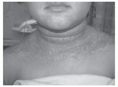
conjunctivitis, rhinitis, or an upper respiratory tract infection. The skin initially becomes erythematous and tender. The eruption usually begins around the nose and mouth and spreads to the trunk. The rash is most prominent in the flexures and can involve the hands and feet. After 1 to 2 days, the skin develops bullae and begins peeling in sheets. After 2 days, the involved skin surfaces become crusted and then begin to develop scaling, which lasts up to 5 days (Figure 64-1).
conjunctivitis, rhinitis, or an upper respiratory tract infection. The skin initially becomes erythematous and tender. The eruption usually begins around the nose and mouth and spreads to the trunk. The rash is most prominent in the flexures and can involve the hands and feet. After 1 to 2 days, the skin develops bullae and begins peeling in sheets. After 2 days, the involved skin surfaces become crusted and then begin to develop scaling, which lasts up to 5 days (Figure 64-1).
Evaluation
Staphylococcus aureus can sometimes be isolated by bacterial culture of the nose, nasopharynx, throat, conjunctiva, or perianal area.
Treatment
Treatment is with a penicillinase-resistant antibiotic, such as nafcillin or oxacillin (parenteral) or dicloxacillin or amoxicillin-clavulanate (oral). Clindamycin reduces the bacterial production of the toxin and is increasingly used because of concern about methicillin-resistant Staphylococcus aureus.
Toxic Shock Syndrome
Etiology
Toxic shock syndrome (TSS) is a multisystem disease caused by exotoxin-producing strains of either S. aureus (TSS) or Streptococcus pyogenes (STSS). These exotoxins—toxic shock syndrome toxin-1 in the case of S. aureus and most commonly
streptococcal pyrogenic exotoxin-A in the case of S. pyogenes— function as superantigens and stimulate the release of massive amounts of proinflammatory cytokines, including tumor necrosis factor-alpha and interleukin-1. Although classic TSS is seen in association with menstruation, nonmenstrual TSS is now more common. STSS is usually seen as a complication of a wound infection.
streptococcal pyrogenic exotoxin-A in the case of S. pyogenes— function as superantigens and stimulate the release of massive amounts of proinflammatory cytokines, including tumor necrosis factor-alpha and interleukin-1. Although classic TSS is seen in association with menstruation, nonmenstrual TSS is now more common. STSS is usually seen as a complication of a wound infection.
Clinical Features
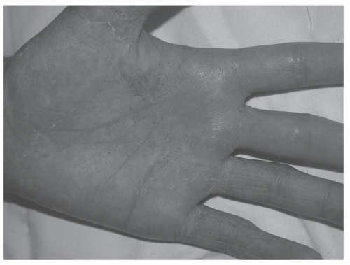
The association of fever, rash, and hypotension is the hallmark of TSS. Involvement of other organ systems may present as myalgias, acute renal failure, encephalopathy, disseminated intravascular coagulopathy, diarrhea, metabolic acidosis, and/or hepatitis. The rash may appear morbilliform (resembling measles) or as confluent erythema (erythroderma). Involvement of the palms and soles is common, and late desquamation of these areas is a key feature (Figure 64-2). A strawberry tongue is also characteristic.
Evaluation
Blood cultures should be sent, although they are not always positive. In TSS only about 5%-15% of cultures are positive and in STSS about 50% are positive. Culture of any cutaneous wounds should also be performed. Evaluation for systemic involvement should include complete blood counts, serum chemistries, liver function tests, urinalysis, creatine kinase, and coagulation studies. Other diagnostic
tests that may be helpful when indicated include electrocardiogram, echocardiography, and chest radiography. Skin biopsy is rarely needed.
tests that may be helpful when indicated include electrocardiogram, echocardiography, and chest radiography. Skin biopsy is rarely needed.
Treatment
Appropriate antibiotic therapy should be promptly initiated. Use of an agent that is effective against both S. aureus and S. pyogenes is required if the causative agent is unknown. Initial choices include use of oxacillin or vancomycin. Addition of clindamycin is recommended as an adjunct to decrease toxin production.
Meningococcemia
Etiology
Meningococcemia is caused by Neisseria meningitidis. Asymptomatic nasopharyngeal carriage occurs and transmission occurs via respiratory droplets.
Clinical Evaluation
The illness begins with symptoms of a mild upper respiratory tract infection, followed by fever, malaise, headache, and the development of a petechial eruption on the skin and mucous membranes. In addition, patients can have erythematous macules, papules, and urticarial lesions. In severe cases, the purpura can involve large areas that eventually necrose.
Many patients develop meningitis, disseminated intravascular coagulation, and hypotension.
Evaluation
Cultures of blood and cerebrospinal fluid are indicated. The organism can sometimes be recovered from a small petechial lesion if a biopsy of the lesion is performed and cultured. A Gram stain of a scraping of a petechial lesion may reveal organisms.
Treatment
Initial treatment is with a broad-spectrum antibiotic; once the diagnosis is confirmed, the patient can be treated with penicillin, ceftriaxone, or cefotaxime.
Rocky Mountain Spotted Fever
Etiology
Rocky Mountain spotted fever is caused by Rickettsia rickettsii, which is transmitted by ticks. Humans are accidental hosts.
Clinical Features
After a prodrome characterized by headache, malaise, photophobia, and joint and muscle pain, the patient develops a fever followed by a rash on the fourth day.
The rash begins on the wrists and ankles as small erythematous macules that eventually become petechial or purpuric. The rash then spreads to the trunk and within 2 days is generalized, with involvement of the palms and soles.
Other findings may include generalized edema (especially affecting the periorbital area in children), severe muscle tenderness, photophobia, and hyponatremia.
Evaluation
Diagnosis is with the detection of rickettsial-specific antibodies. Indirect immunofluorescence antibody, enzyme immunoassay, and indirect hemagglutination are the most sensitive and specific diagnostic tests. Direct immunofluorescence of a skin lesion biopsy specimen and serum polymerase chain reaction (PCR) are useful for confirming a clinical diagnosis. Owing to poor sensitivity and specificity, the Weil-Felix assay is no longer used.
Treatment
The treatment of choice for all patients is a tetracycline derivative.
Herpes Simplex Virus
Herpes simplex virus is discussed in Chapter 70, “Sexually Transmitted Diseases.”
Roseola Infantum
Etiology
Roseola is caused by human herpes virus-6 infection. Transmission occurs through saliva.
Clinical Features
Usually seen in infants, roseola classically presents with 3 days of high fever, followed by rapid defervescence. Febrile seizures are common. After the fever abates, a diffuse, faint, blanchable, erythematous reticulated rash appears. It lasts for several days. An enanthem characterized by erythematous macules on the soft palate and uvula may also develop.
Evaluation
The diagnosis is usually made clinically, although serology and/or serum PCR for human herpes virus-6 DNA may be performed if the diagnosis is unclear.
Treatment
The disease is self-limiting.
Erythema Infectiosum
Etiology
Erythema infectiosum is caused by infection with parvovirus B19. Transmission occurs via respiratory droplets. Clinical signs and symptoms are probably the result of immune complexes in the skin and joints rather than from the virus itself.
Clinical Features
Erythema infectiosum is seen predominantly in school-age children. Prodromal symptoms such as fever, pharyngitis, malaise, or coryza may occur, followed by the development of the classic “slapped cheek” erythema. A lacy, reticulated, erythematous exanthem then develops most commonly on the extremities. It may wax and wane over several weeks. Older children and adults may complain of arthralgias or arthritis.
Evaluation
The diagnosis is usually made clinically. Serologic tests and serum PCR for parvovirus B19 DNA may be performed if the diagnosis is unclear.
Stay updated, free articles. Join our Telegram channel

Full access? Get Clinical Tree


