Postmenopausal Hormone Therapy


Postmenopausal Hormone Therapy


image

Postmenopausal hormone therapy had its beginning in the effort to alleviate specific symptoms associated with the decline in estrogen production at menopause. There is little question that women who suffer from hot flushes or atrophy of reproductive tract tissues can be relieved of their problems by the use of estrogens. In the 1990s, however, the focus of postmenopausal hormone therapy changed from short-term treatment to the preventive health care benefits associated with long-term treatment. It is almost certain that the long-term disabilities of osteoporosis can be largely prevented by therapy with estrogen and progestin. The long-term impact on cognition remains to be documented, but there is reason to believe that there will be a benefit in this area. However, long-term use was challenged by clinical trial data that were interpreted to indicate that hormone therapy did not protect against cardiovascular disease and that the risk of breast cancer was increased. The debate over these issues made decision-making by clinicians and patients very difficult. In this chapter, we offer our own interpretation as a guide for the clinical use of postmenopausal hormone therapy.

The evidence supporting many of the benefits with postmenopausal hormone therapy is also reviewed in Chapter 17, in which the effects of hormonal treatment are considered in
conjunction with the impact of the decrease in estrogen after menopause. In this chapter, we review the clinical aspects of postmenopausal hormone therapy, the impact of clinical trial results, and our methods of patient management.


History1,2,3 and 4

The existence of hormones was unknown 200 years ago. In the last half of the 19th century, a scattering of chemists and physiologists began to produce hormonally active extracts from glands, bile, and urine of animals. Adventuresome clinicians used these extracts to treat patients, for example supplying thyroid hormone to treat severely hypothyroid individuals, and the specialty of endocrinology was born. The word “endocrine” was adopted to designate the “glands of internal secretion,” the multiple sources of hormones.

Charles Edouard Brown-Sequard, the son of a French woman and an American sea captain, was born on the island of Mauritius. Speaking fluent English and French, he practiced medicine and lectured in London and New York before settling in Paris. Brown-Sequard reported in 1889 that he was rejuvenated by the self-administration of extracts from dog testicles, most likely a placebo effect considering the scant amount of testosterone he could have extracted using his aqueous method, and he suggested that ovarian extracts would have the same revitalizing effect in women. Efforts to treat women around the end of the nineteenth century were largely unsuccessful, but in 1897, ovarian extract was reported to be effective for menopausal hot flushing.5

The first American attempt to treat menopausal symptoms is attributed to E.L. Sevringhaus and J. Evans of Madison, Wisconsin, who in 1929 administered a derivative from the amniotic fluid of cattle.2,6 In the 1930s, the ovarian hormones were isolated, and the “estrin” products and the synthetic estrogens, stilbestrol and ethinyl estradiol, were administered to menopausal women. Edgar Allen and Edward Doisy were the first to isolate the ovarian hormone, estrogen. Allen was born in Colorado, educated at Brown University, and served in France during World War I. In 1933, he became the chairman of the Department of Anatomy at Yale University. He died of a heart attack while on patrol off Long Island for the U.S. Coast Guard in February 1943. Doisy was born in Illinois and educated at the University of Illinois and Harvard. During World War I, he was assigned to the Rockefeller Institute in New York City and then to the Walter Reed Hospital in Washington. Doisy was the first chairman of biochemistry at the St. Louis University School of Medicine. He received the Nobel Prize in Medicine, along with Henrik Dam, in 1943 for his isolation and synthesis of vitamin K. Doisy died in 1986 at the age of 92.

In 1919, Allen and Doisy, both discharged from the army after World War I, joined the faculty at the Washington University School of Medicine in St. Louis. They became friends playing on a faculty baseball team and planned their first experiments while driving to work together. In 1922, Allen moved to the University of Missouri to be Professor of Anatomy, and Doisy went to St. Louis University, but they continued their collaboration. Doisy prepared ovarian extracts and mailed them to Allen for experiments. In 1923 and 1924, Allen and Doisy reported the isolation from pig ovaries and the administration to animals of “an ovarian hormone.”

In 1926, Sir Alan S. Parkes and C.W. Bellerby coined the basic word “estrin” to designate the hormone or hormones that induce estrus in animals, the time when female mammals are fertile and receptive to males. Doisy and his students Veler and Thayer in St. Louis isolated a few milligrams of estrogen in crystalline form in 1929 from large amounts of urine from pregnant women. The terminology was extended to include the principal estrogens
in humans, estrone, estradiol, and estriol, in 1932 at the first meeting of the International Conference on the Standardization of Sex Hormones in London, although significant amounts of pure estradiol were not isolated until 1936. At this same meeting, the pioneering chemists were bemoaning the problem of scarcity that limited supplies to milligram amounts when a relatively unknown biochemist, A. Girard from France, offered twenty grams of crystalline estrogen derived by the use of a new reagent to treat mare’s urine.7

In the 1920s, George W. Corner at the University of Rochester invited Willard Myron Allen, an organic chemist who was then a medical student, to join him in the study of the corpus luteum. Within 2 years, they had a pure extract, but it was not until 1934 that crystalline progesterone was isolated almost simultaneously in several countries. It took the corpora lutea of 50,000 pigs to yield a few milligrams. At the Second International Conference on Standardization of Sex Hormones in London, Corner and Allen proposed the name progestin. Others proposed luteosterone, and, at a cocktail party, the various biochemists agreed to call the chemical progesterone.7

Hormones were being administered to patients in the 1940s, but supplies were very limited. And with a scarce supply, hormones were incredibly expensive. Progesterone, for example, cost $200 per gram. “To secure barely enough androsterone to cover the head of a pin, Adolph Butenandt had had to start with nearly four thousand gallons of urine; to obtain less than one hundredth of an ounce of pure testosterone crystals, Ernst Laqueur had had to process nearly a ton of bulls’ testicles. It took a full ton of cholesterol, from the spinal cords or brains of cattle or from the grease of sheep’s wool, to yield just twenty pounds of the starting material from which progesterone ultimately could be obtained. Edward Doisy had had to process the ovaries of more than eighty thousand sows to get just twelve thousandths of a gram of estradiol.”8

In the 1930s, the Ayerst Company was extracting estrogens from the urine of pregnant women. Limited by the problems of supply, low activity, and bad taste and odor, Gordon A. Grant, head of biochemistry for Ayerst, suggested in 1939 that they use urine from horses. The process produced sodium salts from the sulfate esters of the various estrogens, yielding a water-soluble conjugate. Premarin (conjugated estrogens) was approved in Canada in 1941 and in the U.S. in 1942 for the treatment of symptoms associated with menopause.9 The tablets were and are still designated as variations of 1.25 mg, based on the equivalent amounts of Premarin and estrone (1.25 mg) that could produce the same effect in the Allen-Doisy bioassay (amount required to produce an increase in rat uterine weight). It was not until 1972 that the first quantitative analysis of Premarin was performed, based on gas chromatography. Modern studies indicate that there is a large number of steroids in Premarin, even androgens and progestins, but only the 10 estrogens are present in sufficient quantity to have clinical effects. Synthetic conjugated estrogens are available; one mixture, Cenestin, contains 9 estrogens and the other, Enjuvia, 10 estrogens.


Composition of Conjugated Estrogens (Premarin)


Sodium estrone sulfate


49.3%


Sodium equilin sulfate


22.4%


Sodium 17α-dihydroequilin sulfate


13.8%


Sodium 17α-estradiol sulfate


4.5%


Sodium Δ8,9-dehydroestrone sulfate


3.5%


Sodium equilenin sulfate


2.2%


Sodium 17β-dihydroequilin sulfate


1.7%


Sodium 17α-dihydroequilenin sulfate


1.2%


Sodium 17β-estradiol sulfate


0.9%


Sodium 17β-dihydroequilenin sulfate


0.5%


Estrogen Formulations and Routes of Administration


Oral Administration

The relative potencies of commercially available estrogens are of great importance when prescribing estrogen, and the clinician should be familiar with the following potencies:


Relative Estrogen Potencies10,11,12,13,14 and 15


Estrogen


FSH Levels


Liver Proteins


Bone


Conjugated estrogens


1.0 mg


0.625 mg


0.625 mg


Micronized estradiol mg


1.0 mg


1.0 mg


1.0 mg


Estropipate (piperazine estrone sulfate) mg


1.0 mg


1.25 mg


1.25 mg


Ethinyl estradiol


5.0 μg


2-10μg


5.0 μg


Estradiol valerate


1.0 mg


Esterified estrogens mg


0.625 mg


Transdermal estradiol


50 μg


The 17α-ethinyl group of ethinyl estradiol (by resisting metabolism) enhances hepatic effects, because no matter by which route it is administered, liver function is affected.13 The same is true for conjugated equine estrogens. Contrary to the case with estradiol, the liver appears to preferentially extract ethinyl estradiol and conjugated equine estrogens no matter what the route of administration. Thus, the route of administration appears to influence the metabolic responses only in the case of specific estrogens, most notably estradiol.

A major factor in the potency differences among the various estrogens (estradiol, estrone, estriol) is the length of time that the estrogen binds to its receptor. The higher rate of dissociation with the weak estrogen (estriol) can be compensated for by continuous application to allow prolonged binding and activity. Estriol has only 20-30% affinity for the estrogen receptor compared with estradiol; therefore, it is rapidly cleared from a cell. However, if the effective concentration is kept equivalent to that of estradiol, it can produce a similar biologic response.16 At least two studies have been unable to demonstrate prevention of bone loss with the administration of 2 mg estriol daily.17,18 In pregnancy, where the concentration of estriol is very great, it can be an important hormone not just a metabolite. Thus, higher estriol levels are not necessarily protective against potent estrogenic effects. Because estriol protects the rat against breast tumors induced by various chemical carcinogens,19 it has been hypothesized that a higher estriol level protects against the more potent effects of estrone and estradiol. But, antagonism of estradiol occurs only within a vary narrow range of the ratio of estradiol to estriol, a range that is rarely encountered either physiologically or pharmacologically.20 Below this range, estradiol is unimpeded, above this range estriol itself exerts estrogenic activity. The commercial preparation that contains estriol, estradiol, and estrone contains sufficient amounts of estrone and estradiol to produce standard clinical effects.

Esterified estrogens are synthetically prepared from plant precursors and are composed mostly of sodium estrone sulfate with a 6-15% component of sodium equilin sulfate. Estradiol valerate is rapidly hydrolyzed to estradiol; therefore, the pharmacology and effects are comparable at similar dosages.21


Transdermal Patch Administration

The patches first used for transdermal estrogen administration contained an alcohol reservoir; the estrogen was released through a semipermeable membrane attached to the skin with an adhesive. In the current generation of patches, the hormones are dissolved and distributed throughout the adhesive matrix. In a study of women who had previously discontinued patches because of skin irritation (contact dermatitis), skin reactions were less common with the newer matrix patches.22 In addition, the matrix patches are better tolerated in tropical environments.23 The patches are designated according to the amount of estradiol delivered per day: from 14 to 100 μg.

The concentration of estrogen in the hepatic portal system after oral administration is 4-5 times higher than that in the periphery.24 Because of first-pass metabolism in the liver, oral estradiol results in a circulating estrone to estradiol ratio of approximately 3; with transdermal administration the ratio is 1. The first-pass effect may be important for lipoprotein effects. For example, short-term studies (6 weeks) could document increased catabolism of low-density lipoprotein (LDL-cholesterol) and increased production of apoprotein A-I with oral estrogen, but no effect with transdermal estrogen.25,26 A 2-year study in Los Angeles with a transdermal dose (100 μg) detected no significant change in HDL-cholesterol levels.27 However, English data indicate that the transdermal administration of 50 μg estradiol twice a week is as effective as 0.625 mg oral conjugated estrogens, when combined with a progestin in sequential regimens, on bone density and lipids over a duration of 3 years.28 Standard doses of estrogen administered transdermally (50 μg) protect against fractures as well as standard oral doses do.29 As with oral estrogen, lower transdermal doses can produce effects on bone density and menopausal symptoms,30 but a substantial number of women require higher doses.

The critical question is whether the first-pass effect of oral estrogen is clinically important. The different effects of oral and transdermal administration on metabolic parameters have been repeatedly compared over the years, but epidemiologic studies of clinical end points are not abundant, handicapped by the relatively small numbers of women using transdermal estrogen in most countries.

Clotting Factors. First-pass hepatic metabolism affects the synthesis of clotting proteins, markers of coagulation and fibrinolysis that can influence the risk of thrombosis and coronary heart disease events. Oral estrogen increases factor VII and prothrombin 1 and 2 fragment, whereas transdermal estrogen decreases factor VII.31,32,33 and 34 Oral estrogen also increases circulating levels of matrix metalloproteinases, MMP-2 and MMP-9, enzymes that are associated with a tendency for clotting.35 However, what is important is whether the different effects of oral and transdermal delivery on clotting factors translate into clinical differences and cardiovascular risk.

Activated Protein C (APC) Resistance and Risk of VTE. Resistance to APC is an important marker for venous thrombosis in individuals with inherited thrombogenic mutations and even in the absence of these mutations. Oral estrogen increases APC resistance, whereas transdermal estrogen has no significant effect on this marker.36,37 Based on this difference, one would predict that transdermal delivery of estrogen would be less likely than oral delivery of estrogen to be associated with venous thromboembolism (VTE).

A French case-control study (epidemiologic studies of the link between the transdermal route of administration and a relatively rare event are possible in France because of the popularity of the transdermal method) reported no increased risk of VTE in users of transdermal estrogen, as compared with a 4-fold increase in oral estrogen users.38,39 and 40 Estrogen users who carried a factor V Leiden mutation or a prothrombin mutation had a 25-fold higher risk of VTE than did women who did not use estrogen and did not have either mutation. The women with a prothrombotic mutation who used transdermal estrogen had a VTE risk that was similar to that of women with a prothrombotic mutation who did not use estrogen. The French E3N
prospective cohort study also reported an increased risk of venous thromboembolism with current users of oral therapy, a hazard ratio of 1.7 (CI=1.1-2.8), a ratio that is similar to the usual 2-fold increase repeatedly documented in the literature, and no increase with transdermal estrogen.41 Venous thrombosis is discussed in more detail later in this chapter.

Lipids and Hepatic Enzymes. Both oral and transdermal estrogen reduce total cholesterol, low-density lipoprotein cholesterol, and lipoprotein(a). Compared with transdermal estrogen, oral estrogen produces significantly greater elevations in high-density lipoprotein cholesterol and increases triglycerides, whereas transdermal estrogen decreases triglyceride levels.31,33,42,43 and 44 Indeed, triglyceride levels markedly elevated in response to oral therapy return to normal when treatment is changed to transdermal administration.45

Inflammatory Markers. Women on oral estrogen have increased levels of C-reactive protein (CRP), whereas those taking transdermal estrogen do not.31,33,42,46,47,48 and 49 In fact oral hormone therapy while increasing CRP, as discussed in Chapter 17, reduces the circulating levels of other inflammatory markers (E-selectin, intercellular adhesion molecule-1, vascular cell adhesion molecule-1, monocyte chemoattractant protein-1, and tumor necrosis factor-α) with inconsistent effects on interleukin-6.46,47 Transdermal estrogen does not affect levels of these inflammatory markers. It is not certain that the decrease in CRP levels with statins and the increase with oral estrogen are instrumental in clinical outcomes or reflect other effects. Thus raising or lowering CRP levels will not necessarily increase and decrease the risk of clinical disease.

A longitudinal study of 346 postmenopausal women taking oral hormone therapy reported that elevated CRP was a strong predictor of future cardiac events, but only in those with increased IL-6 levels.48 An increase in CRP alone was not associated with an excess of events. The difference in CRP levels between users of oral versus transdermal therapy, especially in younger postmenopausal women, is of little clinical significance. In fact, in the Estrogen Replacement on Progression of Coronary Atherosclerosis trial, estrogen-induced increases in CRP had no effect on disease progression, as measured by serial angiograms.49 A study from the Women’s Health Initiative confirmed the correlation between baseline levels of CRP and an elevated risk of coronary heart disease, but the increase in CRP induced by oral hormone therapy did not further increase the risk!50

Myocardial Infarction Risk. Both oral and transdermal administration of hormone therapy are associated with a decrease in myocardial infarction risk in observational studies.51

Metabolic Syndrome. In a 3-month randomized trial involving 50 obese women with metabolic syndrome, oral estradiol therapy worsened markers of the metabolic syndrome, including insulin resistance, suggesting a worsening of cardiovascular risk, whereas transdermal estradiol had minimal effects.52

Effects in Smokers. Limited evidence suggests that postmenopausal women who smoke may have a better cardiovascular response to transdermal estrogen than to oral estrogen, including greater reductions in total peripheral resistance, vascular sympathetic tone, and norepinephrine levels, and increased vascular responsivity.53 Smokers receiving transdermal estradiol have decreased plasma viscosity and thromboxane B2 levels.54 These results raise the possibility, although the data are limited, that smokers may represent a group of women for whom transdermal estrogen would be an advantage.

Carbohydrate Metabolism. There is little difference between the oral and transdermal methods of delivery on carbohydrate metabolism. Both methods have a beneficial impact on central abdominal fat content, glucose levels and insulin resistance, associated with a reduced risk of developing adult-onset diabetes mellitus.55,56,57,58 and 59

Breast Cancer Risk. Oral conjugated equine estrogens/sequential medroxyprogesterone acetate decreased median levels of insulin-like growth factor-1 (IGF-1) by 26% and
increased median levels of sex hormone-binding globulin (SHBG) by 96% relative to baseline, whereas no change occurred with transdermal estradiol.60 High IGF-1 and low SHBG levels are associated with increased breast cancer risk; however, it is difficult to make clinical conclusions based on these secondary markers. A German case-control study of 3,593 cases found no significantly increased risk of breast cancer with oral or transdermal hormone therapy.61 Thus far, the epidemiologic data comparing oral and transdermal treatment are not sufficient to allow firm conclusions regarding breast cancer risk.

Colorectal Cancer Risk. In a case-control study, both oral and transdermal hormone therapy reduced the risk for developing colorectal cancer.62 When transdermal therapy involved estrogen alone, the benefit was even greater.

Estradiol Levels in Users of Oral Versus Transdermal Estrogen. Studies comparing circulating estradiol levels in women receiving oral or transdermal estrogen reveal therapeutic estradiol levels predictive of a good bone response, but they also contain large standard deviations, indicating substantial variation among individuals.63 Individual women metabolize estrogen differently, depending on the route of administration, liver function, skin absorption, body composition, body size, potential medication interactions, and the presence of binding proteins; all of which contribute to individual variations in serum estradiol levels.64 In the future, measurement of serum estradiol levels may play a role in assessment of adequate treatment. This measurement will be especially useful for users of transdermal estrogen therapy, which produces more consistent estradiol levels than does orally administered therapy.

The only way to accurately compare clinical differences between oral and transdermal estrogen delivery is to establish that the two methods produce similar blood levels and that clinical differences reflect the first-pass effect through the liver. This is difficult to accomplish because the oral first-pass effect raises sex hormone-binding globulin (SHBG) levels such that total serum estradiol levels are greatly affected. A study of 18 women showed that oral estrogen increased SHBG by 67% to 171%, whereas transdermal estrogen did not alter SHGB levels.65 Estrogen-induced changes in SHBG may be clinically significant because estrogen unbound to SHBG determines the estrogen effects of a given regimen. The only study that measured free estradiol levels, compensating for increases in SHBG, indicated at 12 weeks, that serum free estradiol levels in the oral group were similar to those in the transdermal group.32 However, because these results were derived from only 18 women, the effect of oral and transdermal doses on free estradiol levels has not been reliably established. A potential advantage of transdermal treatment because it has no effect on SHGB levels is the absence of a reduction in free, unbound testosterone levels as is observed with oral therapy.63 Thus, the transdermal method may be indicated in women with impaired sexual function.


Oral Versus Transdermal Administration. It is difficult to draw conclusions about clinical differences between oral and transdermal hormone delivery based on secondary markers. Epidemiologic studies on clinical events are needed. However, this is a challenge because of the relatively small number of women receiving transdermal estrogen. In addition, the studies must adjust for individual variability of dosing to ensure that circulating estrogen levels in the patients being studied are similar.


The Vaginal Administration of Estrogen—Very Low-Dose Method

Some patients do not gain full relief from the symptoms of vaginal atrophy with oral or transdermal administration of estrogen. Local vaginal administration makes sense for these patients. Vaginal treatment is especially helpful when a rapid response is desired. In addition, there are many women who desire the genitourinary effects of estrogen but either must or wish to avoid systemic therapy. Overall, there is no evidence that one method or preparation is superior to the others in achieving clinical response. Measurement of vaginal pH from the lateral vaginal wall is a simple and inexpensive way to assess adequate treatment of the vagina. It has been impressive in our experience and others how an acidic pH (<4.5) obtained from the lateral, outer third of the vagina correlates well with good estrogen effects.66,67 and 68

Many clinicians believe that estrogen administered intravaginally is not absorbed, and systemic effects can be avoided. This is not the case. Estrogen in creams is absorbed very readily from a vagina with immature, atrophic mucosa.69 Indeed, the initial absorption is rapid, and relatively high circulating levels of estrogen are easily reached. As the vaginal mucosa matures, absorption decreases.70 This decline takes approximately 3-4 months, after which lesser but still significant absorption takes place.71 Effective treatment of vaginal atrophy with minimal absorption can be achieved with the administration of 0.3 mg conjugated estrogens, 2-3 times per week.72,73 We believe that treatment with a vaginal cream longer than 6-12 months requires endometrial surveillance.

The amount of estradiol delivered in low-dose tablet form or a ring is not sufficient to treat menopausal symptoms, but effectively improves local urogenital atrophy and reduces recurrent urinary tract infections. This has been accepted as a method to relieve atrophic vaginal symptoms in women with contraindications to estrogen treatment; however, systemic effects do occur.

Estring is a 55-mm diameter silicone ring that contains 2 mg estradiol, with a release rate of 7.5 μg/day for 90 days.74 European studies have demonstrated that vaginal maturation can be achieved with this ring that can be left in place for 3 months, with a low-level of systemic absorption.75,76 The subjective symptoms associated with vaginal atrophy are rapidly relieved. No change in endometrial thickness was observed after 1 year of treatment.77

Vagifem is a tablet that contains 25 μg estradiol, and the initial dose of 1 tablet daily produces relief from atrophic symptoms within 2 weeks.78 After the first 2 weeks, the maintenance dose is twice weekly, and endometrial thickness has been reported to not change from baseline; however, the study was only 6 months in duration.79 One 2-month study found no evidence of endometrial stimulation; another reported 1 case of vaginal bleeding with endometrial proliferation.80,81 A smaller dose tablet, 10 μg, also improves vaginal atrophy, but it is not as effective as the larger dose.82

The systemic absorption of estrogen from the low-dose estradiol ring or tablet is very low, especially after the vagina achieves estrogen-induced maturation (about 3 months). Is this low level of absorption free of the risk of endometrial hyperplasia? The problem is that all studies
have been too short (all 1 year or less, except one 2-year study) to determine long-term endometrial safety. Although systemic absorption occurs, the circulating estradiol levels with these low-dose methods remain in the normal postmenopausal range.74,79,83,84,85 and 86 But, because the small increase in circulating estradiol levels causes distant target tissue responses (e.g., an increase in bone density or an improvement in the lipid profile87,88), clinicians cannot assure patients that these methods are totally free of systemic activity. Although the change in blood levels is very slight, and for that reason not effective for the relief of vasomotor symptoms, we believe long-term treatment requires ultrasonographic monitoring of endometrial thickness with biopsy when indicated. This ultrasonographic approach is more preferable than complicating the treatment regimen with the addition of a progestational agent. We further suggest that each patient titrate her dose and schedule of treatment to balance an effective response with minimal dosing. For women who are breast cancer survivors and are considering this treatment, clinicians and patients must accept a small but real unknown risk.


The Vaginal Administration of Estrogen—Standard Dose Method

A vaginal ring (FemRing, MenoRing) that releases estradiol acetate provides 50 or 100 μg estradiol per day over a 3-month time span.89,90 Estradiol acetate is a prohormone, which is rapidly hydrolyzed to estradiol that is reflected in blood estradiol levels similar to those achieved with oral and transdermal methods. The systemic levels achieved effectively suppress hot flushing, and a beneficial impact on bone is to be expected. Endometrial protection requires the addition of a progestin in the presence of a uterus.


Estradiol Implants

Estradiol pellets are available in doses of 25, 50, and 75 mg for subcutaneous administration twice yearly. The 25-mg pellet provides blood levels in the range of 40-60 pg/mL, levels that are comparable with those obtained with standard oral doses.91,92 However, the effect is cumulative, and after several years the blood levels are 2-3 times higher. Significant blood levels of estradiol will persist for up to 2 years after the last insertion. We believe that the estradiol pellets confer no advantages over the usual treatment regimens. We recommend that women receiving pellets be monitored with blood estradiol levels, and levels greater than 200 pg/mL (and preferably, 100 pg/mL) should be avoided by a greater interval between insertions.


Percutaneous Estrogen

Transdermal estradiol can also be administered by a gel, emulsion, or spray. The gel, available in various trade names (Divigel, Elestrin, Estrogel, Estreva Gel), is applied by a metered pump or from a foil packet once daily on an arm, anywhere from the wrist to the shoulder, or the thigh, without rubbing or massaging and alternating sides.93,94 The emulsion, Estrasorb, is packaged in foil pouches; usually two packets are applied daily, one to each thigh, and rubbed in thoroughly. Evamist is the transdermal spray, and the usual dose is one spray daily to the forearm (if more than one dose is required daily, each spray is on a separate site).95 Simultaneous use of sunscreen on the site of administration should be avoided. If dosage is being monitored by blood estradiol levels, blood should be drawn from a site where transdermal estradiol has not been applied for several days. Although comparison studies have not been performed, it is reasonable to expect similar pharmacokinetics for all transdermal methods. As with pellets, we recommend that blood estradiol levels be monitored and maintained at a level below 100-200 pg/mL.


Monitoring Estrogen Dosage with Estradiol Blood Levels

Monitoring the estradiol blood level in postmenopausal women receiving hormone therapy is not as straightforward as it would seem. There are two primary difficulties. First, the clinical assays available differ considerably in their technique and quality (laboratory and antibody variations). Second, the various commercial products represent a diverse collection of estrogenic compounds, ranging from estradiol to unique equine estrogens. Although the body interconverts various estrogens into estrone and estradiol, is this process relatively consistent within and between individuals? A highly specific assay for estradiol will detect very low levels of estradiol in women receiving 0.625 mg conjugated equine estrogens; nevertheless, most clinical assays will report a level of 40-100 pg/mL in these women.

We find measurement of blood estradiol levels very useful in selected patients, such as the patient who requests ever-increasing doses of estrogen for the treatment of symptoms, which in the presence of very high blood levels of estradiol can be confidently diagnosed as psychosomatic. We further advocate titering of estrogen dosage with blood estradiol levels in women who fail to demonstrate a positive bone response on treatment, as discussed in Chapter 17. What each clinician must do is learn what blood level of estradiol as performed by the local laboratory is associated with the standard doses of hormone therapy (0.625 conjugated estrogens, 1 mg estradiol, 50 μg transdermal estradiol) and consistently use the same laboratory. In our laboratory this range is 40-100 pg/mL estradiol when the estrogen is taken the evening before the office visit (with transdermal administration blood sampling should be obtained the day before new patch placement); the range reflects individual variation including the variability from peak to nadir values. Remember that because FSH is regulated by a factor other than estrogen (i.e., inhibin), FSH levels cannot be used to monitor estrogen dosage. Postmenopausal hormone therapy will produce only a 10-20% decrease in FSH and LH, and there is great individual variability in the responses.96

Products containing ethinyl estradiol will not affect the measurement of circulating estradiol levels. Ethinyl estradiol circulates without being changed, and the antibodies in the immunoassays for estradiol will not recognize it. It is for this reason that women on oral contraceptives have very low measurements of estradiol. This problem for the postmenopausal use of ethinyl estradiol is not a major handicap because ethinyl estradiol is slowly metabolized, and blood levels are relatively stable with less variation from individual to individual compared with the other estrogen formulations.


Estrogen-Progestin Sequential and Continuous Regimens

Postmenopausal hormone therapy initially consisted only of sequential regimens that were logical reflections of the cyclic estrogen and progesterone patterns in a premenopausal menstrual cycle. Clinical trials established the doses and durations for progestin administration that would effectively protect the endometrium against unchecked proliferation.97 Progestin withdrawal bleeding occurs in 80-90% of women on a sequential regimen,98,99 and 100 and for this reason the continuous combined method of treatment evolved to improve patient continuance that was adversely affected by bleeding and other symptoms triggered
by the cyclic hormonal changes. The addition of a daily dose of a progestin to the daily administration of estrogen allowed the progestin dose to be smaller, provided effective protection against endometrial hyperplasia, and resulted in amenorrhea within 1 year of treatment in 80-90% of patients.99,101,102 and 103

In the sequential regimen, estrogen is administered daily and progestins for 2 weeks of every month, using the comparable doses of the following progestins100,101,104,105:

5 mg medroxyprogesterone acetate, or 0.7 mg norethindrone, or 1.0 mg norethindrone acetate, or 200 mg micronized progesterone.

In the daily continuous, combined regimen, progestins are combined with estrogen in the following comparable doses102,103,106:

1.5 or 2.5 mg medroxyprogesterone acetate, or

0.35 mg norethindrone, or

0.5 or 1.0 mg norethindrone acetate (0.1 mg dose is available), or

100 mg micronized progesterone or

2 mg drospirenone or

2 mg dienogest.

These hormonal regimens are combined with daily calcium supplementation (500 mg with a meal) and vitamin D (1,000-2,000 IU daily).

There has been a progressive decrease in dose used for postmenopausal hormone therapy. For many years, the standard dose of estrogen was 0.625 mg conjugated estrogens, 1-2 mg micronized estradiol, 1-2 mg estradiol valerate, or equivalent doses of other estrogens such as 5 μg ethinyl estradiol. Lower doses have been proven on the average to be as effective as these “standard” doses, providing clinicians and patients with more options. Conjugated estrogens in a dose of 0.3 or 0.45 mg effectively produce a gain in bone density when combined with 1.5 mg medroxyprogesterone acetate, and a dose of 0.5 mg micronized estradiol produces comparable effects.107,108,109 and 110 The 0.45/1.5 mg and 0.3/1.5 mg conjugated estrogens/medroxyprogesterone acetate combinations improve vaginal atrophy, reduce hot flushing, and improve measures of sexual function in a pattern that is quantitatively and qualitatively similar to the 0.625/2.5 mg combination with less mastalgia.111,112 These lower-dose combinations are associated with less breakthrough bleeding and a higher rate of cumulative amenorrhea compared with older standard doses and retain the favorable changes in the lipid profile.113,114 At these lower doses of conjugated estrogens, the combination with progestin produces an additive effect; therefore, when these lower doses of estrogen are used without progestin, the effect on hot flushing will not be as great. In a dose-response study, the most efficacious dose of oral micronized estradiol was 1 mg/day.115 The lower-dose combination of ethinyl estradiol and norethindrone acetate (2.5 μg/0.5 mg) is nearly as effective in treating hot flushes as the higher dose combination (5.0 μg/1.0 mg).116

Keep in mind our concern that with lower doses there will be more women who respond poorly, probably because of a greater rate of metabolism and clearance (discussed in Chapter 17).

Two metabolites of progesterone, allopregnanolone and pregnanolone, are believed to be responsible for progesterone’s unique sedative effect. Treatment regimens with micronized progesterone should be taken at bedtime, and these estrogen-progesterone combinations are a good choice for women with sleep difficulties. A study in a sleep laboratory has demonstrated a significant improvement in sleep quantity and quality in women using a
sequential regimen of estrogen and micronized progesterone in contrast to no effect in the group using medroxyprogesterone acetate.117


Progestational Side Effects

Many women do not tolerate treatment with progestational hormones. Typical side effects include breast tenderness, bloating, and depression. These reactions are significant detrimental factors with continuance. However, appropriately designed, placebo-controlled studies fail to document adverse physical or psychological effects with short-term treatment utilizing medroxyprogesterone acetate, except for breast discomfort.118,119,120 and 121 This suggests that progestin side effects other than mastalgia are related to duration of treatment or that only studies with large numbers of subjects will detect the small percentage of women who have problems (and both explanations are probably true).

Breast discomfort associated with postmenopausal hormone therapy can be attributed largely to progestins. In the PEPI randomized trial, an increase in mastalgia was observed only in 28.7% of the women receiving estrogen-progestin combinations, containing either medroxyprogesterone acetate or progesterone.120 Comparison studies have not been performed to address whether this symptom is minimized by particular progestins. It has been our experience, that changing to a regimen containing norethindrone or norethindrone acetate has been beneficial (but this may reflect either a placebo response or diminishing severity with time).

Can the progestational agent be administered less frequently? We are secure in our position, supported by clinical data, that a daily combination program effectively prevents endometrial hyperplasia. A sequential regimen that incorporates progestin exposure for less than 14 days has over time an increased risk of endometrial hyperplasia.122,123 In a Finnish study, sequential regimens in standard schedules used for at least 5 years were associated with an increased risk of endometrial cancer.124 Thus, sequential regimens with less than 14 days of progestin monthly or even long-term use of recommended schedules do not match the protection offered by the daily, continuous method of estrogen-progestin treatment.

Experience with extended cycle regimens is very limited. The administration of medroxyprogesterone acetate every 3 months was associated in 1 study with longer, heavier menses and unscheduled bleeding and a 1.5% incidence of hyperplasia at 1 year, whereas in another study, overall bleeding was less, but the incidence of hyperplasia was approximately 4%.125,126 In a Dutch study that was only 12 weeks in length, simple endometrial hyperplasia was encountered at the end of the unopposed estrogen phase.127 In yet another study, there was no endometrial hyperplasia encountered by 143 women who completed 2 years of treatment; however, the progestin administered every 3 months was of high dosage, 20 mg medroxyprogesterone acetate daily for 14 days.128 In Finland, the addition of progestin at 3-month intervals was associated with a striking increase in the risk of endometrial cancer when this regimen was used for many years.124 Most impressively, the Scandinavian Long Cycle Study, a clinical trial scheduled to last 5 years, was canceled after 3 years because of a 12.5% incidence of endometrial pathology and 1 case of endometrial cancer.129 Therefore, if a patient chooses an extended cycle regimen, endometrial monitoring is required. In our view, an annual endometrial biopsy is strongly recommended in estrogen users exposed only intermittently to progestin treatment. Any program that differs from the standard regimen is untested by clinical studies of sufficient length and patient numbers and, therefore, requires periodic surveillance of the endometrium. Even the long-term use of standard sequential regimens is subject to a small increase in the risk for endometrial cancer, and endometrial surveillance should be considered in women using this method.


Progestins Available Worldwide


Estimated Comparable Oral Doses


Progesterone


Oral peanut oil tablet


200 mg


21-Carbon Derivatives:


Medroxyprogesterone acetate


5.0 mg


Megestrol acetate


5.0 mg


Cyproterone acetate


1.0 mg


Dydrogesterone


10.0 mg


Chlormadinone acetate


5-10.0 mg


Medrogestone


10.0 mg


19-Nor Pregnanes:


Trimegestone


0.0625-0.50 mg


Promegestone


0.5 mg


Nomegestrol


5.0 mg


Nomegestrol acetate


3.75-5.0 mg


Demegestone


Nestorone (nonoral)


0.05-0.1 mg


19-Nortestosterone Family:


Ethinylated:


Norethindrone


0.7-1.0 mg


Norethindrone acetate


1.0 mg


Levonorgestrel


0.075 mg


Desogestrel


0.15 mg


Norgestimate


0.09 mg


Gestodene


0.20 mg


Norethynodrel


Lynestrenol


Ethnynodiol diacetate


Nonethinylated:


Dienogest


2.0 mg


Derived from Spironolactone and Nonethinylated:


Drospirenone


2.0 mg


Some patients are very sensitive to medroxyprogesterone acetate. In our experience, these patients are often relieved of their symptoms by switching to norethindrone. In a sequential regimen, the dose of norethindrone is 0.7 mg (available in the progestin-only, minipill oral contraceptive; each pill contains 0.35 mg norethindrone). In the continuous, combined regimen, the dose of norethindrone is 0.35 mg daily. Commercial combination products are available containing estradiol and norethindrone acetate.

Progesterone can be administered in a vaginal gel that allows the delivery of very low doses that can effectively protect the endometrium with low systemic levels because of a first-pass effect on the uterus.130 The administration of 90 mg every 2 days produces secretory changes in the endometrium.131 An application of the 4% commercial preparation twice weekly protects the endometrium and is associated with amenorrhea in most patients. In a sequential regimen, the 4% preparation should be applied daily for at least 14 days each month. No long-term studies are available that document endometrial safety and metabolic effects.


The transdermal estrogen-progestin combinations incorporate norethindrone acetate in a daily dose of 0.140 or 0.250 mg; or levonorgestrel in daily doses of 0.007, 0.015, 0.030, and 0.040 mg/day; and in a sequential regimen, norethindrone acetate, 0.250 mg, or levonorgestrel, 0.010 mg.132,133 and 134


The Progestin Intrauterine Device

The contraceptive levonorgestrel-releasing intrauterine system (IUS) has been reconfigured in a smaller model (not yet available) that releases 10 μg of levonorgestrel per 24 hours; however, the larger, contraceptive levonorgestrel IUS (Mirena) can also be used in postmenopausal women.135,136,137,138 and 139 The intrauterine presence of the progestin effectively protects the endometrium against hyperplasia and cancer.140 The local site of action provides endometrial protection and escapes systemic progestin side effects; for example, estrogen’s favorable lipid effects are not attenuated.141 As with the oral continuous, combined regimens, there is irregular breakthrough bleeding in the first 6 months, and after 1 year, approximately 60-70% of the women are amenorrheic. The levonorgestrel system has the advantage of a 10-year duration of use. The frameless IUD has also been designed for postmenopausal use (FibroPlant-LNG), delivering 14 μg of levonorgestrel per 24 hours.142, 143 These methods provide treatment options that minimize, if not totally eliminate, the systemic effects of progestins. See Chapter 25 for a complete discussion of advantages and problems.


Progestins for Hysterectomized Women

There are some special conditions that warrant the use of a combined estrogen-progestin regimen in hysterectomized women.


  • Because adenocarcinoma has been reported in patients with pelvic endometriosis who are treated with unopposed estrogen,144,145,146,147,148 and 149 the combined estrogen-progestin program is strongly advised in patients with a past history of endometriosis. In addition, we have encountered a case of hydronephrosis secondary to ureteral obstruction caused by endometriosis (with atypia) in a woman on unopposed estrogen for years after hysterectomy and bilateral salpingo-oophorectomy for endometriosis.


  • Patients who have undergone procedures that have the potential to leave residual endometrium (e.g., a supracervical hysterectomy) should be treated with an estrogen-progestin combination. Responsive endometrium may be sequestered in patients who have undergone endometrial ablation,150,151 and combined estrogen-progestin treatment is recommended for these women.


  • It has been reported that patients who have had adenocarcinoma of the endometrium can take estrogen without fear of recurrence (discussed later in this chapter), but the combination of estrogen-progestin is recommended in view of the potential protective action of the progestational agent. Treatment can be initiated immediately postoperatively.


  • The combined estrogen-progestin approach makes sense for patients previously treated for endometrioid tumors of the ovary.152


Treatment with Androgens

The total amount of testosterone produced after menopause is decreased because the amount of the primary source, peripheral conversion of androstenedione, is reduced. The early postmenopausal circulating level of androstenedione decreases approximately 62% from young adult life.153 Nevertheless, the menopausal decline in the circulating levels of testosterone is not great, from no change in many women to as much as 15% in others.153,154,155 and 156 In an excellent longitudinal Australian study from 5 years before menopause to 7 years after menopause, the circulating levels of testosterone did not change.157 Indeed, because of a decrease in sex hormone-binding globulin, this Australian study calculated an increase in free androgens. The total amount of testosterone produced per day, however, is slightly decreased because the primary source, the peripheral conversion of androstenedione, is reduced. Because of this decrease, some argue that androgen treatment is indicated in the postmenopausal period.

The potential benefits of androgen treatment include improvement in psychological wellbeing and an increase in sexually motivated behavior. Hypoactive sexual desire disorder is defined as a decrease in sexual activity sufficient to cause distress. Beneficial effects of androgen treatment have been reported with the administration of relatively large doses of androgen.158 In a well-designed, placebo-controlled study, lower doses of androgen (but still very pharmacologic, 5 mg methyltestosterone) contributed little to actual sexual behavior, although an increase in sexual fantasies and masturbation could be documented.159 The transdermal testosterone treatment of women improved sexual function compared with a placebo group only in the dose that raised circulating testosterone levels to about 100 ng/dL (the upper limit of normal for reproductive-age women is 80 ng/dL in most laboratories).160

Any benefit must be balanced by the unwanted effects, in particular, virilization (acne, alopecia, and hirsutism) and a negative impact on the cholesterol-lipoprotein profile. In a short-term study comparing a product with estrogen and a relatively low oral dose of testosterone (1.25 mg methyltestosterone) to estrogen alone, a negative impact on the lipid profile was apparent within 3 months.161 Over a 2-year period, the administration of estrogen (1.25 mg) combined with 2.5 mg methyltestosterone produced a significant overall adverse impact on the cholesterol-lipoprotein profile.162 In addition, 30% of the patients experienced acne, and 36% developed facial hirsutism. A lower dose of this combination (0.625 mg esterified estrogens and 1.25 mg methyltestosterone) also significantly lowered HDL-cholesterol.163 The adverse impact on the lipid profile is less (and may even be avoided) by the parenteral administration of testosterone.164 Of course, the clinical effects of these metabolic changes are not known.

It should be remembered that androgens do not protect the endometrium, and the addition of a progestin is still necessary. It is uncertain (and unstudied) how much aromatization, especially local aromatization in target tissues, of the administered testosterone increases the estrogen impact and whether this might increase the risk of endometrial and/or breast cancer. The addition of androgen does not reduce the amount of breakthrough bleeding women experience with a continuous combination regimen.165 Adding testosterone to an estrogen therapy program has been reported to provide no additional beneficial impact on bone or on relief from hot flushes.162,166 On the other hand, others have demonstrated a greater increase in bone density with an estrogen-androgen combination compared with estrogen alone, although the blood levels achieved were higher than those associated with standard postmenopausal hormone therapy.164 In another study, only a very pharmacologic dose of methyltestosterone added to the bone density achieved with estrogen alone.167 A greater effect on bone associated with androgen treatment may be indirect, reflecting higher free estrogen levels because of a reduction in sex hormone-binding globulin and/or androgen-induced changes in muscle mass.


There is no doubt that pharmacologic amounts of androgen can increase libido, but these same doses produce unwanted effects.168 In addition, patients on high doses of androgens often are somewhat addicted to this therapy. Small amounts of androgen supplementation can be provided in situations in which the patient and clinician are convinced that a depressed libido cannot be explained by psychosocial circumstances. In these cases, the lipid profile should be carefully monitored. Any positive clinical response may well be a placebo effect. Our preferred method is to use a testosterone product that can be titered by measuring the total testosterone blood level and maintaining the concentration in the range of 20-80 ng/dL. The products that are available in various parts of the world include testosterone undecanoate (used orally), sublingual micronized testosterone, intramuscular injections, subcutaneous implants, and transdermal preparations.169 For example, the testosterone transdermal gel marketed for use in men (AndroGel), 5 gm/day, can be used at a starting dose of about 1 gm/day. Testosterone undecanoate produces very high testosterone levels with great variability and is not recommended.170 A transdermal testosterone patch for women is available in many countries (Intrinsa, applied twice a week), but, in our view, monitoring with testosterone blood levels will still be important.

The initial clinical trials concluding that the 300 μg transdermal dose of testosterone was effective for low libido consisted of women with either surgical (1,172 women) or natural menopause (549 women) who were being also treated with estrogen.171,172 A 1-year, randomized, placebo-controlled clinical trial of 814 women with hypoactive sexual desire disorder and not on estrogen therapy from 65 centers in the U.S., Canada, Australia, the U.K., and Sweden assessed the impact of transdermal testosterone that delivered 150 or 300 μg/day.173 The higher dose of testosterone increased sexuality (including desire, arousal, orgasm, and pleasure) by 1.4 episodes per month compared with placebo. This increase appeared as early as the second month of treatment. The lower dose did not differ from placebo. In the higher dose group, 30% reported unwanted androgenic effects (essentially an increase in facial hair). In addition 1 woman in the low-dose group and 3 women in the high-dose group developed clitoral enlargement (the enlargement resolved in the woman receiving the low dose, but not in the high-dose women).The frequency of acne, alopecia, and voice deepening was the same in all groups. It is certainly plausible that with longer exposure to the high dose, more and more women would develop androgenic side effects. There were four cases of breast cancer in the treatment groups and none in the placebo group; however, one was diagnosed after only 4 months of treatment and one had a bloody nipple discharge before the trial started.

In the Nurses’ Health Study, the risk of invasive breast cancer associated with the use of combined estrogen and testosterone was nearly 2-fold increased.174 This report from the Nurses’ Health Study is complicated by the same problem in other breast cancer reports from this cohort: the hormone users (in this case, estrogen and testosterone) differ substantially from never users. This requires multiple statistical adjustments, a process that is further influenced by the number of cases involved. The analysis is limited by relatively small numbers; there were only 29 cases of breast cancer among the estrogen-testosterone users. Nevertheless, the results should raise caution regarding the postmenopausal use of androgens.

If testosterone affects breast tissue, does it do so directly or is it aromatized locally into estrogen? The majority of studies indicate that testosterone inhibits proliferation of breast cancer cell lines in vitro, as well as in vivo markers of breast epithelial proliferation in animals and women,175,176 suggesting that aromatization is of greater concern. Testosterone preparations such as implants and transdermal applications do carry the risk of target tissue aromatization, perhaps raising local estrogen levels to high levels in breast tissue. Perhaps an argument against this possibility was the failure to demonstrate any increase in breast density associated with transdermal testosterone treatment, even with the higher dose.177 However, the mean age of the women in this study was 54.6, and an increase in breast density with estrogen-progestin therapy is largely observed in women over age 55.


In women younger than age 55, it is difficult to find any differences between hormone users and nonusers.178

In the transdermal testosterone trial with women not on estrogen treatment, there was a 10.6% incidence of vaginal bleeding in the women who had not undergone hysterectomy and were receiving the higher dose, compared with 2.6% in the placebo group and 2.7% in the low-dose group.173 Was this due to aromatization of testosterone in the endometrium? There were no cases of endometrial hyperplasia or cancer in this trial, but again, a longer duration of exposure might have unwanted consequences. This issue cannot be resolved without long-term data. In addition, the long-term effects on the cardiovascular system are unknown.

Response in the clinical trials with transdermal testosterone did not correlate with testosterone levels at baseline, and higher levels during treatment did not predict androgenic side effects. This is not surprising because measurement of free and bioavailable testosterone is subject to considerable inaccuracy and variability. For this reason, testosterone levels cannot be used to diagnose the hypoactive sexual desire disorder.179 The transdermal clinical trials have reported that all testosterone levels remained within the premenopausal ranges. However, the mean level of free testosterone was relatively high at 6.8 pg/mL, although within the reference range. According to the data in the supplemental appendix, available only online, the mean levels were at or above the upper end of the reference age. In addition, because of individual variability, there was a wide range of testosterone levels, with a significant number of values elevated above normal. For many women these are not physiological levels! Isn’t the fact that 30% of the women receiving the high dose reported an increase in androgenic effects evidence of a pharmacologic effect? We don’t know if it is possible to avoid unwanted consequences by careful monitoring of blood levels.

There is little doubt that the administration of pharmacologic amounts of testosterone can produce favorable effects on sexuality, but it remains doubtful that maintaining testosterone levels within the normal physiologic range can have a beneficial impact on health. Some women receiving pharmacologic amounts of testosterone develop very high circulating levels. The fundamental problem is that the long-term consequences of pharmacologic amounts of testosterone are unknown; however, long-term safety trials are underway.

If a clinician and a patient choose to use supplemental androgens, our advice is to select a treatment that can be monitored with measurements of total testosterone in serum. The choices include the testosterone transdermal patch, a testosterone skin gel (on the market for use in men), and testosterone compounded for individual use by a pharmacist. We are left with this question: Is a modest increase of 1 or 2 episodes per month sufficient to offset the unanswered question of long-term safety? Some women would say yes, but the clinician has an obligation to avoid excessive doses and to educate the patient regarding the unanswered questions.


Selective Estrogen Agonists/Antagonists (Selective Estrogen Receptor Modulators)

A greater understanding of the estrogen receptor mechanism (Chapter 2) allows us to understand how mixed estrogen agonists/antagonists can have selective actions on specific target tissues. New agents are being developed in an effort to isolate desired actions from unwanted side effects. Indeed, in time we can expect to see new products with progressively better agonist/antagonist profiles, yielding increasingly user-friendly drugs.


Raloxifene

Raloxifene exerts no proliferative effect on the endometrium but produces favorable responses in bone and lipids.180,181,182 and 183 The MORE (Multiple Outcomes of Raloxifene Evaluation) study of raloxifene administration to osteoporotic women reported results from 8 years of follow-up.184,185 Women with low T-scores had approximately a 50% reduction in vertebral fractures with raloxifene treatment, and with previous vertebral fractures, approximately 35%. However, there has been no evidence of a reduction in hip or wrist fractures. The major side effect was about a 3-fold increase in venous thromboembolism. Raloxifene (and tamoxifen) share with estrogen an increased risk for venous thromboembolism.186 The size of the risk is comparable for all three drugs, and nearly all the cases occur in the first 1 or 2 years of exposure. A small number of women experience hot flushing with raloxifene. Raloxifene treatment in the MORE trial had neither a positive nor a negative effect on cognition.187

Women who received raloxifene in the MORE trial had about an 80% reduction in the incidence of estrogen receptor-positive breast cancers. The CORE study, the Continuing Outcomes Relevant to Evista trial, was designed to measure the impact of four additional years of raloxifene (60 mg/day), beginning during the fourth year of the MORE trial.188 Of the 7,705 participants initially randomized in the MORE trial, 3,510 women elected to continue raloxifene treatment (2,336 completed the CORE trial) and 1,703 continued on placebo (1,106 completed the trial). During the 4-year CORE study, raloxifene treatment was associated with a 66% (HR=0.34; CI=0.18-0.66) reduction of estrogen receptor-positive invasive breast cancer in the treated group. There was no difference in estrogen receptor-negative tumors. Over the entire 8-year period, the reduction in estrogen receptor-positive cancers reached 76%. In the 8-year period, there was no difference in the number of deaths in the two groups.

The Study of Tamoxifen and Raloxifene (STAR) trial enrolled 19,747 women at increased risk of breast cancer who were randomized to treatment with either raloxifene, 60 mg daily, or tamoxifen, 20 mg daily, in more than 500 centers in the U.S., Canada, and Puerto Rico.189 After an average treatment period of almost 4 years, the numbers of invasive breast cancers were identical in the two groups of women. It was estimated that these results were equivalent to about a 50% reduction (based on the previous results in the tamoxifen prevention trial),190,191 but without a placebo arm, an accurate assessment was impossible. Thus, raloxifene appears to achieve the same reduction as tamoxifen in invasive breast cancers with a lesser increase in venous thrombosis, and perhaps no increase in cataracts and uterine cancer. Fractures, as well as strokes and heart attacks, were equally prevalent in the two groups. “Quality of life” was said to be the same for both drugs.

Tamoxifen has been demonstrated to reduce the incidence of both lobular carcinoma-in-situ and ductal carcinoma-in-situ.190,191 In the 7-year follow-up report of the tamoxifen for prevention study, the risk for breast cancer was 0.57 (CI=0.46-0.79), a 43% reduction, not the 50% cited in the results above, and the risk for in-situ disease was 0.63 (CI=0.45-0.89), a 37% reduction.190 Not only did raloxifene not yield a reduction in in-situ cancers, the number with raloxifene in the STAR trial was greater. If raloxifene is truly preventing breast cancer, this should produce a reduction in in-situ disease. Perhaps with longer follow-up, a difference between the two treatment groups will no longer be apparent.

In a 2-year randomized trial in monkeys, raloxifene exerted no protection against coronary artery atherosclerosis despite changes in circulating lipids similar to those achieved in women.192 The Raloxifene Use for the Heart (RUTH) study included more than 10,000 women from 26 countries, either at high risk for myocardial infarction or with known coronary heart disease.193,194 The participants were randomized to placebo or raloxifene, 60 mg daily, and followed for up to 7 years. There was no effect of raloxifene treatment on coronary heart disease events; however, there was a small increase in stroke mortality.


The results of the RUTH trial are not surprising. The known favorable impact of raloxifene on the cholesterol-lipid profile was not robust enough to prevent coronary events.

Because the Women’s Health Initiative and the Nurses’ Health Study reported a reduction in coronary events associated with estrogen therapy administered to young postmenopausal women, the RUTH trial performed a post hoc analysis of the impact of raloxifene according to age of the women at entry to the study as well as subgroups such as the use of medications, including statins.194 Overall, raloxifene did not increase or decrease coronary events in either of the treated groups. The only category demonstrating a significant difference, a 40% reduction in coronary events, consisted of women less than 60 years of age. Despite the statistically significant reduction in coronary events in women under age 60, there was no relationship in any subgroup according to years since menopause, even in the group less than 10 years postmenopausal. The women who were less than 60 years of age were an average of 9.9 years since menopause, compared with 19.4 years for the overall study population. The finding of a beneficial effect of Raloxifene in the youngest postmenopausal women in the RUTH trial does not jibe with the failure to find a relationship with years since menopause. Out of the 10,101 women, there were only 360 to 460 women (14-18%) in each of the patient groups who were under age 60, and only 134 women younger than age 60 experienced a coronary event. Only one subgroup demonstrated a statistically significant different conclusion than the overall finding, out of 51 analyzed subgroups. A decision to use raloxifene should not be influenced by its effects on the cardiovascular system. This is a bone and breast decision.

In our view, raloxifene is an option for prevention of osteoporosis-related spinal fractures, especially for patients reluctant to use hormone therapy or in those wanting to combine some bone protection with a reduction in the risk of breast cancer. We recommend, however, periodic evaluation of bone density in the hip, and if bone loss occurs, patient and clinician should consider another treatment option.


Arzoxifene

Arzoxifene is an estrogen agonist-antagonist similar to raloxifene, originally studied for the treatment of breast cancer. Preclinical studies indicated that arzoxifene is an estrogen agonist in bone and on lipids, but an estrogen antagonist in endometrial and breast tissue. Arzoxifene, therefore, had the potential to be as effective as tamoxifen but be free of the risk of endometrial stimulation, and perhaps, venous thrombosis.

A Phase III clinical trial comparing arzoxifene and tamoxifen in the treatment for advanced local breast cancer or metastatic tumors was disappointing.195 The trial was terminated when it became apparent that the results with arzoxifene were inferior to tamoxifen. Two other members of this drug family, droloxifene and idoxifene, have also failed to yield superior results to tamoxifen for the treatment of breast cancer. For this reason, attention was turned to another use for these agents.

A 2-year, randomized trial compared the bone density responses in 331 postmenopausal women treated with either arzoxifene, 20 mg/day, or placebo.196 Bone density was slightly increased in the spine and the hip in the treated group compared with placebo. There was no evidence of endometrial stimulation in the treated group either on biopsied specimens or by measurement of endometrial thickness by transvaginal ultrasonography. Three patients in the placebo group and none in the treated group developed breast cancer. However, the initial results from a projected 5-year, phase III trial of 9,354 postmenopausal women indicated an increase in venous thromboembolic events, endometrial changes, and hot flushes, and no decrease in nonvertebral fractures in the treatment arm.197 The overall impact was deemed insufficient to achieve a competitive edge, and development for osteoporosis prevention and treatment was terminated.


Ospemifene

Ospemifene (Ophena), another estrogen agonist-antagonist, is equally effective as ralo xifene, in a dose of 90 mg/day, in suppressing bone turnover and avoiding endometrial and breast stimulation.198 However, it is being primarily developed in a dose of 60 mg/day given orally for the treatment of vulvar and vaginal atrophy, sites where ospemifene exerts a significant estrogenic impact.199 There is no effect on vasomotor symptoms. In preclinical studies, ospemifene suppressed the development of breast cancer in the drug-induced mouse model.


Drugs in Development

Several selective estrogen agonists/antagonists that were first tested for the treatment of breast cancer have potential for the prevention and treatment of osteoporosis. One possibility is droloxifene. Other members of this family with potential use are lasofoxifene and ormeloxifene. Preclinical studies have indicated the possibility of greater efficacy and potency in bone effects, but clinical use will depend on the outcomes of clinical trials.

The results from a 5-year, international, randomized trial with lasofoxifene given to postmenopausal women with osteoporosis indicated a reduction in both vertebral and nonvertebral fractures with a dose of 0.5 mg daily.200 However, the reduction in nonvertebral fractures was small and consisted of forearm and wrist fractures; there was no significant reduction in hip fractures. There was a substantial decrease in the risk of estrogen receptorpositive breast cancer, a modest reduction in coronary heart disease and stroke, and no adverse effects on the endometrium. Treatment was associated with a 2-fold increase in venous thrombosis, hot flushing, and leg cramps. A clear-cut superiority over other treatment options was not apparent.

Keep two important points in mind as new data emerge in this slow and expensive process:


  • Comparison Phase III clinical trials are essential. Preclinical studies indicate potential, but only head-to-head comparisons tell us if a new drug is any better than what we already have. The comparison of these agonist-antagonist drugs with tamoxifen is a good example. Hoped-for superiority of the new drugs failed to emerge. In addition, the new drugs will have to perform better than the aromatase inhibitors. Comparison data are also required to determine whether one of the new drugs is superior in avoiding hot flushing and venous thrombosis.


  • Fracture data for both the hip and spine are necessary. These drugs differ in potency as measured by bone density and biochemical markers of bone metabolism. Greater potency, therefore, gives some hope that one of these new drugs will overcome the serious drawback of raloxifene treatment, a lack of effect in preventing hip fractures.


Bazedoxifene

Bazedoxifene belongs to the estrogen agonist-antagonist family of drugs. It has favorable effects on bone and lipids, but does not affect the endometrium or the breast.201,202 Bazedoxifene in a dose of 20 mg daily decreased the risk of all clinical fractures in a randomized
clinical trial, with a potency comparable to other anti-resorptive agents in postmenopausal women at high risk for fractures.203,204 In a subgroup of women at higher risk for fractures, bazedoxifene had a reduced risk of nonvertebral fractures (50% reduction with 20 mg), compared with both raloxifene and placebo. The only adverse event that differed with treatment was an increase in venous thrombosis with treatment compared with placebo; there was neither a beneficial nor an adverse effect on coronary heart disease or stroke. Treatment for 2 years had no effect on mammographic breast density.205 The results of this trial indicate that the effect of bazedoxifene on bone should be comparable to that of estrogen and bisphosphonates.

The reduction of nonvertebral fractures with bazedoxifene compared with raloxifene should not be ignored. We have known for some time that even with 8 years of follow-up, raloxifene has no impact on the risk of hip fractures. This is likely because raloxifene is less potent, and thus the hip with a mixture of cortical and trabecular bone is more resistant to raloxifene’s effects, compared with the spinal column that is composed of sensitive, trabecular bone. Bazedoxifene partnered with estrogen is called TSEC (tissue-selective estrogen complex). The idea is to gain the benefits of estrogen (bazedoxifene by itself has little impact on hot flushes), protect the endometrium and possibly the breast, and enhance some actions of estrogen, such as a reduction in fractures.206 Bazedoxifene combined with conjugated estrogens effectively suppresses hot flushing, improves vaginal atrophy and lipids, prevents bone loss, and does not cause breast tenderness.207,208 and 209 The combination of 20 mg bazedoxifene with conjugated estrogens prevents endometrial hyperplasia and has an extremely high rate of amenorrhea.210,211 This approach to postmenopausal hormone therapy may eliminate the need for progestational agents.


Tibolone

Tibolone is marketed for postmenopausal hormone therapy in many countries throughout the world, but not in the U.S. It was first introduced in the Netherlands, the home country of its manufacturer Organon (now Schering-Plough), which initiated research on this product in the 1960s. Although tibolone was specifically developed as a drug to treat osteoporosis, the clinical performance of tibolone led rapidly to its approval for the treatment of menopausal symptoms as well as prevention of osteoporosis. Various chemists and clinicians have tried to link tibolone with a popular acronym. In our view, this is unnecessary and further compounds the confusion created by multiple trade names (Livial, Liviel, Liviella, Livifem, Boltin, and Tibofem). Tibolone, the generic name, is a good name that is well established in history and deserves retention. Because of its unique metabolism, tibolone can exert different hormonal activities at different sites. This unique characteristic is precisely what makes the drug difficult to understand.


The Chemistry of Tibolone

Tibolone is structurally related to the 19-nortestosterone progestins that are used clinically in oral contraceptives; however, its activity depends on its metabolism. Tibolone {7-α,17α-17-hydroxy-7-methyl-19-norpregn-5(10)-en-20-yn-3-one} is metabolized among human and nonhuman primates into three biologically active metabolites; the 3β-hydroxy (3β-OH) metabolite and the 3(3-hydroxy (3(3-OH) metabolite have estrogen agonist properties, whereas the Δ-4 ketoisomer has progestogenic and androgenic effects.212,213


Although tibolone itself binds to the estrogen receptor, in vivo the activity of the 3-hydroxy metabolites is 100 times greater, with a greater affinity for the estrogen receptor-alpha than for estrogen receptor-beta.213 The loss of the hydroxyl group at position 3 of the A ring eliminates estrogenic activity in the δ-4 isomer. The δ-4 isomer exerts its androgenic effects primarily in the liver and brain.


image

The conversion of tibolone into metabolites occurs chiefly in the liver and intestine. The metabolism of the parent compound is rapid and very near total, yielding mainly the 3α-OH and the 3β-OH metabolites in the circulation; the level of the 3α-OH metabolite is 3-fold higher compared with the 3β-OH metabolite.214,215 Tibolone and the Δ-4 isomer can be detected only at peak levels 2 h after ingestion, and, even then, the levels are very low, at the limit of detection. The half-life of the metabolites that predominate in the circulation (the 3α-OH and 3β-OH metabolites) is approximately 7 to 8 h, and a steady state is attained by day 5.216 Eating does not affect the metabolism, and tibolone can be taken at any time of the day.214 The pharmacokinetics of tibolone are not affected by impaired renal function. There is a very weak effect of the metabolites on cytochrome P450 enzymes, and no interference is to be expected with coadministered drugs.215 After the initial metabolism of tibolone, the products are rapidly sulfated, and more than 75% of the metabolites circulate as the sulfates to be activated by tissue sulfatases.215

Tibolone is available in 2 daily doses, 1.25 and 2.5 mg. There is considerable variability (about 30-40%) within and between subjects, but the 1.25 and 2.5-mg doses produce the same bioequivalence as measured by maximum levels and areas under the curve for the 3a-OH and 3(3-OH metabolites.216 However, there are differences in clinical responses, which influence the choice of dose.

The metabolism of tibolone is not limited to the liver and intestine. Important effects are explained by specific local tissue metabolism. For example, the A-4 isomer is primarily produced within the endometrium, binds to the progesterone receptor, and protects the endometrium from the agonist effects of the two estrogenic metabolites.217,218,219 and 220


The Effect of Tibolone on Menopausal Symptoms

Menopausal symptoms provide the main motivation for women to use postmenopausal hormonal therapy. Tibolone must perform well in this category in order for it to be an attractive option for clinicians and patients. Clinical studies have established without question that tibolone exerts an estrogenic beneficial impact on hot flushing and vaginal dryness. Appropriate studies have documented that tibolone in a daily dose of 2.5 mg is as effective as standard postmenopausal hormone regimens in treating hot flushing.221,222,223,224,225,226 and 227 The 1.25-mg dose takes longer to be effective, and this dose also has a higher incidence of persistent flushing.228 In addition, tibolone effectively treats the side effect of hot flushing associated with gonadotropin-releasing hormone (GnRH) agonist therapy.229 Fortunately, tibolone treatment provides an estrogenic effect on the vagina. Tibolone, 2.5 mg daily, relieves vaginal dryness and dyspareunia, and in most studies, tibolone is as effective as estrogen treatment.223,224,225,226 and 227,230,231,232 and 233

A decided advantage for tibolone can be found in studies examining sexuality. In prospective, randomized trials comparing tibolone with estrogen or estrogen-progestin therapy, tibolone has been associated with a better sexual response.224,227,234,235,236 and 237 An increase in libido has been reported in studies comparing tibolone with placebo,233,238 and the response has been greater than that with estrogen therapy, comparable to that associated with androgen treatment.239 The overall effect has included an increase in sexual interest and sexual performance, specifically fantasies, arousal, and orgasm.

There are two possible mechanisms for tibolone’s effect on sexuality: a direct androgenic effect of the δ-4 isomer and/or an increase in the circulating level of free testosterone. Tibolone is associated with a profound change in the circulating levels of sex hormonebinding globulin, about a 50% decrease.233,240 This is undoubtedly due to the δ-4 isomer and an androgenic effect on the liver. Tibolone treatment, therefore, produces a decrease in the concentration of total testosterone (bound and unbound) but a substantial increase in the amount of free, unbound testosterone. This hormonal profile is a striking contrast to that associated with estrogen therapy, which increases sex hormone-binding globulin and decreases both total and free testosterone levels. The androgen side effects of acne and hirsutism, however, have not been reported with tibolone treatment.

When women who had been on tibolone for 10 years were compared with a matched group, the treated women were less clumsy, less anxious in response to mild stress, and demonstrated better memory for facts, although there was no difference in memory for events and worse performance on sustained attention and planning.241 Overall, tibolone exerts a positive effect on mood that is modest in impact.240,242 However, this is an area in which it is not easy to achieve consistent effects, a problem often due to the differences in measurement tools and definitions. The study of cognition is difficult because of the need to match treated and control groups for intelligence, age, occupation, education, and mental state (e.g., depression). Because of this difficulty, the literature reporting the effects of hormone therapy on cognition provides an inconsistent picture. This is further complicated by the sensitivity and appropriateness of the assessment tools that are used. This is an area that requires standardization and new approaches for research, not only for tibolone but for all pharmacologic treatments that affect the central nervous system.


The Effect of Tibolone on the Cardiovascular System

Consistent with reports of tibolone’s effects on HDL-cholesterol in postmenopausal women, tibolone-treated monkeys have much lower HDL-cholesterol compared with control
monkeys.243,244 Although tibolone treatment resulted in lower circulating HDL-cholesterol, coronary artery atherosclerosis extent in monkeys was not significantly different from the control group. Similar results were observed in the carotid arteries.244 That observation prompted the question of whether the HDL-cholesterol reductions noted among the animals treated with tibolone were associated with physiologically meaningful reductions in HDL-cholesterol function. HDL-cholesterol has a critical role in reverse cholesterol transport, the mechanism by which cell cholesterol (i.e., artery wall cholesterol) can be returned through the plasma to the liver for excretion.245 Further, it has been found that cholesterol efflux capacity predicts the severity and extent of coronary artery disease in human patients.246 Postmenopausal monkeys treated with tibolone had no reduction in cholesterol efflux.247 This disassociation between reductions in circulating concentrations of HDLcholesterol and the lack of change in HDL-cholesterol function suggests the likelihood that this may account in large part for the finding that coronary artery atherosclerosis was not increased or decreased in the monkey model.

Short-term clinical studies uniformly document that tibolone treatment, 2.5 mg/day, reduces HDL-cholesterol levels in women by about 20%; however, there is also a reduction in total cholesterol (about 10%) and triglycerides (about 20%) and a slight decrease or no change in LDL-cholesterol levels.235,248,249,250,251,252,253 and 254 In women, therefore, tibolone does not increase LDLcholesterol, and the reduction in HDL-cholesterol is less that that recorded in monkeys. In addition, tibolone decreases LDL-cholesterol oxidation and produces a shift away from small dense LDL-cholesterol (which is more atherogenic); both changes would be beneficial.254 The potential harmful effects associated with reductions in HDL-cholesterol are further balanced by tibolone-associated reductions in endothelin and lipoprotein(a), antiischemic effects detected in women with angina, and an improvement in insulin sensitivity.253,255,256,257 and 258 In longer term studies, HDL-cholesterol levels did not come back to baseline at the end of 2 years of treatment, but did return to baseline at the end of 3 years.253,259,260 and 261 And other studies have found that the decrease in HDL-cholesterol is statistically insignificant.262,263

The recognition that reductions in HDL-cholesterol are potentially harmful is based on the important roles for HDL-cholesterol in the mediation of cholesterol movement from lipid-laden cells and inhibition of LDL-cholesterol oxidation. However, the experimental results in the monkey model indicate that reductions in HDL-cholesterol concentrations are not directly paralleled by reductions in important HDL-cholesterol functions. At least one reason for this lack of direct correlation is the complex nature of HDL-cholesterol lipoproteins, a heterogenous collection of particles that differ in their activities.264 The overall change in HDL-cholesterol levels will not reflect specific changes in particles that can affect specific biologic activities. Similar to results in the monkey model, a randomized trial in women demonstrated that significant reductions in HDL-cholesterol levels (average 27%) caused by tibolone treatment, 2.5 mg/day, were due to a decrease in one subclass of HDLcholesterol particles, and measures of HDL-cholesterol antiatherogenic functions (reverse cholesterol transport and inhibition of LDL-cholesterol oxidation) were not impaired.258 The study was limited by the short, 12-week duration of treatment; however, the findings are consistent with those obtained in the 2-year monkey experiment. These human results were confirmed and strengthened by a study of 68 postmenopausal women randomized to daily treatment for 3 months with either 2.5 mg tibolone or placebo.265 Changes in HDLcholesterol were associated with an increase in hepatic lipase activity, an androgenic effect, again without impairing the ability of plasma to maintain cholesterol efflux.

Results in the monkey model are consistent with an overall neutral impact on the cardiovascular system.244 A long-term (average of 7.5 years) follow-up of women treated with tibolone found no increase in carotid artery intimal media thickness and the number of atherosclerotic plaques, results that are consistent with the monkey model.266 This neutral impact is further supported by failing to find any effect of tibolone on experimentally induced brachial artery dilation or on vascular resistance measured in the carotid and middle cerebral arteries.262,267 On the other hand, a method studying venous dilation in the hand found an improvement in
endothelium-dependent responses after tibolone treatment.268 Myocardial infarction and heart failure have been reported to be associated with overactivity of the sympathetic component of the cardiac autonomic nervous system, and tibolone treatment decreases plasma levels of free fatty acids, an effect that results in an improved ratio of cardiac sympathetic tone to parasympathetic tone.269 Another favorable effect connected to tibolone and its metabolites is a direct impact on endothelial cells that results in a beneficial decrease in endothelial-leukocyte adhesion molecules, another human finding similar to that in the monkey trial.270

The OPAL study (Osteoporosis Prevention and Arterial effects of tiboLone) was a 3-year, randomized, double-blind trial in six U.S. centers and five European centers, treating 866 postmenopausal women with either 2.5 mg tibolone daily, 0.625/2.5 mg daily of conjugated estrogens/medroxyprogesterone acetate, or placebo.271 The arterial endpoint of the study was carotid intima-media thickness measured by ultrasonography every 6 months. Both the tibolone-treated group and the estrogen/progestin-treated group demonstrated an increase in intima media thickness over the time period of the study, at a rate significantly greater than the placebo group, leading to the conclusion that both tibolone and estrogen/progestin treatment increased atherosclerosis compared with the placebo group.

In the OPAL trial, European women differed from American women in multiple ways: higher lipids, higher blood pressure levels, more smokers. Hysterectomized women were excluded in the U.S., but not in Europe (28% of the study population). The overall mean results, indeed, indicated a difference comparing both treatment groups to placebo. But in the European women, atherosclerosis, measured by intima-media thickness, improved in the placebo group, making it easy to calculate a significant difference compared to the treated groups! In American women, there were no differences comparing the three treatment groups, all demonstrated progression of thickness. Thus the overall conclusion was inordinately influenced by the results in the European women. The investigators could not explain these differences. Unfortunately, the OPAL trial did not achieve its goal of providing robust data on cardiovascular effects, due to the older age of the women and the notably different results in American and European women. There continues to be good reason to believe that tibolone has a neutral effect on the cardiovascular system. In addition, tibolone does not adversely affect the blood pressure in women with established hypertension.251

A case-control study assessed the risk of venous thromboembolism in a very large population of postmenopausal women (23,505 cases and 231,562 controls) derived from the U.K. General Practice Research Database.272 No increase in risk was observed with the current use of either tibolone or transdermal estrogen compared with a significant increase associated with the current use of oral estrogen.


The Effect of Tibolone on Diabetes

The administration of tibolone, 2.5 mg/day, to older women with type 2 diabetes mellitus produced no significant changes in the lipid profile.273 Tibolone treatment is associated with an increase in insulin sensitivity in women with insulin resistance, although some have reported no effect in normal women.250,258,274,275 Therefore, tibolone is an attractive option for postmenopausal women with diabetes mellitus.


The Effect of Tibolone on the Uterus

Tibolone does not stimulate endometrial proliferation. This is because the predominant if not exclusive tibolone metabolite produced within the endometrium is the Δ-4 isomer,
which binds to the progesterone receptor and protects the endometrium from the agonist effects of the two estrogenic metabolites.217,218,219 and 220 This protective effect has been documented in long-term (up to 8 years) human studies.219,220,223,231,232,276,277 and 278 Isolated cases of endometrial proliferation have been reported, for example, 4 of 150 women treated with 2.5 mg daily for 2 years.279 In a 5-year follow-up, 47 of 434 women experienced bleeding, and of these 11 had endometrial polyps or fibroids, but there were two with simple hyperplasia and two with endometrial carcinoma in situ.280 This underscores the standard clinical principle to investigate persistent vaginal bleeding in any postmenopausal woman. In the major U.S. clinical trial, three cases of endometrial cancer were observed, but, in each case, pre-existing carcinoma was later detected when the initial biopsy samples were more extensively examined.253 Nevertheless, a second large 2-year trial was conducted, the THEBES Study, and no endometrial hyperplasia or cancer occurred in the tibolonetreated groups.281

The reported breakthrough bleeding rates with tibolone treatment have been comparable to treatment with combined, continuous estrogen-progestin therapy224,249,276 and 277; but welldesigned comparison clinical trials indicate that the rate is less with tibolone.225,227,233,282,283 In addition, amenorrhea is achieved more rapidly; 90% of tibolone-treated women are amenorrheic by 6 months.225,280,284 Bleeding is less in older women and can be greater with the 2.5-mg dose compared with the 1.25-mg dose but the difference is too small to be detected in some studies.228,253,285 Importantly, a lack of correlation has been observed between bleeding and endometrial thickness measured by ultrasonography.285,286 This again emphasizes the need to biopsy tibolone-treated women with persistent bleeding.

Careful evaluations of women with fibroids who have been treated with tibolone have revealed no evidence of myoma growth, with up to 3 years of follow-up.287,288 and 289 Furthermore, add-back treatment with tibolone effectively prevents flushing and bone loss and does not impair the fibroid response to therapy with GnRH analogs.290


The Effect of Tibolone on the Breast

The breast is a complicated estrogen factory. Breast tissue, normal and abnormal, contains all the enzymes necessary for the formation of estrogens (sulfatase, aromatase, and 17β-hydroxysteroid dehydrogenase) and the conversion of estrogens into their sulfates (sulfotransferase). Estrone sulfate concentrations are high in the breast (higher than in plasma) and even higher in cancer tissue. This state is achieved in postmenopausal women with very low systemic levels of estrogen, indicating that a local mechanism is operative.

The major pathway of estrogen synthesis in human breast tumor cells is by conversion of estrone sulfate to estrone by estrone sulfatase, a pathway that is more important that the aromatase pathway.291 Aromatase is an enzyme complex that produces the irreversible conversion of androgens to estrogens. The location of aromatase activity is predominantly in the stromal tissue of the breast. Comparing normal to tumor tissue, the levels of estrone sulfate and estradiol were higher in the tumor tissue.292 Sulfatase activity is higher (130-200 times) than aromatase activity in all breast tissues examined, and the sulfatase and aromatase activity was higher in the tumor tissue than in normal tissue. Thus, estrogen concentrations in the breast are higher in women with breast cancer, and formation of estradiol from sulfated estrogen is the primary pathway. Most importantly, this increase in estrogen activity is independent of the estrogen receptor status of the tissue.

Tibolone and its metabolites inhibit estrone sulfatase and 17β-hydroxysteroid dehydrogenase in normal stromal cells and in hormone-dependent breast cancer cells (MCF-7 and T-47D).293,294,295 and 296 This inhibits conversion of estrone sulfate to estradiol. In addition, tibolone
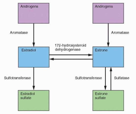
and its 3-hydroxymetabolites increase the conversion of estrone back to estrone sulfate by increasing the activity of sulfotransferase.297 Tibolone and all three metabolites inhibit the conversion of estrone to estradiol by 17β-hydroxysteroid dehydrogenase.294 Although these effects resemble progestin activity, tibolone is more potent. Tibolone increases aromatase activity in stromal cells but only at high concentrations that are beyond in vivo levels.295 These tibolone-induced enzyme changes would lower the active estrogen concentrations in breast tissue.


image

In the rat and mouse breast cancer models (cancer induced by 7,12-dimethylbena{a} anthracene, DMBA), tibolone exerts protective effects to the same degree as tamoxifen.298 However, tibolone is not antiestrogenic and does not inhibit aromatase. Therefore, the mechanism is explained by the enzyme effects summarized previously, inhibition of sulfatase and 17β-hydroxysteroid dehydrogenase and stimulation of sulfotransferase to increase the production of inactive sulfates.295 In addition, tibolone increases cellular differentiation and stimulates apoptosis, at least with normal breast cells in vitro.299 An increase in apoptosis is an action of the parent tibolone and its δ-4 isomer. Thus, tibolone acts like progestins and weak androgens in breast cell line studies examining proliferation, differentiation, and apoptosis.

Tibolone and its metabolites do not display the same activity directed toward the sulfatase enzyme in all tissues. Strong inhibition of sulfatase is a major feature in breast cells, but tibolone and its metabolites inhibit sulfatase only moderately in the endometrium (contributing to an antiestrogenic action) and provide no inhibition in bone (allowing a greater estrogenic impact).300

Postmenopausal hormone therapy increases breast density on mammography in about 10-20% of estrogen users and about 20-35% of estrogen-progestin users, an effect that occurs within the first months of treatment. In contrast, tibolone does not increase breast density and causes far less mastalgia than that seen with estrogen treatment.225,237,253,283,301,302,303,304 and 305 It is logical to conclude that these favorable responses are a consequence of the tibolone effects on the breast tissue enzymes involved in local estrogen production.

The Livial Intervention following Breast cancer: Efficacy, Recurrence, And Tolerability Endpoints (LIBERATE) trial was a multinational, placebo-controlled, randomized study of women with vasomotor symptoms who had had breast cancer surgically treated within
the previous 5 years.306 The study was designed to demonstrate that tibolone was superior to placebo, but when the drug monitoring safety board notified the sponsor that there appeared to be an excess of breast cancers in the treated group, the trial was canceled July 31, 2007, 5 months before its scheduled end. The median duration of participation and treatment was about 3 years, with a wide range from a few weeks to almost 5 years. The participants used a variety of adjuvant treatments, mostly tamoxifen, 66.8%; 6.5% used aromatase inhibitors. The dose of tibolone was 2.5 mg daily. Final numbers for analysis were 1,556 women in the treated group and 1,542 in the placebo group. The women ranged in age from under 40 to 79, with a mean age of 52.7 years. 57.8% had positive lymph nodes and 70% had a tumor stage of IIA or higher. Estrogen receptor status was known in 2,808 women in whom the tumors were estrogen receptor positive in 77.8%. In the intent-to-treat analysis, the hazard ratio for recurrent breast cancer in the tibolone-treated women was 1.40 (CI=1.14-1.70). The absolute risk for tibolone was 51 cancers per 1,000 women per year, and 36 in the placebo group. The increase occurred only in women with estrogen receptor-positive tumors. There was no difference in mortality rates between the two groups during the 5-year study period. There were no differences in cardiovascular events or gynecologic cancers, and not surprisingly, vasomotor symptoms, quality of life measures, and bone density improved with tibolone treatment.

How do the LIBERATE results that indicate an estrogenic action of tibolone in breast cancer survivors jibe with the literature indicating that tibolone exerts a non-estrogenic effect on breast tissue? Indeed, it was realistic to expect tibolone to have a salutary effect on the breast. It is well documented that the breast responds to tibolone with less stimulation compared with estrogen, judged by changes in mammographic breast density and the characteristics of tissue obtained by fine-needle aspiration. In the LIFT clinical trial (discussed under bone) that had vertebral fractures as the primary endpoint and breast cancer as a secondary endpoint, the risk of breast cancer after 3 years was significantly 68% reduced with tibolone treatment, although the dose was lower, 1.25 mg daily.307

The previous literature documenting beneficial actions of tibolone on the breast reflected, however, the impact of tibolone on normal breast tissue, and tibolone’s activity to lower local bioactive estrogen levels in target tissues might be lost in cancer cells. The contrary results in the LIFT trial could reflect its older population of women at high risk for fractures, a population that also differed by having lower body weights, no history of tamoxifen treatment, and lower risk factors for breast cancer.

Although the LIBERATE trial may apply to all breast cancer survivors, speaking strictly in a scientific sense, the results were derived mainly from tamoxifen users with 10-fold fewer users of aromatase inhibitors. The possibility that estrogen or tibolone would interfere with the beneficial effects of tamoxifen or aromatase inhibitors has always been one of the objections to treating breast cancer survivors with estrogenic hormones. In a subgroup analysis of the LIBERATE trial, the group of women who had used aromatase inhibitors had a greater risk of recurrent breast cancer compared with tamoxifen; however, the confidence interval was wide because of relatively small numbers. Possibly the estrogenic effect of tibolone would be more pronounced on an occult breast cancer in estrogen-depleted tissue compared with tissue where tamoxifen was bound to the estrogen receptor and prevented estrogenic stimulation. We don’t know if the LIBERATE data are meaningful for future treatment regimens. Nevertheless, until there are new data, the use of tibolone in women with a history of breast cancer remains relatively contraindicated.


The Effect of Tibolone on Bone

Tibolone prevents bone loss in postmenopausal women as effectively as estrogen or estrogen-progestin therapy.235,253,308,309,310,311 and 312 The beneficial impact on bone can be attributed to the
estrogenic metabolites acting through the estrogen receptor because it is blocked by an antiestrogen but not by an antiandrogen or an antiprogestin.313 In a large, U.S. dose-response study with doses ranging from 0.3 to 2.5 mg daily, only the 1.25 and 2.5-mg doses produced progressive bone density increases in the femoral neck.253 Indeed, the impact on bone was essentially the same for the 2 highest doses, 1.25 and 2.5 mg. Although the 1.25-mg dose is acceptable for the prevention of bone loss, the 2.5-mg dose is more effective for the alleviation of hot flushes.228 Tibolone prevents the bone loss associated with GnRH agonist treatment (and the side effect of hot flushing).290,314

The LIFT study (Long-term Intervention on Fractures with Tibolone) was a randomized, placebo-controlled multicenter trial in 22 countries of tibolone, 1.25 mg, given daily over 3 years.307 The 4,538 women who participated in the trial were age 60 to 85, all at high risk of fractures because of osteoporosis, and all treated with calcium and vitamin D supplementation. The study was stopped in February 2006 after a mean treatment of 34 months because of an increased risk of stroke. The risks of all events were assessed after 5 years of follow-up. Tibolone treatment reduced the number of vertebral fractures by about 45%, and nonvertebral fractures by about 25%. The reduction of fractures was about 4 times as great in women who already had a vertebral fracture upon entry to the study compared with women who had not had a fracture at baseline. It is noteworthy that the number of falls in the treated group was 25% less.

Based on previous bone density studies, the results of the LIFT trial on fracture reduction were not unexpected. The magnitude of the effect is roughly comparable to those with estrogen, bisphosphonates, and raloxifene (with the important exception being a lack of effect of raloxifene on hip fractures). The reduction of breast cancer was comparable to that reported with tamoxifen and raloxifene, but this was not a primary endpoint of the study. Although the difference was not statistically significant, there were four cases of endometrial cancer in the tibolone group and none in the placebo group.

The reported 2-fold increase in stroke in the LIFT trial was greater in the oldest women (over age 70), similar to that observed with estrogen. It is best to avoid the use of tibolone in elderly women and in women who are at risk for stroke (specifically those with hypertension, smoking, diabetes, or atrial fibrillation).


Treatment Options for Hot Flushes

The treatment of choice for vasomotor symptoms is hormone therapy. However there exists a substantial number of women who either cannot or will not accept hormone therapy. The choices other than hormone therapy that have been available in the past offered only a modest benefit. Transdermal clonidine, applied with the 100-μg dose once weekly was a common choice, but the reduction in hot flushing was only slightly better than that obtained with placebo treatment.317,318 Clonidine, bromocriptine, and naloxone given orally are only partially effective for the relief of hot flushes and require high doses with a high rate of side effects such as drowsiness and dry mouth. Bellergal treatment (a combination of belladonna alkaloids, ergotamine tartrate, and phenobarbital) is slightly better than a placebo and a potent sedative in the short-term; however, one study documented a similar response with bellergal and placebo after 8 weeks.319,320 Veralipride, a dopamine antagonist that is active in the hypothalamus, is relatively effective in inhibiting flushing at a dose of 100 mg daily, but is associated with major side effects, including mastodynia and galactorrhea.321,322 and 323 Medroxyprogesterone acetate (10-20 mg daily) and megestrol acetate (20 mg daily) are also effective (as effective as estrogen), but concerns regarding exogenous steroids, especially in patients who have had breast cancer, would apply to progestins as well.324,325 and 326 Vitamin E, 800 IU daily, is barely more effective than placebo.327 Dong quai, ginseng, black cohosh, isoflavones (including soy protein), yoga, and acupuncture all have little clinical difference compared with placebo treatment.328,329,330,331,332,333,334,335,336,337 and 338

In the last few years, selective serotonin reuptake inhibitors (SSRIs) have gained a reputation for significant efficacy in treating hot flushing. The drugs that have been studied include citalopram (Celexa), fluoxetine (Prozac), sertraline (Zoloft), paroxetine (Paxil), and serotonin and norepinephrine reuptake inhibitors, venlafaxine (Effexor) and desvenlafaxine succinate (Pristiq). In addition, an antiseizure medication, gabapentin (Neurontin), has been demonstrated to reduce vasomotor symptoms.


Drugs For Hot Flushing-Randomized Clinical Trial Results


Drug


Dose


Reduction in Flushing


Citalopram (Celexa) placebo


20 mg/d


50%, same as placebo


Fluoxetine (Prozac)


20 mg/d


50%, same as placebo


Sertraline (Zoloft)


50,100 mg/d


40%, same as placebo


Paroxetine (Paxil)


12.5 mg/d


62%


25 mg/d


65%


Venlafaxine (Effexor)


37.5 mg/d


37%


75 mg/d


61%


150 mg/d


61%


Desvenlafaxine succinate (Pristiq)


100 mg/d


64%


Gabapentin (Neurontin)


900 mg/d


50%


Pregabalin (Lyrica)


150 mg/d


65%


In the study with paroxetine (the controlled release product), 61% of the treated group (a general population of postmenopausal women with only 12 individuals who were breast cancer survivors) at the end of the study had at least a 50% reduction in frequency and severity of flushing, an effect that was about 2.5 times better than placebo with the higher dose.339 Venlafaxine was studied in breast cancer survivors; although the optimal dose was 75 mg, an appreciable response with 37.5 mg indicated that it would be worthwhile to begin treatment with the lower dose.340,341 The response was very rapid, within days, and therefore the dose can be increased in 1-2 weeks. The main side effects were mouth dryness, anorexia, nausea, and constipation. The efficacy of venlafaxine was demonstrated to be the same in women taking or not taking tamoxifen. Desvenlafaxine succinate is a metabolite of venlafaxine and equally effective as the parent compound.342,343 The effects of citalopram, fluoxetine and sertraline are no more effective than placebo, about a 50% reduction in short-term trials.344,345,346,347 and 348

Gabapentin (Neurontin) is a γ-aminobutyric acid analogue that has been used for seizures since 1994. It is also effective for migraine headaches, tremors, and panic disorder. In a gabapentin clinical trial, 67% of the treated women experienced more than a 50% reduction in flushes at week 12, compared with 38% in the placebo group.349 The most common side effects were somnolence (20%) and dizziness (13%). Peripheral edema occurs occasionally because of an induced decrease in serum protein. The potency of this agent appears to be more modest than the SSRIs in a dose of 900 mg/day.350,351 At higher doses, gabapentin was as effective as estrogen (about a 70% reduction in flushing); however, side effects are common at higher doses.352

Pregabalin (Lyrica), a more potent form of gabapentin, is an anticonvulsant drug that has been used in doses of 150-300 mg daily to treat anxiety and neuropathic pain, e.g., diabetic neurogenic pain, pain after shingles, and fibromyalgia. Side effects include dizziness, drowsiness, visual disturbances, tremor, weight gain, and a decrease in libido. In a phase III randomized trial, a dose of 75 mg b.i.d. (the recommended dose because of more side effects with higher doses), hot flushing was reduced by 65% after 6 weeks of treatment, an impact that was only 15% greater than placebo.353 Although the data are limited to this short-term, small clinical trial, the effect of pregabalin appears to be comparable to gabapentin.

The SSRIs are the best choice after hormone therapy, although the reduction in hot flushing is considerably less than what can be achieved with estrogen treatment. It is worth trying to titer the dose down to its lowest effective level because of a bothersome incidence of decreased libido. In addition, clinical experience indicates that it is best to slowly titrate upward to the recommended dose and, likewise, to wean the patient slowly when discontinuing treatment. SSRIs are effective for flushing secondary to both tamoxifen and hypoestrogenemia, and the efficacy is similar in women with and without breast cancer.354 An added advantage of the SSRIs is the fact that the clinical studies have also reported improvements in depression, anxiety, and sleep.

Tamoxifen is converted to an active metabolite by enzymes that are inhibited by certain SSRIs. Paroxetine coadministration decreases plasma concentrations of the active tamoxifen metabolite.355,356 A lesser effect is associated with fluoxetine and sertraline. In a retrospective cohort study, only paroxetine use during tamoxifen therapy was associated with an increased risk of death due to breast cancer.357 Paroxetine, fluoxetine, and sertraline are best avoided in women being treated with tamoxifen.


Bioidentical Hormones

The adverse publicity following the publications from the Women’s Health Initiative was a multibillion dollar bonanza for compounding pharmacies providing postmenopausal
hormones. Bioidentical hormones are now the focus of a political, financial, and legal conflict. Bruce Patsner, research professor in the Health Law & Policy Institute at the University of Houston Law Center, has written what is, in our view, a masterful analysis of the problem, with suggestions for its resolution.358


The History of the Conflict

The operations of a pharmacy are regulated in the individual states by state boards of pharmacy, in a system similar to the regulation of medical practice. The first federal law regulating drugs, the Federal Food, Drug and Cosmetic Act, was passed in 1938, at a time when most drugs were compounded according to a doctor’s prescription. The American Pharmaceutical Association defines pharmacy compounding as the preparation of a prescription drug that is “individualized” to the needs of the patient. This changed after World War II with the development and growth of the pharmaceutical industry. The Kefauver-Harris Amendment in 1964 extended the role of the Food and Drug Administration (FDA) to include safety and efficacy.

In the 1990s, the FDA began to regard the drugs coming from compounding pharmacies as falling under the “new drug” regulations, and therefore, the FDA had jurisdiction over the marketing and promotion of those drugs. The pharmacy world was immediately challenged; there was no way that an individual pharmacy could carry out the kind of clinical studies required for the approval of new drugs. Thus, the pharmacists immediately realized that all compounded drugs would be illegal. At the same time, the FDA was a bit ambivalent, acknowledging that there were examples where the individual needs of a patient required the compounding of a drug, e.g., the creation of a liquid preparation when none was available. This was before compounding took to the Internet for marketing and promotion.

In 1992, the FDA issued its Compliance Policy Guide on Compounding, reserving a right for “selective enforcement,” as a compromise between believing it was correct in assuming that compounded drugs represented new drugs and admitting that some patients required compounding. The pharmacy profession immediately rejected the idea that the FDA had any regulatory jurisdiction over pharmacies. The 1997 Food and Drug Modernization Act attempted to clarify the situation. An amendment was added to the existing laws stating that compounded drugs were not “new drugs,” but at the same time the 1997 act prohibited the marketing of compounded drugs.

The pharmacy profession sued the FDA, arguing that a restriction on advertising and promotion of compounded drugs was unconstitutional, a restriction of free speech. The District Court ruled against the FDA in 1999. The FDA appealed and lost again in the 9th Circuit Court of Appeals, which further invalidated the entire 1997 act. In 2002, the U.S. Supreme Court upheld the Circuit Court decision.

The FDA issued a new Compliance Policy Guide in 2002, stating that selective enforcement would hinge on 3 major factors: (1). A potential adverse effect of a drug, (2). Whether drugs were compounded from non-FDA-approved components, and (3). Whether compounded drugs were similar to drugs already removed from the market for safety reasons. At this point, the FDA affirmed that it did not want to infringe on the traditional practice of compounding, the preparation of a drug according to a doctor’s prescription to fit an individual patient’s requirements.

Wyeth Pharmaceuticals filed a Citizen Petition with the FDA in October 2005 requesting that the FDA take action against several compounding pharmacies that were primarily
Internet-based. The Petition’s major allegation was that these pharmacies were essentially manufacturing new drugs and should be subject to new drug regulations. On January 9, 2008, the FDA announced it would take action against seven pharmacies providing prescription bioidentical hormones, and issued warning letters that potentially could be followed by seizures of drugs and injunctions against production.

The FDA would like to regard compounded drugs as “new drugs,” but the legal precedent has now been set by the courts: compounded drugs are not “new drugs.” This was reaffirmed in a 2006 decision in the U.S. District Court for Texas. The FDA would further like to regard the giant compounding pharmacies, especially those operating over the Internet, as manufacturers, like pharmaceutical companies, but again, the court decisions have prevented the FDA from requiring compounding pharmacies to meet “new drug” standards. The “new drug” argument doesn’t work.

“Bioidentical” and “natural” are often used in concert. Strictly defined, the hormones must be precisely the same as normal endogenous estrone, estradiol, and estriol, the three endogenous estrogens; progesterone, the progestational agent synthesized by the ovarian corpus luteum after ovulation, and testosterone and dehydroepiandrosterone, androgens made by the human body. To argue that products are not “artificial,” begs the issue because even if the source is a steroid molecule derived from plants, chemical and manufacturing processing is still required. These terms obviously have marketing value, and the terms are used to imply greater safety, even greater efficacy. The situation is further compounded (pun intended) because it is likely most patients assume that the marketed bioidentical and natural hormones have been demonstrated by appropriate studies to be effective and safe. Of course this is not the case, although it seems like an obvious conclusion to view product A and product B to be the same if they are the same molecule. The problem is that compounding pharmacies are not required to compare the formulation with the performance of an approved product, nor is there any way for a patient to be assured that the dosage is correct (that the drug contains what it is supposed to contain).

The large compounding pharmacies meet the “individualization” requirement that usually comes from a clinician-patient interaction, by promoting salivary measurements of hormones, as interpreted by one of their employed clinicians to produce tailored hormone choices and doses. Assessment of this approach by researchers, as well as organizations such as the American College of Obstetricians and Gynecologists and the Endocrine Society, have concluded that the variations in salivary sex steroid levels from individual to individual and from specimen to specimen preclude clinical interpretation.359,360 and 361 For most patients, laboratory testing is not necessary in hormone decision-making.


A Better Approach

The American Pharmacists Association and the National Association of Boards of Pharmacies define compounding as the steps required in order to provide a drug in response to a clinician’s prescription according to an individual patient’s needs, and the preparation of drugs in anticipation of a demand. Therefore, there are three people involved: the patient, the clinician, and the pharmacist. This is in contrast to the production of large amounts of a drug for a national market of unknown users. The American Pharmacists Association further says that if an FDA-approved product is commercially available that meets a patient’s needs, it should be the drug provided.

The key to the position of the pharmacists is the contention that there are circumstances, decided by the patient that makes the use of commercial products not a good choice. This seems reasonable, but it is also reasonable that this decision requires the involvement of the
clinician because ultimately a prescription is still required. The traditional view of compounding, therefore, is one of personal relationships with patient, clinician, and pharmacists. This becomes a totally different story when a large Internet pharmacy responds to thousands of prescriptions with no knowledge of the patients. What happened to the “individualization” aspect of compounding? Is it practicing medicine by the pharmacy to have a registered clinician employed by the pharmacy to interpret hormone levels and adjust doses?

Clinicians are appropriately frustrated by the claims made that bioidentical compounded drugs have greater efficacy and safety. Bruce Patsner argues that it should be accepted that bioidentical drugs from the big compounding pharmacies do not meet the definition of compounding supported by the pharmacist’s own organization, the American Pharmacists Association—a personal relationship of patient, clinician, and pharmacist addressing an individual’s needs.358 The FDA can argue that the big operations are not legitimate compounding, but big commercial operations directed to unknown consumers.

Patsner also argues that the most vulnerable point is the false safety and efficacy claims. The contention should not be that the safety and efficacy claims are inaccurate, because the pharmacies can always compose their words to avoid legal assaults. The point of emphasis should go back to the pharmacist’s published credo: if a commercial, approved product is available to meet the patient’s needs, a compounded product is not indicated. Replacing a commercial product with a “natural,” untested, unregulated product is not the same thing as prescribing a compounded product when no commercial product will meet the individual’s needs.

Pastner summarizes his argument by saying that the large compounding pharmacies are not true compounders because they advertise and promote their products as replacements for commercially available, approved and tested drugs, and that the attempt at “individualization” uses an unsubstantiated method that marginalizes clinicians.358

The bioidentical hormones and the various commercial female hormone products are produced by pharmaceutical companies using similar methods that start from the same raw material, usually soy or yams. Some of the commercial products available consist of estradiol, testosterone, and progesterone, the exact same steroid drugs provided by compounding pharmacies. A major difference between the commercial products and products from compounding pharmacies is the important fact that commercial products are federally regulated and tested for purity and potency; compounding pharmacies have not been regulated in this fashion.

The compounded estrogen formulations that contain combinations of estrone, estriol, and estradiol contain sufficient estradiol to produce the same biologic effects associated with commercial preparations. There are no clinical studies documenting that these combinations confer better results or safety. Whether the presence of estriol reduces the risk of breast cancer has not been tested in appropriate clinical studies. Case-control data indicate that estriol used without a progestational agent increases the risk of endometrial cancer, thus its biologic behavior is similar to that of other estrogens.362

Custom-compounded formulations have not been proven to be safer or better, and should be regarded as having similar risks and benefits as commercial products. Given bioequivalent doses of various estrogens and progestational agents, one should expect the same biologic results. Claims for custom-compounded products have not been scientifically tested. For these reasons, unless well-designed studies document differences for a specific product, the same risks and benefits apply to equivalent doses of all products.

Salivary sex steroid levels vary widely between individuals, and from measurement to measurement within the same individual. Most importantly, the appropriate clinical studies to document correlation of salivary hormone levels with clinical state or responses have not been performed. Tailor-making a hormone therapy regimen according to
salivary testing is an appealing idea that has not been addressed scientifically, and given the variability in salivary hormone levels, it is unlikely that clinical studies would yield useful information.

Patients who wish to use products similar to endogenous hormones should be made aware of the content of available commercial formulations. Books and pharmacies promoting their own products should be viewed with caution; don’t confuse marketing with science.


“Natural” (Alternative) Therapies

The business of selling alternative therapies is now a worldwide phenomenon. The promotion of many of these treatments relies on a network of alternative providers, authors, and compounding pharmacies. Why are herbs and botanicals not regulated? In the U.S., the Dietary Supplement Health and Education Act of 1994 deregulated the industry by classifying dietary supplements as neither foods nor drugs. Thus, manufacturers of dietary supplements are not required to demonstrate that they are safe or effective. In addition to a lack of regulation, there are many other problems associated with herbs and botanicals. The products vary in the amount and purity of active ingredients; indeed, products on the shelf often are adulterated and contaminated with drugs or metals.363 And very importantly, there is enormous variation in the plants themselves because of genetic, harvest year, and processing differences and in individual metabolism of the products.


Phytoestrogens

“Phytoestrogens” is a descriptive term applied to nonsteroidal compounds that have estrogenic activity or are metabolized into compounds with estrogen activity. Phytoestrogens are classified into three groups: isoflavones, lignans, and coumestans.364,365 They are present in about 300 plants, especially legumes, and bind to the estrogen receptor. Soybeans, a rich source of phytoestrogens, contain isoflavones, the most common form of phytoestrogens, mainly genistein and daidzein, and a little glycitin.

PHYTOESTROGENS


  • Isoflavones (Genistein, Daidzein, Glycitin) soybeans, lentils, chickpeas (garbanzo beans)


  • Lignans

    flaxseed, cereals, vegetables, fruits


  • Coumestans

    sunflower seeds, bean sprouts

Isoflavones exist in plants bound as glycoside conjugates attached at the 3 position, called glycones. The carbohydrate component requires gut bacteria to remove the sugar moiety to produce active compounds, the aglycones. Individual variability in gastrointestinal microflora, as well as absorption, influences the bioavailability of isoflavones. Biochanin and formononetin are methylated precursors that are metabolized to genistein and daidzein. Red clover and lentils contain significant amounts of these precursors. The isoflavones are in the active, deconjugated forms in fermented soy foods like miso and tempeh. The concentration of isoflavones in tofu is highly variable.


The phytoestrogens are characterized by mixed estrogenic and antiestrogenic actions, depending on the target tissue and local estrogen concentration. Variations in activity may also be due to the fact that the soy phytoestrogens have a greater affinity for the estrogen receptor-β compared with estrogen receptor-α, although the affinity for the beta receptor is still only 35% that of estradiol.366 Despite a low affinity for the alpha receptor, circulating levels many times that of steroidal estrogens produce the potential for biologic activity.

You can eat soybeans every day and never see a bean. Soybeans are defatted to produce soy flour. Soy flour is then prepared to remove the carbohydrates. 95% of soy flour is toasted and used as animal feed. Alcohol washing is used to get a taste-free product, but alcohol extraction removes the phytoestrogens.367 SUPRO, known as “isolated soy protein,” from Protein Technologies International, the major supplier for commercial products and research, is extracted by aqueous washing and retains the isoflavones.

The reason why most of the soybean crop is devoted to animal feed is because what is left after removing lipids is totally bland. The solution is to mix soybeans with other foods, e.g., beans and soups. Unfortunately they require standing in water for about 12 h and simmering for 2-3 h to be cooked. The average Japanese intake of isoflavones is about 50 mg/day.368 The rest of Asia has an average consumption of about 25-45 mg/day, and Western consumption is less than 5 mg/day.369,370


Alternative Therapies for Flushing

A belief that Asian women report fewer menopausal symptoms has been an underlying force in the promotion of isoflavones. However, this apparent difference in the prevalence of symptoms comparing Asia and the West may reflect cultural differences and not actual experience. An Italian study found a 45% reduction in flushing with 60 g of isolated soy protein daily (76 mg isoflavones), compared with a 30% reduction in the placebo group.371 Two other studies, both with 50 mg/day of isoflavones, found a similar 15% reduction in the number of flushes compared with placebo.372,373 Another placebo-controlled short-term trial found a greater reduction in flushes with 70 mg isoflavones daily.374 In a randomized, crossover study of a high dose of isoflavones, 150 mg/day, for flushes in breast cancer survivors, the treated group and the placebo group demonstrated equal effects.331 The dose of 150 mg isoflavones per day was similar to three glasses of soy milk daily. Two Italian randomized trials found the same response to placebo and 72 or 80 mg/day isoflavones.375, 376 An Australian study randomized women to 118 mg/day isoflavones or placebo and could detect no difference after 3 months in hot flushing, libido, vaginal dryness, or any of a long list of symptoms, and a Finnish randomized trial using 114 mg isoflavones found no effects on the vagina or on menopausal symptoms.377,378 In a randomized study in Iowa, no differences were found in hot flush frequency comparing isoflavone-rich soy protein to a whey protein control.332 And finally, another randomized trial of breast cancer survivors found no difference comparing placebo with 90 mg isoflavones daily.333


Red Clover

Promensil is an extract of red clover (Trifolium pratense) containing formononetin, biochanin, daidzein, and genistein. Formononetin and biochanin are metabolized to daidzein and genistein, respectively. Red clover is a legume used to enrich nitrogen levels in soils. Promensil is produced by Novogen in Australia and marketed by Solvay in the U.S. A 500 mg tablet contains 200-230 mg of dried extract, which contains 40 mg of isoflavones. Two randomized, placebo-controlled studies of the effect of Promensil on hot flushes were
reported in 1999.379,380 Neither demonstrated a significant difference compared with the placebo group. In 1 of the reports, 4 times the recommended dose (4 tablets daily) also had no effect.380 On the other hand, an appropriately designed Dutch study, 2 tablets daily, detected a significant reduction of flushing in a 12-week period of time.381 A large placebo-controlled trial randomized 252 women with severe hot flushing to either Promensil (2 tablets daily) or another red clover extract Rimostil (2 tablets daily for an intake of 57 mg isoflavones).382 The quantitative reduction in flushing (about 41% in 12 weeks) was identical in the Promensil, Rimostil, and placebo groups, although Promensil had a very slightly faster response. In another randomized clinical trial, the effect of red clover (a daily intake of 128 mg isoflavones) on vasomotor symptoms was no better than placebo treatment.383 The best evidence indicates that the impact of red clover on vasomotor symptoms is the same as placebo treatment.

Why do these randomized, blinded, and placebo-controlled trials lack agreement? One reasonable explanation is that isoflavones have a mild impact on hot flushing, detectable only in women with frequent and severe flushing. A major clinical response should not be expected. Another possibility is the role of equol (see later discussion).


Other Alternative Treatments

One randomized, placebo-controlled trial examined the effect of dong quai on hot flushing.328 No estrogenic effects could be detected on flushing, endometrium, or vagina. Ginseng has the same impact on menopausal symptoms as placebo treatment.329 Similarly, vitamin E supplementation is ineffective for hot flushing.327


Evening Primrose

Evening primrose is often recommended for mastalgia, premenstrual syndrome, and menopausal symptoms. Evening primrose oil is extracted from the seed of the evening primrose; it provides linoleic and gamma-linoleic acids (precursors of prostaglandin E). Appropriately blinded and controlled studies have failed to find any differences comparing primrose oil with placebo.384,385 and 386


Black Cohosh

Black cohosh (Cimicifuga racemosa) is also called black snakeroot and bugbane. “Remifemin” is commercially available as an alcoholic extract of the root. A tablet contains 2 mg; the dose is 2 tablets b.i.d. or 40 drops of liquid extract b.i.d. Black cohosh has been heavily promoted, especially by German clinicians, for the treatment of menopausal hot flushes. Keep in mind that the study of hot flushing requires randomization to placebo treatment because placebo treatment is associated with an average 51% reduction in hot flush frequency.387 Unfortunately, most of the early reports supporting the efficacy of black cohosh were case series or studies without placebo control groups or the studies did not directly and quantitatively measure hot flushing. Black cohosh has been reported to contain formononetin, a methylated precursor that is metabolized to the two primary phytoestrogens, genistein and daidzein. More sophisticated analysis, however, using liquid chromatography methods, has failed to detect the presence of formononetin in various black cohosh preparations, nor in black cohosh roots and rhizomes.388


An older clinical trial was noteworthy and alone in finding a similar impact on hot flushing with black cohosh and placebo treatment.330 Well-designed trials are confirming that early study and providing us with a uniform story. The Herbal Alternatives for Menopause (HALT) Study is centered in Seattle, Washington. This double-blind trial randomized 351 women to placebo or one of four treatment groups: (1) black cohosh 160 mg daily (note the higher dose); (2) a multibotanical treatment containing 50 mg black cohosh, alfalfa, chaste tree, dong quae, false unicorn, licorice, oats, pomegranate, and Siberian ginseng, 4 capsules daily; (3) the multibotanical plus counseling to increase dietary soy intake; (4) conjugated estrogens 0.625 mg with or without 2.5 mg medroxyprogesterone acetate.389 After 1 year, no differences were observed in hot flushing comparing any of the three herbal treatment groups to placebo.389 The herbal remedies also had no effect on sleep quality as reported after 3 months.390

A randomized trial in Chicago compared black cohosh, 128 mg, and red clover, 120 mg, to standard hormone therapy and placebo treatment.383 Over a period of 1 year, only hormone therapy reduced vasomotor symptoms greater than placebo. In this same clinical trial, neither black cohosh nor red clover had an impact on measures of cognition.391 A Mayo Clinic study reported the results of a double-blind, randomized, cross-over clinical trial to assess the efficacy of black cohosh for the treatment of menopausal hot flushes.334 The dose was 20 mg b.i.d., the dose of the most commonly marketed black cohosh product in the U.S. The similarity of the studied product with Remifemin was confirmed by high performance liquid chromatography and proton nuclear magnetic resonance analysis. 132 patients were treated for two 4-week crossover periods. Black cohosh reduced hot flushing scores by 20% in the fourth treatment week compared with 27% in the placebo group; and frequency was reduced 17% on black cohosh and 26% on placebo. A randomized trial in Australia found no difference between placebo and a combination of black cohosh with Chinese herbs.392

Black cohosh is not estrogenic, and black cohosh has no effect on menopausal symptoms.

An expert committee of the U.S. Pharmacopoeia concluded that black cohosh may be associated with hepatotoxicity; however, a European review of cases with hepatoxicity emphasized the difficulty in establishing a cause-effect relationship.393,394 Hepatoxicity remains a concern, awaiting the accumulation of definitive data.


Ginkgo Biloba

Ginkgo biloba is an extract prepared from the leaves of the G. biloba tree. It contains flavonoids and unique terpene lactones. Ginkgo biloba is a multimillion dollar herb sold in the U.S. for the preservation of memory. In vitro studies suggested that ginkgo had antioxidant (from the flavonoids) and anti-amyloid (from the lactones) effects. Indeed, the biologic studies provided a rationale for the use of ginkgo to prevent dementia.


A randomized, double-blind, placebo-controlled trial comparing Ginkgo biloba with placebo for the prevention of dementia enrolled 3,069 elderly individuals (over age 75) in five academic centers in the U.S.395,396 The participants were randomized to b.i.d. doses of 120 mg ginkgo or placebo (45% female in the ginkgo group and 47% female in the placebo group). The ginkgo formulation and dosage were that used in many of the brands sold in the U.S. The dementia rate steadily increased in both groups over a 7-year period of follow-up, accumulating 277 cases (17.9%) in the treatment group and 246 cases (16.1%) in the placebo group. The rate of dementia did not differ between the two groups, nor did the rate of Alzheimer’s disease. In addition, treatment with Ginkgo biloba did not produce less cognitive decline in either adults with normal cognition or with mild cognitive impairment. Other randomized trials have failed to demonstrate any beneficial effects on Alzheimer’s, learning, memory, attention, verbal fluency, or concentration.397,398

The American trial robustly demonstrated that Ginkgo biloba in the tested and commonly used dose did not delay the onset of dementia or cognitive decline.395,396 The concept of “delay” is important. A treatment that could delay the onset of dementia by 5 years would reduce the number of dementia cases by 50%. In fact, this clinical trial found a statistically significant increase in the risk for developing dementia with ginkgo treatment in the 25% of participants who had cardiovascular disease prior to enrollment. However, the authors appropriately urged caution in interpreting this subgroup analysis. A Cochrane review in 2007 of 35 clinical trials with 4,247 participants concluded that there was no convincing evidence that ginkgo treatment benefited individuals who already had dementia or cognitive impairment.399


St. John’s Wort

St. John’s wort has been reported to be comparable to tricyclic antidepressants in treating mild to moderate depression, based on eight appropriate trials.400 This is the conclusion of two meta-analyses.401,402 All studies were short-term, about 4-6 weeks in duration, and with small numbers. The treatment consisted of a 300-mg plant extract in tablet form, administered t.i.d. However, 2 large, American 8-week trials found no difference between treatment and placebo.403,404

The U.S. Food and Drug Administration (FDA) issued an alert in February 2000 that St. John’s wort may interact with drugs known to be metabolized by the cytochrome P450 pathway: theophylline, digoxin, immune suppressants, and oral contraceptives.405 St. John’s wort activates an orphan receptor that induces the expression of metabolic enzymes.406 In clinically depressed individuals being treated with prescription antidepressants, manic reactions can result (the central serotonergic syndrome).


Phytoestrogens to Prevent Cardiovascular Disease

The cardiovascular story with phytoestrogens received a large boost in 1995, when a metaanalysis concluded that an intake of an average of 47 g soy protein/day lowered total cholesterol and LDL-cholesterol.407 This was supported by studies in the monkey indicating that isoflavone increased HDL-cholesterol, enhanced vasodilation, and decreased atherosclerosis.408

Only intact soy protein has a beneficial effect on lipids. Separation of the protein component from dietary soy protein loses the effect. This effect depends on the inhibition of cholesterol absorption by the non-isoflavone protein.409,410 The mechanism involves upregulation of the LDL-cholesterol receptor and catabolism of LDL-cholesterol, leading
to an increase in bile excretion. The soy peptide binds bile acids and prevents resorption. Alcohol extraction removes the isoflavones from soy protein and causes a loss of the beneficial effect on atherosclerosis in monkeys.411 Thus, both the isoflavone portion and the protein component are required for a full cardiovascular effect. Non-alcohol-washed soy protein extract has been extensively studied in monkeys. This preparation lowers total cholesterol and LDL-cholesterol, and raises HDL-cholesterol,412,413 produces coronary artery vasodilation,414 inhibits reduction in coronary flow after collagen induced platelet aggregation and serotonin release,415 and inhibits atherosclerosis but not as robustly as estrogen.408,413

In women, soy protein reduces total and LDL-cholesterol and does not affect triglycerides or HDL-cholesterol; ethanol-extracted soy protein has no effect.416,417,418 and 419 The minimal dose is about 60 mg isoflavones daily, which is present in 25 g soy protein/day.420 LDL-cholesterol must be above 130 mg/dL in order to have an effect. Studies of healthy men and women could detect no effect of phytoestrogens (25 to 80 mg isoflavones per day) on lipids or brachial vasodilation.421,422 and 423 In a 12-week study of women with type 2 diabetes mellitus, dietary supplementation of 30 g soy protein (132 mg isoflavones) daily improved insulin resistance and glucose control in addition to lowering total cholesterol and LDL-cholesterol levels.424 In addition, soy intake prevents LDL-cholesterol oxidation in hyperlipidemic men and women even when circulating LDL-cholesterol levels are unaffected.425 Promensil, in a 10-week study, had no effect on lipids (it only contains isoflavones, no protein) but did improve arterial compliance.426

The U.S. FDA, in October 1999, authorized the use in food labeling of health claims related to the association between soy protein and reduced risk of coronary heart disease, “based on the totality of publicly available scientific evidence, soy protein included in a diet low in saturated fat and cholesterol may reduce the risk of CHD by lowering blood cholesterol levels.”427

Remember that both protein and isoflavones are needed for a cardiovascular effect. Isoflavones by themselves have no effect on lipids.373,426,428 Protein without isoflavones has no effect on vasodilation and atherosclerosis.411 The FDA has stated that there is insufficient evidence to allow them to exclude alcohol-washed products from the health claim, but it makes sense that a combined protein-isoflavone product is best. Even in older women with moderate hypercholesterolemia, a high intake of soy phytoestrogens (purified isoflavones without protein) had no effect on the lipid profile.429 And also remember, that there is no effect on the lipids in individuals who already have a normal profile. Even in individuals with high cholesterol levels, the beneficial impact of soy protein intake is modest and likely to have little clinical effect.

It will require appropriate clinical trials to determine how phytoestrogens compare in the cardiovascular system with estrogens and to determine the efficacy, safety, and correct dosage (studies thus far recommend a daily intake of 60 g soy protein). In addition, the intake of sufficient soy to produce a clinical response is not easy; intake is handicapped by gastrointestinal symptoms, a major alteration in diet or the use of an unpalatable supplement, and great variability in plant contents and products (due to processing). A dietary intake to match the isoflavone dose used in the studies on the lipid profile, for example, would require about 1 lb daily of tofu! In addition, individuals demonstrate great variability in absorption and metabolism. A user-friendly preparation must be developed that minimizes individual variability in response.


Phytoestrogens to Prevent Bone Loss

Phytoestrogens are effective in preventing bone loss in rats but not in monkeys.430,431 and 432 In women, most studies have demonstrated at best a slight effect on spinal bone but no effect on hip bone.30,417,433,434 One 3-year, randomized trial demonstrated no effect on bone loss
in the spine and femur, with perhaps a modest bone-sparing at the femoral neck with 120 mg/day of isoflavones after adjustment for age and body fat.435 A 1-year clinical trial could detect no impact of soy intake on bone mineral density in either equol producers or nonproducers.436 Flaxseed supplementation had no effect on biomarkers of bone metabolism.437 The difference between hip fracture incidence in Japanese and American women may be due to structural and/or genetic differences not dietary intake.438

Ipriflavone is a synthetic isoflavone; it is methylated dehyroxydaidzein, which is metabolized to daidzein. It was developed by Chiesi Pharmaceuticals in Italy. It is marketed in the U.S., and each tablet contains 150-mg ipriflavone combined with calcium (375 mg), vitamin D (187 IU), soy isoflavones (40 mg), and 3 mg boron. The Italian product is pure ipriflavone. The recommended dose is 600 mg/day, 2 tablets b.i.d. taken with meals. Studies with ipriflavone have demonstrated prevention of bone loss over a year.439,440,441 and 442 Overall the effect on bone is not as great as that observed with standard doses of estrogen or bisphosphonates, perhaps not great enough to yield a benefit. A 4-year randomized trial in Europe assessed the effect of ipriflavone on bone density, urinary markers, and vertebral fractures in 474 women and could find no difference in the treated group compared with the placebo group.443


Phytoestrogens and Cognition

Phytoestrogens up-regulate cognition markers and improve memory in rats equally when compared with estrogen444,445 There is one human study that is disturbing. Men, in a National Institutes of Health study that began in 1965, reported their tofu consumption.446 Cognition was tested in 1991-1993 when the men were age 71-93. Higher midlife tofu consumption (two or more servings per week) was associated with poor cognitive test performance, enlargement of ventricles, and low brain weight. Soy supplementation has been reported to improve measurements of memory and attention in postmenopausal women.447,448 On the other hand, randomized trials detected no effects of soy protein, red clover, or black cohosh on tests of memory, executive function, language, visual perception, cognition, or measures of quality of life.391,423,449,450


Phytoestrogens and the Breast

In the parts of the world where soy intake is high, there is a lower incidence of breast, endometrial, and prostate cancers.451 For example, a case-control study concluded that there was a 54% reduced risk of endometrial cancer, and another case-control study indicated a reduction in the risk of breast cancer in women with a high consumption of soy and other legumes.452,453 Daidzein and genistein urinary excretion are lower in Australian women who develop breast cancer.454 High soy and tofu consumption and high urinary excretion of isoflavones have been reported to be associated with a lower risk of breast cancer in Singapore, China, Australia, and even in American women consuming a diet rich in isoflavones.453,455,456,457,458 and 459 These studies have supported the belief that high phytoestrogen intake protects against breast cancer. It is by no means certain, however, that there is a direct effect of soy intake.460 Indeed, a 6-month study of the impact of administered soy protein on breast secretions in premenopausal and postmenopausal women revealed increased breast secretions with the appearance of hyperplastic epithelial cells.461 Epithelial hyperplasia based on cytology in breast secretions was demonstrated in 7 of 24 (29.2%) of the subjects. Swedish and English cohort studies could not detect a relationship between dietary phytoestrogens and the risk of breast cancer.462,463


Genistein increases epidermal growth factor in immature rat mammary tissue, and it has been hypothesized that earlier exposure to genistein promotes early cell differentiation leading to breast glands that are more resistant to the development of cancer.464 On the other hand, using the chemically induced rat breast cancer model, no evidence of isoflavone inhibition on tumor development has been detected.465 In the monkey, treated for 6 months, no proliferation was reported in either endometrium or mammary tissue.466,467

One hypothesis speculates that phytoestrogens protect the breast by decreasing exposure to the more potent endogenous estrogens. The evidence does not support this idea. Highdose treatment (100 mg of daidzein plus 100 mg genistein) does lower estradiol and dehydroepiandrosterone sulfate levels in premenopausal women and increases cycle length.468 However, these are extremely high doses. One study reported that treatment with Asian soy foods (approximately 32 mg isoflavones per day) was associated with a 9.3% significant decrease in luteal serum estradiol levels, but there were no other changes, including follicular-phase estradiol, progesterone levels, and sex hormone-binding globulin levels, or cycle length.469 Interestingly, the reduction in luteal estradiol was observed only in Asian participants in whom urinary excretion of isoflavones was higher than nonAsians.469 These same investigators reported that a high intake of the soy protein alone (with the isoflavones removed) reduced estradiol and progesterone levels throughout the cyle.470 Other studies have found no effects on estradiol, FSH, LH, or sex hormone-binding globulin in premenopausal women,471 and, most importantly, no effects on circulating hormones in postmenopausal women.472,473 The lack of an effect on gonadotropin and steroid levels is important, depriving the clinician of a method to assess dosage.

Catecholestrogens (2-hydroxy and 4-hydroxy estrogens) have long been proposed as a metabolite pathway that could be protective, or at least antiestrogenic. Hydroxylation in the 2 or 4 position produces inactive metabolites. In 1 study, 8 premenopausal women treated with a soy milk supplement increased their urinary excretion of 2-hydroxy estrone by an average of 47%.474 Another study could detect no change in 2-hydroxyestrogens.471 A study limited to Asian-American women also was unable to identify an impact of soy intake on overall estrogen metabolite excretion; however, an increase in catecholestrogens was observed with greater soy intake, balanced by a decrease in 16-hydroxylation.475

In response to soy, no significant increase in nipple aspirate levels of genistein and daidzein could be detected.476 However an indication of estrogenic stimulation occurred, as measured by pS2 (a protein up-regulated by estrogen) levels, but there was no evidence of an effect on epithelial cell proliferation, estrogen and progesterone receptors, apoptosis, or mitosis. Thus, no antiestrogenic effect could be detected, and at best there was a very weak estrogen effect. In another study, 48 women with normal breasts received a 60 g soy supplement for 14 days, and in these women lobular epithelial proliferation and progesterone receptor expression increased, an indication of estrogen stimulation.477 Some argue that the key to a beneficial impact on breast may be early exposure, and a sudden increase late in life of dietary phytoestrogens may be harmful. On the other hand, a Chinese cohort study of 5,042 breast cancer survivors documented a reduced risk of recurrence and death associated with increasing levels of soy intake, evident among women with either estrogen receptor-positive or receptor-negative disease and among either tamoxifen users or nonusers.478 In an American cohort of 1,954 breast cancer survivors, a 60% greater decrease in breast cancer recurrence was observed in postmenopausal women using tamoxifen comparing the highest level of soy intake with the lowest.479 Most of the evidence indicates that a high intake of phytoestrogens is associated with a reduced risk of breast cancer, including recurrence in breast cancer survivors. It is not known whether this effect is a marker for beneficial metabolic responses to phytoestrogens or whether there is a direct impact on breast tissue. The evidence also indicates that phytoestrogen consumption does not adversely interfere with tamoxifen’s mechanism of action.


The Role of Equol

Equol is a bacterial metabolite and the only hormonally active metabolite of the soy phytoestrogen, daidzein. It is one of the estrogenic compounds in pregnant mare’s urine, hence its name. At least in vitro, equol stimulates gene transcription with both estrogen receptors and with a greater potency than any other isoflavone.481 Equol formation is totally dependent on intestinal microflora; therefore, strictly speaking it is not a phytoestrogen. To be accurate, equol is a nonsteroidal estrogen, a member of the isoflavone family, and exclusively a metabolic product of intestinal bacteria.

The most important observation regarding equol is that 30-50% of adults do not produce equol, even when challenged with high doses of soy.482 This is in contrast to nonhuman primates and other animals; all that have been studied produce high levels of equol. Thus, there are two human populations: equol producers and equol nonproducers. The key question is whether equol producers receive greater clinical effects from phytoestrogens than nonequol producers. As noted, thus far the clinical effects of isoflavones on bone have not been impressive. In a 2-year randomized trial of postmenopausal women, isoflavone-rich soy milk increased spinal bone mass in the 45% of the subjects who were equol producers, with essentially no effect in equol nonproducers.483 More profound beneficial effects on the lipid profile have been reported in equol-producing women.482 Therefore, the population destined to receive a benefit from soy intake may be limited to equol producers. Studies need to be repeated measuring the responses in individuals who are identified as equol producers or equol nonproducers. If the population destined to receive a benefit from soy intake is limited to equol producers, a convenient, inexpensive method must be developed to identify equol production. It may be possible to convert nonproducers to producers.


An emerging approach is to administer equol itself. Daidzein yields two forms of equol, the R-equol inactive isomer and S-equol, the active isomer that binds to estrogen receptor-β. S-equol has been synthesized and its administration is effective for the treatment of menopausal symptoms.484 Another alternative is the S-equol supplement made by incubating equol-producing bacteria with soy isoflavones.485,486 S-equol also blocks the activity of dihydrotestosterone, and thus, it has potential to treat androgenic effects such as acne, hirsutism, male pattern baldness, and prostate cancer.487


Estriol

Interest in estriol can be traced to Lemon’s report in 1975 that estriol limited the growth of breast tumors in the chemical-induced rat tumor model.19 However, it is usually overlooked that estradiol worked equally well in that model. Estriol treatment of postmenopausal women has no overall effect on lipids and no effect in the prevention of myocardial infarction.488,489 Estriol, without concomitant progestin treatment, does increase the risk of endometrial cancer with the long-term oral use of 1-2 mg/day.362 At least two studies have been unable to demonstrate prevention of bone loss with the administration of 2 mg estriol daily.17,18 And one case-control study found no reduction in hip fractures with estriol compared with a lower risk with estradiol.489 There is no evidence indicating any beneficial effects unique to estriol.


Transdermal Progesterone

Transdermal (or percutaneous) progesterone cream has been promoted to have multiple benefits. In order to achieve widespread effects, absorption must yield adequate blood levels. Two English randomized, blinded, placebo-controlled studies used 2-4 times the recommended dose and reported blood levels of about 1 ng/mL, supported by very low urinary pregnanediol levels.490,491 An American study achieved progesterone blood levels of 2-3 ng/mL with application twice daily.492 An Italian 1-year study did not measure blood levels but could detect no effects on bone density, lipid profiles, or depression scores.493

These studies indicate very little systemic absorption of progesterone from the cream product (the levels do not reach normal luteal phase concentrations), and there is great variability.

An English randomized clinical trial using transdermal doses of 5, 20, 40, and 60 mg progesterone cream could detect no differences in measures of psychological, somatic, and vasomotor symptoms compared with placebo.494 An Australian study of 16, 32, or 64 mg transdermal progesterone cream administered daily could detect no significant absorption and, most importantly, no endometrial response and no effect on flushes, lipids, bone, moods, or sexuality.495,496 Incidentally, this study found salivary progesterone levels to be so variable that they had no meaning. Progesterone cream can produce high salivary levels, without a significant change in serum or urinary levels (the mechanism is unknown).497,498 Red cell levels reflect serum levels and do not indicate preferential transport or sequestration.498 Clinicians and patients should be aware that transdermal progesterone cream will not reduce hot flashes more than a placebo response, but most importantly, this treatment will not protect the endometrium against the risk of endometrial cancer associated with estrogen therapy.

Wild yam creams are marketed as progesterone precursors or “balancing” formulas. Yam contains diosgenin, a plant steroid that can be converted to progesterone in a chemical laboratory but not in the human body. Predictably, a wild yam cream has no effects on a wide range of measurements in postmenopausal women.499 Some do contain progesterone, added by the manufacturer. Creams with less than 0.016% progesterone can be sold over-the-counter. There is no evidence to indicate that these preparations produce systemic effects.


Better absorption is provided by progesterone gels, an alcoholic solution with hydroxypropyl methylcellulose and water.500 With a 100 mg dose of a progesterone gel, serum progesterone levels are well into the luteal phase range, but clinical use awaits studies documenting the impact on endometrium.


Dehydroepiandrosterone (DHEA)

Adrenal androgen production decreases dramatically with aging. The mechanism is not known, but it is not due to the loss of estrogen at menopause nor can it be reversed with estrogen treatment.501 The impressive decline (75-85%) in circulating levels of DHEA that occur with aging (greater in men than in women) has stimulated a search for a beneficial impact of DHEA supplementation.502

The only proven function of DHEA and its sulfate, DHEAS, is to provide a pool of prohormone for conversion to androgens and ultimately estrogens. By age 70 or 80, the circulating levels in men and women are about 10% of peak levels that occur between 20-30 years of age. DHEA supplementation does not produce improvements in menopausal symptoms, mood, libido, cognition, or memory, but it does increase testosterone and decrease HDLcholesterol.503 The acute administration of DHEA did produce a modest effect on sexual response in postmenopausal women, but the dose was enormous, 300 mg.504

Although low levels of DHEA and DHEAS have been reported to be associated with increased risk of cardiovascular disease in men, in women conflicting results are found in cross-sectional data. In a longitudinal study of 236 women, higher levels of DHEA and DHEAS in middle-aged women correlated with an increased risk of cardiovascular disease.505

DHEA supplementation, 50 mg/day, produced reproductive levels of DHEAS in elderly men, did not change levels of testosterone and dihydrotestosterone, and raised estradiol and estrone levels, although still within normal range.506 In women, 25 or 50 mg/day increased testosterone levels, decreased sex hormone-binding globulin levels, and produced adverse effects on the lipid profile.507,508 Exogenously administered DHEA is converted to potent androgens and estrogens. Potential long-term effects include hirsutism, alopecia, voice changes, prostate and breast effects, and an increased risk of coronary heart disease. Supplementation with DHEA requires titering of dosage using the circulating level of total testosterone and keeping the concentration below 80 ng/dL. This is difficult because the U.S. Food and Drug Administration measured the DHEA content of 45 commercial products, and assayed values varied from 0 to 109.5%!509

The daily intravaginal use of DHEA in low doses is reported to improve vaginal atrophy and sexuality, with little change in the serum levels of DHEA, estradiol, and testosterone.510,511,512 and 513 Presumably, the DHEA is converted locally to estrogen and testosterone. Studies documenting the effects on target tissues, such as endometrium, bone, and liver, will be required to assess the long-term safety of this treatment.


Managing Bleeding During Postmenopausal Hormone Therapy

With sequential therapy, approximately 80-90% of women experience monthly withdrawal bleeding. With continuous, combined estrogen-progestin therapy, one can expect 40-60% of patients to experience breakthrough bleeding during the first 6 months of treatment; however, this percentage decreases to 10-20% after 1 year.99,100,514 Although this percentage of amenorrhea with continuous, combined therapy is a gratifying accomplishment, the number of women who experience breakthrough bleeding is considerable, and it is a difficult management problem. Indeed, the single most aggravating and worrisome problem with daily, continuous therapy is this breakthrough bleeding.

Why call it breakthrough bleeding? The bleeding experienced by women on continuous, combined therapy is similar to that seen with oral contraceptives. It originates from an endometrium dominated by progestational influence; hence the endometrium is usually atrophic and yields little, if anything, to the exploring biopsy instrument. Breakthrough bleeding is due to a progestational effect on vascular strength and integrity, producing a fragility that is prone to breakdown and bleeding. It is helpful to explain to patients that this bleeding represents tissue breakdown as the endometrium adjusts to its new hormonal stimulation. From our experience with oral contraceptives, we have learned to be comfortable with this type of bleeding. We have learned, that for most patients, the incidence of breakthrough bleeding with oral contraceptives is greatest in the first few months of treatment and usually disappears in the majority of women. Indeed, this is the same pattern exhibited by postmenopausal women on continuous, combined therapy, and, therefore, the most effective management strategy is patient education and support.

There is no effective method supported by clinical studies, or a large experience, of drug alteration or substitution to manage this breakthrough bleeding. The breakthrough bleeding rate is only slightly better with a higher dose of progestin (5.0 mg medroxyprogesterone acetate) than with a lower dose (2.5 mg).99,514 Therefore, there is not a strong reason to use the higher dose, thus minimizing side effects. The best approach is to gain time, because most patients will cease bleeding. This means good educational preparation of the patient beforehand and frequent telephone or Internet contact to allay anxiety and encourage persistence. Estrogen-progestin combinations that contain a 19-nortestosterone progestin (e.g., norethindrone acetate) demonstrate the same pattern of bleeding, but fewer patients bleed in the first 6 months and the amenorrhea rate by 1 year is higher.515,516 and 517

OPTIONS FOR PERSISTENT BLEEDING

Sequential therapy

Vaginal hysterectomy

Endometrial ablation

The progestin IUD


There is a hard core of patients (10-20% at the end of 1 year) who continue to bleed. The closer a patient is to having been bleeding (either to her premenopausal state or to having been on a sequential method with withdrawal bleeding), the more likely that patient will experience breakthrough bleeding. Some clinicians, therefore, prefer to start patients near the menopause on the sequential method and convert to the continuous method some years later. We prefer to start with the continuous method because those women who achieve amenorrhea are highly appreciative. For the patients who persist in having breakthrough bleeding, it is better to return to the sequential program in order to have expected and orderly withdrawal bleeding instead of the irregularity of breakthrough bleeding.

Some patients may choose to undergo endometrial ablation to overcome the problem of breakthrough bleeding. But remember that concern still exists regarding the potential for isolated, residual endometrium to progress to carcinoma without recognition. Another option deserving of consideration is the progestin intrauterine device (IUD), discussed in detail in Chapter 25. The local release of progestin is effective in suppressing endometrial response and preventing bleeding, although there is a significant amount of breakthrough bleeding in the first year of use. The levonorgestrel-releasing IUS can be left in place for 10 years, a decided advantage.136,518 Finally, for some patients, vaginal hysterectomy will prove to be an acceptable alternative.

INDICATIONS FOR PRETREATMENT BIOPSY

Characteristics associated with a high risk of pathology Previous unopposed estrogen therapy

INDICATIONS FOR ENDOMETRIAL BIOPSY DURING TREATMENT

Clinician anxiety

Patient anxiety

Treatment with unopposed estrogen

Endometrial thickness greater than 4 mm

Past history of unopposed estrogen therapy

It is not essential to routinely perform endometrial biopsies prior to treatment. Endometrial abnormalities in asymptomatic postmenopausal women are very rare.514,519,520 A reasonable economic moderation would be to limit pretreatment biopsies (using a plastic endometrial suction device in the office) to patients at higher risk for endometrial changes: those women with conditions associated with chronic estrogen exposure (obesity, dysfunctional uterine bleeding, anovulation and infertility, hirsutism, high alcohol intake, hepatic disease, metabolic problems such as diabetes mellitus and hypothyroidism) and those women in whom irregular bleeding occurs while on estrogen-progestin therapy. In the absence of abnormal bleeding, a certain amount of trust in the protective effects of the progestin is justified, and routine, periodic biopsies are not necessary. However, women who elect to be treated with unopposed estrogen require endometrial surveillance at least once a year.

It is appropriate to perform an endometrial aspiration biopsy when the patient’s anxiety over the possibility of pathology requires this response. It is also appropriate to perform a biopsy when the clinician is concerned; with increasing experience with this method, it takes more and more to be concerned. If bleeding persists for 6 months, consider an office hysteroscopy; an impressive number of polyps and intrauterine fibroids will be discovered.

Abnormal endometrium is more frequently encountered in patients on combination estrogen-progestin when the patients have previously been treated for a period of time with unopposed estrogen. Breakthrough bleeding or unscheduled bleeding in these patients requires endometrial surveillance because an increased risk for endometrial cancer persists beyond the period of exposure to unopposed estrogen, and it is unknown
how effective the subsequent protective exposure to a progestin will be.521,522 and 523 It is prudent to assess the endometrium in these patients prior to changing from unopposed to combined therapy. Clinicians should maintain a highly anxious state of mind with patients who have been treated previously with unopposed estrogen.

A combined estrogen-progestin program will not totally prevent endometrial cancer.522 Vigilance on the part of the clinician, however, will detect endometrial cancer at an early stage, a stage that can be treated with excellent results.

It is common for women on a sequential regimen to begin bleeding while in the midst of progestin administration. The timing of withdrawal bleeding in women on a sequential estrogen-progestin program was suggested as a screening method for biopsy decision making. In women taking a variety of progestins for 12 days each month, bleeding on or before day 10 after the addition of the progestin was associated with proliferative endometrium. Bleeding beginning on day 11 or later was associated with secretory endometrium, presumably indicating less need for biopsy.524 But does this correlate with the risk of hyperplasia and cancer? According to a study of 413 postmenopausal women, the day of bleeding did NOT predict endometrial safety.525 Late regular withdrawal bleeding on a sequential program does not give 100% assurance that there is no hyperplasia and perhaps endometrial cancer. This uncertainty with the sequential program is another reason to turn to the daily, combined method where irregular bleeding and sonographic measurement of increased endometrial thickness provide good indications for endometrial biopsy.

If a patient has recurrent bleeding despite repeated medical therapy, submucous myomas or endometrial polyps must be suspected. Thorough curettage can miss such pathology, and further diagnostic study can be helpful. Either hysterosalpingography with slow instillation of dye and careful fluoroscopic examination or ultrasonography with instillation of saline into the uterine cavity or hysteroscopy may reveal a myoma or polyp. Hysteroscopy can also direct a more accurate biopsy of the endometrium.


Measurement of Endometrial Thickness by Transvaginal Ultrasonography

The thickness of the postmenopausal endometrium as measured by transvaginal ultrasonography in postmenopausal women correlates with the presence or absence of pathology. However, the severity of pathologic change does not correlate with the measured thickness.526 Endometrial thickness (the two layers of the anterior and posterior walls in the longitudinal axis) under 5 mm is reassuring and allows conservative management.527, 528 Endometrial thickness greater than 4 mm requires biopsy; it is estimated that 50-75% of bleeding patients on hormone therapy and evaluated by ultrasonography will require biopsy.526,529 An endometrial thickness less than 5 mm in women receiving hormone therapy, either a sequential regimen or a daily combination of estrogen-progestin, is reassuring.528,530,531 It seems logical that endometrial thickness by ultrasonography in patients on a sequential regimen can be affected by day in the treatment cycle, and for that reason, ultrasonography assessment should be obtained toward the end of the progestin phase or at the beginning of the cycle.532,533 and 534 An Italian study concluded that endometrial thickness measured soon after withdrawal bleeding in women on a sequential regimen was comparable to thickness in women on a continuous, combined program of estrogen-progestin treatment.535 When a thick endometrium is associated with atrophic endometrium on biopsy, polyps are often present. Greater accuracy can be gained by the instillation of saline into the uterine cavity during ultrasonography.536 Doppler velocimetry does not improve the accuracy of discriminating between normal and abnormal endometrium.537 A clinician should not be satisfied with “normal” findings on ultrasonography if a patient has persistent bleeding. The pursuit of abnormal bleeding despite “normal”
findings should reduce missed cases of pathology to nearly zero.538 In this circumstance, hysteroscopy is recommended.


The Progestin Challenge Test

The administration of a progestational agent (e.g., 10 mg medroxyprogesterone acetate for 2 weeks) was developed by R. Don Gambrell Jr. as a means of detecting the presence of estrogen-dependent endometrium in postmenopausal women.539 A withdrawal bleed would indicate that an endometrial response has occurred to the progestin, a response that requires previous endometrial stimulation by estrogen and indicates the need for endometrial assessment. In other words, the lack of a withdrawal bleed is reassuring for clinician and patient. Concern with this clinical maneuver has focused on whether there are falsenegative and false-positive responses. Several studies are now available regarding the efficacy and validity of this method.540,541 and 542 The published data indicate that most, but perhaps not all, women with endometrial proliferation, hyperplasia, and even cancer will respond with a withdrawal bleed after a progestin challenge, and ultrasonography measurement of endometrial thickness will be greater than 4 mm.543,544 In an experiment in monkeys, 1 of 14 animals treated with estrogen did not bleed and 5 of 13 placebo-treated animals did bleed.545 The problem is that the studies thus far do not consist of very large numbers, and there is a lingering question whether a patient with abnormal endometrium will always bleed in response to progestin treatment and withdrawal.


Risks and Benefits of Estrogen-Progestin Therapy


Cardiovascular Disease—Evidence from Basic Science


A Favorable Impact on Lipids and Lipoproteins

The most important lipid effects of postmenopausal estrogen treatment are the reduction in LDL-cholesterol and the increase in HDL-cholesterol. Estrogen increases triglyceride levels and LDL-cholesterol catabolism as well as lipoprotein receptor numbers and activity, resulting in decreasing LDL-cholesterol levels.546,547 and 548 Estrogen induces a change in LDLcholesterol toward a smaller more dense particle, but it is in a form with a more rapid turnover in the circulation, allowing less time for oxidation and acquisition of cholesterol.549,550 The increase in HDL-cholesterol levels, particularly due to the HDL2 subfraction, is to an important degree the consequence of the inhibition of hepatic lipase activity, which converts HDL2 to HDL3. Postmenopausal estrogen therapy with or without added progestin also produces a reduction in the circulating levels of lipoprotein(a).551,552

The changes in circulating apoprotein levels mirror those of the lipoproteins: apolipoprotein B (the principal surface protein of LDL-cholesterol) levels diminish in response to estrogen, and apolipoprotein A-I (the principal apolipoprotein of HDL-cholesterol) increases. The HDL-cholesterol and triglyceride increases induced by estrogen treatment are attenuated if progestins are added in sufficient doses.100,102,553,554,555,556,557 and 558 The concomitant administration of estrogen and an HMG-CoA reductase inhibitor (pravastatin) produced a more favorable change in the lipid profile in hypercholesterolemic women than either treatment alone.559


Direct Antiatherosclerotic Effects

Important studies in monkeys support the protective action of estrogen against atherosclerosis, emphasizing mechanisms independent of the cholesterol-lipoprotein profile. Oral administration of a combination of estrogen and a high dose of progestin to monkeys fed a high-cholesterol diet decreased the extent of coronary atherosclerosis despite a reduction in HDL-cholesterol levels.560,561 and 562 In somewhat similar experiments, estrogen treatment markedly prevented arterial lesion development in rabbits, and this effect was not reduced by adding progestin to the treatment regimen.563,564,565 and 566 These findings of a direct effect against atherosclerosis suggest that women with already favorable cholesterol profiles would benefit through this additional action. And, in considering the impact of progestational agents, lowering of HDL-cholesterol is not necessarily atherogenic if accompanied by an increased estrogen impact.

The monkey studies were extended to a postmenopausal model (ovariectomized monkeys). Compared with no hormone treatment, treatment with either estrogen alone or estrogen with progesterone in a sequential manner significantly reduced atherosclerosis, once again independently of the circulating lipid and lipoprotein profile.567,568 A direct inhibition of LDL-cholesterol accumulation and an increase in LDL-cholesterol metabolism in arterial vessels could be demonstrated in these monkeys being fed a highly atherogenic diet.569 The daily administration of medroxyprogesterone acetate in this monkey model did not prevent the beneficial effect of conjugated estrogen on coronary artery atherosclerosis.408

Estradiol fatty acid esters are present in low concentrations in the circulation, transported in lipoproteins. These esters are potent estrogens and protect against the oxidation of LDL-cholesterol; indeed, the antioxidant efficacy of estradiol may require esterification and incorporation into LDL-cholesterol.570 Estradiol fatty acid ester concentrations are increased by oral estrogen but not by transdermal administration.571


Endothelium-Dependent Vasodilation and Antiplatelet Aggregation

Endothelium modulates the degree of contraction and function of the surrounding smooth muscle, primarily by the release of endothelium-derived relaxing and contracting factors. In hypertension and other cardiovascular diseases, the release of relaxing factors (which is probably one factor, nitric oxide) is blunted, and the release of contracting factors (the most important being endothelin-1) is augmented. The endothelins are a family of peptides that act in a paracrine fashion on smooth muscle cells. Endothelin-1 appears to be exclusively synthesized by endothelial cells. Endothelin-induced vasoconstriction is a consequence of a direct action on vascular smooth muscle cells, an action that is reversed by nitric oxide. Impaired release of nitric oxide, therefore, enhances endothelin action. Hypertension and atherosclerosis are believed to be influenced by the balance among these factors. Women have lower circulating levels of endothelin, and the levels are even lower during pregnancy and decrease in response to oral and transdermal estrogen treatment.572,573

Nitric oxide (and estrogen) also inhibits the adhesion and aggregation of platelets in a synergistic manner with prostacyclin (also a potent vasodilator derived from the endothelium).574,575 Increased blood flow due to vasodilation and decreased peripheral resistance can be observed to occur rapidly following the administration of estrogen. This response can be produced by both transdermal and oral administration.576,577 and 578 The synthesis and secretion of nitric oxide (the potent endothelial vasodilating product) can be directly stimulated by estrogen in in vitro experimental preparations of coronary arteries.579 In both normal postmenopausal women and women with hypertension, hypercholesterolemia, diabetes mellitus, or coronary artery disease, the intraarterial infusion of physiologic amounts
of estradiol into the forearm potentiates endothelium-dependent vasodilation, and there is a dose-response effect.580,581 Similar brachial artery dilation has been reported with 0.3 and 0.625 mg conjugated estrogens.582 Comparing brachial artery responses in women who are long-term hormone users (with or without progestin) with nonusers, improved endothelium-dependent vasodilation could be observed with standard doses.583 In careful, randomized studies, the addition of norethindrone acetate or medroxyprogesterone acetate did not reduce the beneficial effect of estrogen on peripheral artery blood flow.584,585 However, not all studies agree; a Danish assessment of brachial artery responses demonstrated no difference between postmenopausal women on long-term combined estrogen-progestin therapy compared with postmenopausal women receiving no treatment.586


image

The synthesis of nitric oxide is involved in the regulation of vascular (and gastrointestinal) tone and in neuronal activity. A family of isozymes (nitric oxide synthases) catalyzes the oxidation of l-arginine to nitric oxide and citrulline. The action of nitric oxide synthase in the endothelium is calcium dependent, and its synthesis is mediated specifically by estrogen.587 In animal experiments, the endothelial basal release of nitric oxide is greater in females, a gender difference that is mediated by estrogen.579,588 In women treated with postmenopausal estrogen and either cyproterone acetate or medroxyprogesterone acetate, circulating nitric oxide (as reflected in nitrite-nitrate levels) is increased, a consequence of estrogen-induced nitric oxide production in the endothelium.589,590 In contrast, longterm treatment with estradiol and norethindrone acetate was not associated with changes in nitric oxide, endothelin-1, prostacyclin, or thromboxane A2.591

Acetylcholine induces vasoconstriction in coronary arteries; however, the direct administration of estradiol in physiologic doses into the coronary arteries of postmenopausal women with and without coronary heart disease converts acetylcholine-induced vasoconstriction into vasodilation with increased flow.592 This favorable vasomotor response to acetylcholine can also be demonstrated in acute experiments with the transdermal administration of estradiol (achieving blood levels of 67-89 pg/mL).593 This same estrogen-associated response is observed in women with coronary atherosclerosis comparing estrogen users to nonusers.594 This is an endothelium-dependent response, mediated to a significant degree by an increase in nitric oxide.595 The administration of standard doses of estrogen (with or without daily progestin) to women with coronary artery disease reduces the degree of ischemia and delays the onset of signs of myocardial ischemia on electrocardiograms and increases exercise tolerance.596,597,598,599 and 600 This electrocardiographic response was not observed in women who presented with unstable angina.601 In normal women, the standard oral 0.625 mg
dose of conjugated estrogens had no effect on hemodynamic responses to treadmill exercise.602 In the monkey, the vasodilatory response to acetylcholine required a blood level of estradiol higher than 60 pg/mL.603 Studies with transdermal estrogen treatment that indicate no effects on endothelial function need to be standardized according to blood levels of estradiol.


Endothelium-Independent Vasodilation

Estrogen causes relaxation in coronary arteries that are denuded of epithelium.604 This response is not prevented by the presence of inhibitors of nitric oxide synthase or prostaglandin synthase. Thus this vasodilation is achieved through a mechanism independent of the vascular endothelium, perhaps acting on calcium-mediated events.605 The vasodilation produced by sodium nitroprusside is endothelium-independent. In normal postmenopausal women and postmenopausal women with risk factors for atherosclerosis (hypertension, hypercholesterolemia, diabetes mellitus, coronary artery disease), the administration of physiologic levels of estradiol increased forearm vasodilation induced by sodium nitroprusside.580 However, others have reported no effect of estrogen administration on endothelium-independent vasodilation.578


Actions on the Heart and Large Blood Vessels

Estrogen treatment increased left ventricular diastolic filling and stroke volume.606,607 and 608,578 This effect is probably a direct inotropic action of estrogen that delays the age-related change in compliance that impairs cardiac relaxation.609 In a 3-month study, medroxyprogesterone acetate (5 mg daily for 10 days each month) did not attenuate the increase in left ventricular output (systolic flow velocity) observed with estrogen treatment.610 On the other hand, others have detected attenuation of estrogen’s beneficial effects on compliance (stiffness) associated with combined estrogen-progestin treatment,609,611 And others have not been able to demonstrate an effect of short-term oral estrogen or long-term transdermal estrogen treatment on cardiac structure and function.612,613 The reasons for these differences are not apparent.


Improvement in Glucose Metabolism

An age-related decline in the basal metabolic rate is accentuated at menopause, associated with an increase in body fat, especially central (android) body fat.614,615 Insulin resistance and circulating insulin levels increase in women after menopause, and impaired glucose tolerance predicts an increased risk of coronary heart disease.616,617 Estrogen (with or without progestin) prevents the tendency to increase central body fat with aging.618,619,620 and 621 This would inhibit the interaction among abdominal adiposity, hormones, insulin resistance, hyperinsulinemia, blood pressure, and an atherogenic lipid profile. Indeed, the Women’s Health Initiative randomized, clinical trial documented improvements in fasting glucose and insulin levels in the estrogen-progestin treated group.622 Hyperinsulinemia also has a direct atherogenic effect on blood vessels, perhaps secondary to insulin propeptides. In addition to its vasoconstrictive properties, endothelin-1 exerts a mitogenic effect and, therefore, contributes to the atherosclerotic process. Insulin directly stimulates the secretion of endothelin-1 in endothelial cells, and the circulating levels of endothelin-1 are correlated with insulin levels.623

Postmenopausal women being treated with oral estrogen have lower fasting insulin levels and a lesser insulin response to glucose.555,622,624,625,626 and 627 In a 1-year randomized trial comparing
unopposed conjugated estrogens to the usual sequential and continuous regimens of conjugated estrogens and medroxyprogesterone acetate, no differences in the treatment groups were observed in the favorable decreases in fasting insulin levels.555 Nonoral administration of estrogen has little effect on insulin metabolism, unless a dose is administered that is equivalent to 1.25 mg conjugated estrogens.625,628 Because a lower oral dose produces a beneficial impact, this suggests that the hepatic first-pass effect is important in this response, at least in normal women; reports with transdermal hormone therapy have indicated improvements in insulin resistance and hyperinsulinemia, but no effect in women with normal insulin sensitivity.629,630 In double-blind, cross-over, placebo-controlled studies of postmenopausal women with type 2, noninsulin-dependent diabetes mellitus, estrogen treatment improved all glucose metabolic parameters (including insulin resistance), the lipoprotein profile, and measurements of androgenicity.631,632

The evidence strongly indicates that postmenopausal estrogen therapy improves glucose metabolism. Epidemiologic studies impressively document that this beneficial metabolic effect associated with estrogen lowers the incidence of adult-onset, type II diabetes mellitus. Three large cohort studies, the Nurses’ Health Study, the Finnish Kuopio Osteoporosis Risk Factor and Prevention Study, and the French E3N study reported decreases in newonset diabetes associated with estrogen therapy, 20% in American nurses who were everusers, 18% in French ever users, and 69% in Finnish women who were current users.633,634 and 635 In the French cohort, no effect of progestins was observed, and the reduction in the incidence of diabetes (32%) was greater with oral administration of estrogen compared with the transdermal method.635 Clinical trial results are in agreement. In the HERS trial, the hormone-treated group developed diabetes at a rate that was 35% lower compared with the placebo group.627 The Women’s Health Initiative found a 21% significant reduction in estrogen-progestin users and a 12% reduction in estrogen-only users that did not achieve statistical significance.57,636


Inhibition of Lipoprotein Oxidation

The oxidation of LDL-cholesterol particles is a step (perhaps the initial step) in the formation of atherosclerosis, and smoking is associated with a high level of lipoprotein oxidation. In animal experiments the administration of large amounts of antioxidants inhibits the formation of atherosclerosis and causes the regression of existing lesions. Estrogen is an antioxidant. Estradiol directly inhibits LDL-cholesterol oxidation in response to copper and decreases the overall formation of lipid oxides.637,638 Importantly, this antioxidant action of estradiol is associated with physiologic blood levels.639 In addition, estrogen may regenerate circulating antioxidants (tocopherols and beta-carotene) and preserve these antioxidants within LDL-cholesterol particles. This antioxidant action of estrogen preserves endothelial-dependent vasodilator function by preventing the deleterious effect that oxidized LDL-cholesterol has on endothelial production of vasoactive agents.640 In an assessment of peroxide formation by platelets, women treated with both estrogen and medroxyprogesterone acetate in a sequential regimen had greater antioxidant activity compared with the days on estrogen alone.641 In a 1-year study, the presence of levonorgestrel did not attenuate the antioxidant activity of estradiol.642


A Favorable Impact on Fibrinolysis

Menopause is followed by increases in factor VII, fibrinogen, and plasminogen activator inhibitor-1 (PAI-1).643,644 These changes produce a relatively hypercoaguable state and are associated with an increased risk of cardiovascular events. Postmenopausal women treated with estrogen have lower fibrinogen and plasminogen levels. Reduced levels of
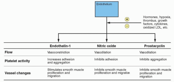
fibrinogen, factor VII, and PAI-1 have been observed in premenopausal women compared with postmenopausal women, and oral estrogen alone or combined with a progestin prevents the usual increase in these clotting factors associated with menopause.645,646,647,648 and 649 This would be consistent with increased fibrinolytic activity, a possible cardioprotective mechanism probably mediated, at least partially, by nitric oxide and prostacyclin. Platelet aggregation is also reduced by postmenopausal estrogen treatment, and this response is slightly attenuated by medroxyprogesterone acetate.574 In a randomized 1-year trial, the addition of medroxyprogesterone acetate, either sequentially or continuously, produced a more favorable change in coagulation factors compared with unopposed estrogen.650

The transdermal and oral routes of administration of estrogen (combined with medroxyprogesterone acetate) have puzzling differences in the reported effects on most hemostatic risk factors, such as factor VII, fibrinogen, PAI-1, and antithrombin III. In at least one study, however, antithrombin III levels were reduced by oral estrogen but not transdermal administration; however the values remained within the normal range.651 In regards to PAI-1, studies with transdermal estrogen have provided conflicting data; for example, favorable changes in PAI-1 levels as well as no effect.32,652,653 However, in a crossover study designed to compare 100 μg transdermal estradiol with 0.625 mg oral conjugated estrogens (both combined with 2.5 mg medroxyprogesterone acetate daily), only the oral estrogen had a favorable reduction in PAI-1 levels.653 Appropriate doses of hormone therapy have been reported to not have an adverse impact on clotting factors.646,654,655 One study found slightly increased clotting activation with transdermal administration of estradiol, but no change with oral conjugated estrogens.656 Fibrinopeptide A is an indicator of thrombin generation, and, in 3-month studies, no significant alteration was produced by 0.625 mg conjugated estrogens in 1 and an increase in another.657,658 The clotting story is difficult to unravel. Perhaps one contributor to the uncertainty is a possible difference between short-term and long-term effects.651,654,657

Overall, estrogen treatment is associated with favorable changes consistent with an increase in fibrinolysis.659,660 How can there be favorable changes indicating an increase in fibrinolysis and at the same time an increased risk of venous thrombosis, and why in elderly women, especially those with clinically apparent coronary heart disease, does estrogen seem to have a prothrombotic effect? Decreases in antithrombin III and protein S associated with estrogen treatment, a hypercoagulable change, may have a greater impact on the venous system.658 There also may be subtle variations of inherited susceptibilities that tilt the balance towards thrombosis; for example, concentrations of factors that favor arterial thrombosis have been reported (tissue factor pathway coagulation inhibitor and thrombin activatable fibrinolysis inhibitor) in women treated with estrogen.661 Another possibility is that the fibrinolysis is a response to coagulation activity, and, therefore, not necessarily a beneficial response.

Estrogen has adverse effects on already established atherosclerosis. Matrix metalloproteinase enzymes are secreted by inflammatory cells and smooth muscle cells. These enzymes digest the proteins in the fibrous cap of an atherosclerotic plaque, making the plaque unstable and predisposed to rupture. Estrogen induces matrix metalloproteinase enzymes and decreases their specific inhibitors (TIMP); this is a mechanism involved in the prothrombotic effects of estrogen in the presence of established atherosclerosis. This effect of estrogen may be dose-related and might be avoided with transdermal administration.662


Inhibition of Intimal Thickening

Hypertension and atherosclerosis are associated with increased proliferation of vascular smooth muscle cells. This growth of smooth muscle cells is also characterized by migration into the intima. Arterial intimal thickening is an early indicator of atherosclerosis.
The proliferation and migration of human aortic smooth muscle cells in response to growth factors are inhibited by estradiol, and. importantly, this inhibition is not prevented by the presence of progestins.663,664 Nitric oxide, which is regulated by estrogen, also inhibits smooth muscle proliferation and migration.665 Imaging studies have documented a reduction in intimal thickening in postmenopausal women who are estrogen users compared with nonusers, and this beneficial effect is not compromised by the addition of a progestational agent to the treatment regimen.611,666,667 and 668 Thus, postmenopausal hormonal therapy can bring about a reduction in atherosclerosis, and this effect is comparable with that produced by a lipid-lowering drug.666,669


Protection of Endothelial Cells

Endothelial cells can respond to injury by initiating the clotting process. Animals studies indicate that estrogen accelerates healing and recovery of the endothelium in response to injury.670 This is correlated with inhibition of intimal thickening and recovery of important functions such as nitric oxide production. In vitro studies of human endothelial cells demonstrate that estrogen can inhibit cytokine-induced apoptosis.671 In the rat, medroxyprogesterone acetate blocked the estrogen-induced healing response after carotid artery injuries.672


Inhibition of Macrophage Foam Cell Formation

A feature of atherosclerotic plaque formation is monocytic infiltration into the arterial wall and the formation of macrophage foam cells. In a nonantioxidant activity, estrogen inhibits macrophage foam cell accumulation in atherosclerotic lesions.673


Reduction of ACE and Renin Levels

Although oral estrogen, but not transdermal estrogen, increases angiotensinogen levels, ACE (angiotensin-converting enzyme) and renin levels are decreased (with or without progestin) by both routes of administration.674,675 The angiotensin II receptor (the AT1 receptor) is involved in vasoconstriction, aldosterone release, sodium and water retention, and growth and proliferation of myocardial and vascular cells. Estrogen induces down-regulation of the AT1 receptor, and hypercholesterolemia is associated with AT1 upregulation and function.676,677


Reduction of Adhesion Molecules

Adhesion molecules recruit leukocytes to the endothelium and play a role in attaching platelets to endothelium. Studies with multiple markers report that oral estrogen therapy increases only C-reactive protein (CRP), the only marker synthesized in the liver. In fact, oral hormone therapy although it increases CRP, reduces the circulating levels of other markers (E-selectin, P-selectin, intercellular adhesion molecule-1, vascular cell adhesion molecule-1, monocyte chemoattractant protein-1, and tumor necrosis factor-a) with inconsistent effects on interleukin-6.46,47,678,679 An increase in CRP levels may be due to estrogen’s well-known effect to stimulate the hepatic synthesis of proteins, especially because of the first-pass phenomenon with oral administration. For this reason, transdermal estrogen treatment reduces adhesion markers but does not change CRP levels.680,681 and 682


Reduction of Homocysteine

Increased circulating levels of homocysteine are correlated with increased risks of atherosclerosis and thrombosis. Homocysteine levels increase after menopause and are associated with hypertension and degree of atherosclerosis.683 Homocysteine levels are significantly lowered by estrogen or estrogen-progestin treatment, administered either orally or transdermally.684,685

Only gold members can continue reading. Log In or Register to continue

Stay updated, free articles. Join our Telegram channel

Jul 5, 2016 | Posted by in GYNECOLOGY | Comments Off on Postmenopausal Hormone Therapy

Full access? Get Clinical Tree

Get Clinical Tree app for offline access