Collagen vascular diseases
Neonatal lupus erythematosus
Childhood systemic lupus erythematosus
Childhood discoid lupus erythematosus
Juvenile dermatomyositis
Idiopathic photodermatoses
Actinic prurigo
Hydroa vacciniforme
Polymorphous light eruption
Solar urticaria
Genetic and metabolic conditions
Hermansky–Pudlak syndrome
Bloom syndrome
Rothmund–Thomson syndrome
Kindler syndrome
Trichothiodystrophy
Cockayne syndrome
Xeroderma pigmentosum
Ultraviolet sensitive syndrome
Albinism
Hartnup disease
Smith–Lemli–Opitz syndrome
Phenylketonuria
Hepatic porphyrias
– Porphyria cutanea tarda
– Variegate porphyria
– Hereditary coproporphyria
– Hepatoerythrocytic porphyria
Erythropoietic porphyrias
– Congenital erythropoietic porphyria
– Erythropoietic protoporphyria
Nutritional deficiencies
Pellagra
Exogenous causes
Drug-induced photosensitivity
Phytophotodermatitis
Photoaggravated conditions
Atopic dermatitis
Psoriasis
Seborrheic dermatitis
Darier’s disease
Herpes simplex
Acne vulgaris
Rosacea
Pityriasis rubra pilaris
Dermatitis herpetiformis
This chapter provides an overview of these disorders, with a focus on photosensitivities with the strongest association to children of color. These include lupus erythematosus (both systemic and cutaneous), actinic prurigo, hydroa vacciniforme, polymorphous light eruption (PMLE), Hermansky–Pudlak syndrome (HPS), and some subtypes of oculocutaneous albinism. The epidemiology and demographics, clinical findings, therapeutic options, prognosis, and areas of future research are discussed.
Collagen Vascular Diseases
The collagen vascular diseases associated with photosensitivity in childhood include neonatal lupus erythematosus (NLE), childhood systemic lupus erythematosus (SLE), childhood discoid lupus erythematosus (DLE), and juvenile dermatomyositis (JDMS) [1–3]. Here NLE, SLE and DLE are discussed in further detail, given their increased prevalence in children of color.
While JDMS is not fully reviewed in this chapter because it is not necessarily more common in children of color, two facts pertaining to that disorder in patients who are African-American and Hispanic will be noted. First, patients of color with dermatomyositis have linkage to the HLA-DQA1 allele DQA1*0501 [4] and second, the clinical presentation of the skin lesions and heliotrope rash show more hyperpigmentation than violaceous lesions [5].
Neonatal Lupus Erythematosus
Epidemiology/Demographics
Reported in African-Americans, Caucasians, Hispanics and Asians in the United States, and in numerous ethnicities internationally.
No consensus regarding male or female predominance, although a study from the United States National Lupus Registry demonstrates an equal sex distribution.
Incidence of 1:20,000 births, with greater associated morbidity and mortality in children of color.
NLE is caused by transplacental transfer of maternal anti-SSA/Ro-SSB/La IgG antibodies, although in rare cases anti-U1 RNP antibodies have been implicated [14]. Mothers generally have Sjogren syndrome, SLE, rheumatoid arthritis, or other connective tissue disorders, although in the majority of cases the mother is unaware of her disease [2]. The affected neonate may present with cutaneous, cardiac, hepatobiliary, or hematologic abnormalities. In a prospective study of 128 neonates with neonatal lupus, there was a 16 % incidence of skin involvement [15], while other studies note cutaneous findings in half to three-quarters of patients [2, 16, 17].
In the multiethnic/racial Research Registry for Neonatal Lupus (RRNL), the demographics of mothers of 297 children with anti-SSA/Ro-related cardiac NLE were as follows: Caucasian (75.1 %), Black (9.1 %), Hispanic (8.8 %), Asian (4.7 %), and mixed race (2.4 %). Demographics for the RRNL as a whole are not provided in the publication [6]. There are reports from the United States [7], China [8], and Japan that assert female infant predominance [9], whereas others have demonstrated male infant predominance [10]. An incidence of 1:20,000 births has been reported [11]. Non-Caucasian neonates have a greater risk of associated morbidity and mortality [12].
Clinical Presentation
Neonatal lupus is associated with cutaneous, cardiac, hematologic, and hepatobiliary abnormalities.
Cutaneous findings have been variably reported to occur in 16–75 % of patients in different series.
Skin findings characterized as erythematous, arcuate, or annular plaques with elevated active margins and mild central atrophy, often on the scalp or periorbitally although other areas can be involved.
The sequelae of NLE can be long-term hyperpigmentation or hypopigmentation [13].
Clinically, NLE is associated with cutaneous findings, similar to subacute cutaneous lupus, cardiomyopathy, congenital heart block (CHB), hematologic irregularities, and hepatobiliary disorders [18, 19]. Erythematous arcuate or annular plaques (Fig. 40.1) with elevated active margins and mild central atrophy generally present periorbitally and on the scalp although other areas of the body can be involved. As stated earlier there are variable reports of skin manifestations in 16 to 75 % of NLE patients [2, 16, 17]. Other cutaneous findings may include congenital erosive lesions, periocular lesions termed raccoon eyes (Fig. 40.2), and, in lighter skinned children, telangiectases can be noted.
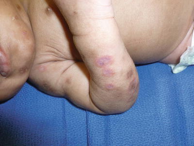
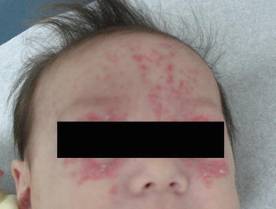
Fig. 40.1
Classical subacute cutaneous lupus like plaques in NLE (photo courtesy of Nanette Silverberg, MD)
Fig. 40.2
Raccoon eyes with periocular telangiectases in NLE (photo courtesy of Nanette Silverberg, MD). From Springer from Atlas of Cutaneous Biodiversity, Fig. 9.3
Cardiac screening is important in all children with NLE, however, the incidence of cardiac lesions in children who present with cutaneous lesions is not high when neonatal heart block is not detected. As there may be an undetected first-degree heart block that could evolve in the postnatal period, an electrocardiogram and at times an echocardiogram are performed, in addition to appropriate rheumatologic (ANA, SS-A, SS-B, and U1RNP antibodies), hematologic, and hepatic screening [7, 20].
Treatment, Prognosis, and Ongoing Research
Photoprotection with broad-spectrum sunscreen, photoprotective clothing, and sun avoidance.
Low-potency corticosteroids or calcineurin inhibitors may be tried, but it is unknown whether they lead to superior outcomes.
Coordination of care with other pediatric, including cardiology, hematology, and gastroenterology, where needed.
The mean age of presentation and mean duration of skin lesions according to a National Registry in the United States were respectively 6 and 17 weeks. In that study there were no significant sequelae in 65 % of children while 25 % had resultant postinflammatory dyspigmentation and telangiectasias and a few children developed autoimmune disorders, such as juvenile rheumatoid arthritis and Hashimoto’s thyroiditis. This elevated risk of autoimmune disease has been demonstrated in other studies, although there does not appear to be a higher risk of developing SLE. Photoprotection and topical regimens such as low-potency corticosteroids or calcineurin inhibitors may be tried but it is not known if they lead to superior long-term cosmetic outcomes [16, 21] (Table 40.2).
Photoprotection | Broad-spectrum sunscreen, photoprotective clothing and sun avoidance |
Dermatologic therapies | Topical regimens such as low-potency corticosteroids or calcineurin inhibitors may be tried but do not lead to superior outcomes |
Other subspecialties | Given the risk of congenital heart block, cardiomyopathy, hematologic and hepatobiliary disorders, involvement of other pediatric subspecialties and screening for these complications with appropriate therapies are indicated |
CHB can be detected during the pregnancy, and monitoring with Doppler echocardiograms or other modalities is recommended in women with anti-SSA/Ro-SSB/La antibodies to ensure that arrhythmia is identified and treated. The 3-month mortality rate in children with complete CHB has been reported as 15 % [22, 25]. Treatment for cardiac complications of neonatal lupus involves a pediatric cardiologist with close echocardiographic monitoring and often glucocorticosteroids for neonates with incomplete heart block, hydrops fetalis, ascites, or pleural effusions [20]. Early introduction of hydroxychloroquine is recommended in pregnancies with anti-SSA/Ro antibodies if there was a prior childbirth associated with cardiac manifestations of neonatal lupus. These medications, however, do carry risks during pregnancy [11, 23, 26]. Despite cardiac pacing and other interventions, dilated cardiomyopathy can develop in children with CHB, which may lead to congestive heart failure or a need for cardiac transplant [24]. A better understanding of the pathogenesis of NLE is necessary for developing therapeutics for this serious disorder.
Experimental
The role of L-type calcium channels in the cardiac manifestation of NLE in animal models may afford gene-targeted therapies [23].
Childhood Systemic Lupus Erythematosus
Epidemiology/Demographics
Higher incidence in African-American, Hispanic, and Asian children with a lower reported incidence in Caucasians and Native Americans.
1:1 prepubertal female:male ratio with a 4.5:1 female predominance after puberty.
Incidence of 1:10,000 with 15–20 % of SLE cases presenting by age 20 with the following age distribution: 11–15 years (60 %), 5–10 years (35 %), and <5 (5 %).
Ethnic and Racial Groups Affected [27] |
African-American, Caucasian, Asian, Native American |
SLE is a condition that can present with cutaneous manifestations such as a malar rash, photosensitivity, or discoid lesions [28, 29]. In 15–20 % of patients, the disease presents by age 20. In that subset, the age range for 60 % of those patients is 11–15 years, 35 % from 5 to 10 years, and 5 % in those less than 5 years [2]. The female:male ratio has been reported as 1:1 in prepubertal children with a 4.5:1 female predominance after puberty [2, 30]. In a study examining over 30 million children with Medicaid from 2000 to 2004, the prevalence was approximately 1:10,000 84 % of whom were females, with the following demographic data: African-American (40.9 % of total cases, incidence 2.73), Hispanic (23.7 %, incidence 2.45), Caucasian (22.1 %, incidence 1.33), Asian (6.1 %, incidence 4.16), and Native American (0.9 %, incidence 1.61) [27]. There is a higher risk of nephritis in children as opposed to adults with the highest incidence in non-Caucasian children. African-American children are often younger at diagnosis [31].
Clinical Presentation
Diagnosis is based on meeting 4 of 11 criteria in the guidelines of the American College of Rheumatology, which includes three cutaneous features—a malar rash, a discoid rash, and photosensitivity.
The malar rash of SLE presents as thin erythematous plaques with scale on the bilateral cheeks, sparing the nasolabial folds.
The presence of discoid lesions on more than one body segment has an association with systemic disease and there is a higher association and progression of systemic involvement in children with DLE than in their adult counterparts.
The diagnosis of SLE is based on the American College of Rheumatology guidelines, for which 4 of 11 criteria must be met. These criteria include malar or discoid rash, photosensitivity, mucocutaneous ulcerations, serositis, arthritis, immunologic, hematologic, renal or neurologic disorders, or elevated antinuclear antibody (ANA). Constitutional symptoms such as fatigue, myalgias, and fevers may be presenting symptoms [28, 29]. The malar rash of SLE presents as thin erythematous plaques with scale on bilateral cheeks sparing the nasolabial folds (Fig. 40.3). On the dorsal hands, there are periungual telangiectasias and sparing of the knuckles as opposed to JDMS where hyperpigmented lichenoid to flat plaques can be noted over the joints (corresponding to violaceous lichenoid papules seen in Caucasian patients). Discoid lesions may also be present. The presence of discoid lesions on greater than a single body segment in children is strongly associated with systemic disease. Often discoid lesions are an early cutaneous manifestation of SLE and there is a higher association with systemic involvement in children than in adults with discoid lupus [2]. Children with DLE should be tested for SLE and observed closely over time when the latter cannot be diagnosed.
Treatment, Prognosis, and Ongoing Research
Use of NSAIDs for arthritis and other musculoskeletal involvement.
Systemic glucocorticosteroids in conjunction with steroid-sparing disease-modifying antirheumatic drugs (DMARDs) such as hydroxychloroquine for skin and systemic involvement.
Use of agents like methotrexate for arthritis and azathioprine and cyclophosphamide for lupus-associated nephritis with rituximab as a combination therapy in treatment failure for patients with severe systemic disease.
Therapies for SLE are targeted to treat the affected organ systems and are usually coordinated by a rheumatologist. NSAIDs are used for arthritis and other musculoskeletal involvement. Systemic glucocorticosteroids are used in conjunction with DMARDs such as hydroxychloroquine to reduce the risk of side effects from steroid use in children, i.e., prednisone exceeding 0.2 mg/kg/day has been shown to result in growth retardation [32]. Hydroxychloroquine can cause ocular toxicity, more so in younger patients and when paired with other antimalarials. Methotrexate has shown benefit for arthritis and azathioprine and cyclophosphamide for lupus-associated nephritis [28]. Rituximab, an anti-CD20 monoclonal antibody, has been used in combination with some of the aforementioned therapies for treatment failures [33] (Table 40.3). Systemic corticosteroids in children with SLE often produce striae, both from the medication and from the associated weight gain (Fig. 40.3).
Drug | Indication |
|---|---|
NSAIDs (i.e. ibuprofen) | Arthritis and myalgias |
Prednisone | Skin disease, serositis, arthritis or acute moderate-severe disease |
Methylprednisolone | Acute severe renal, neurologic or hematologic disease |
Hydroxychloroquine | Prevention of flares and constitutional symptoms, arthritis, skin involvement, improves lipids |
Methotrexate | Arthritis, monitor kidney function due to renal metabolism |
Azathioprine | Renal, neurologic, hematologic or vasculitic involvement |
Cyclophosphamide | Best recorded success in severe neurologic or renal disease |
Mycophenolate mofetil | Severe renal disease |
IV immunoglobulin | Severe hematologic involvement |
Rituximab | Severe disease refractory to treatment with cyclophosphamide |
Fig. 40.3
Striae in an otherwise thin Hispanic girl receiving high dose oral steroid medication (photo courtesy of Nanette Silverberg, MD)
SLE in children is more severe than SLE in adults. SLE-associated nephritis causes 3 % of renal transplants in children, with a 5-year mortality of 22 % in those with end-stage renal disease, primarily due to cardiopulmonary complication or infections. While SLE was once universally fatal for children, now the mortality rate in developed countries is less than 10 % at 5 years, 14 % at 10 years, and 21 % at 15 years. However, in some developing nations such as Thailand, the mortality rate at 5 years is 27 % and at 10 years 36 %.
Experimental
Belimumab, a monoclonal antibody that acts as a B-cell activating factor inhibitor, was introduced in 2012 as a promising therapeutic agent in adults with SLE with autoantibodies. Currently, many other therapies are under development for adults and children suffering from SLE [36].
Childhood Discoid Lupus Erythematosus
Epidemiology/Demographics
Cases have been reported in multiple ethnicities including African-American, Caucasian, Hispanic, and Middle-Eastern patients.
Equal incidence in men and women.
Rare disorder with only 80 reported cases reported in the literature as of 2008.
Clinical Presentation
Discoid lesions are erythematous plaques with follicular plugging often found on the face and scalp, which can result in atrophic scarring.
The presence of discoid lesions on more than one body segment correlates with the development of systemic disease.
A low incidence of photosensitivity has been reported in many cases, although a Tunisian study cited an 81 % incidence.
DLE (Fig. 40.4) may be present with erythematous plaques with follicular plugging, most often found on the face and scalp, and can resolve with atrophic scarring [38]. Discoid lesions presenting on more than one body segment in children is associated with systemic disease. Discoid lesions can be an early cutaneous manifestation of SLE. There is a higher rate of systemic involvement in children with DLE than in adults with DLE [2]. In a Tunisian study, there was a high incidence of photosensitivity in 81 % of cases while low incidences have been reported in other studies [39, 40]. The age of presentation of the reported 80 patients in the 2008 review was less than 10 years in half of patients and between 10 and 16 years in the other half [37]. Diagnosis by biopsy on the scalp with or without direct immunofluorescence can differentiate this form of alopecia from other scarring alopecias, including kerion and morphea, and in teenagers, central centrifugal cicatricial alopecia (CCCA), a condition of follicular inflammation and degeneration noted to relate to aggressive styling techniques.
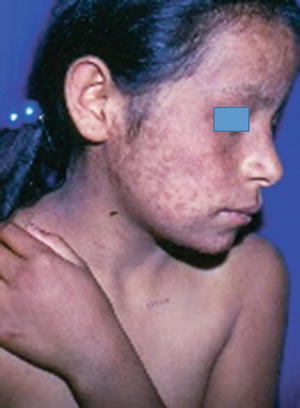
Fig. 40.4
Actinic prurigo (photo courtesy of Carola Duran-McKinster/Ramon Ruiz-Maldonado)
Treatment, Prognosis, and Ongoing Research
Photoprotection with broad-spectrum sunscreen, photoprotective clothing, and sun avoidance.
First-line therapy with high-potency topical or intralesional corticosteroids.
Second-line therapy with hydroxychloroquine or oral corticosteroids.
Treatment for DLE without systemic involvement involves photoprotection and high-potency topical or intralesional corticosteroids. If necessary, oral corticosteroids or hydroxychloroquine can be tried, although clinicians should be aware of potential retinal toxicity and other complications [38, 41]. In other studies, thalidomide [42] and dapsone have been tried although hematologic and liver function abnormalities have been reported with dapsone [43]. In adults, years of treatment leads to resolution of lesions in half of patients [38]. A quarter of children with DLE develop SLE, as opposed to 6 % for adults with DLE. Long-term serologic monitoring for development of SLE is required in this subset of patients.
Experimental
In light of the success of calcineurin inhibitors in adults with DLE, these medications may have benefit in children but future studies are necessary [37] (Table 40.4).
First-line | Broad-spectrum sunscreen, photoprotective clothing and sun avoidance with high-potency topical or intralesional corticosteroids |
Second-line | Oral corticosteroids or hydroxychloroquine; there are case reports with success using thalidomide or dapsone |
Idiopathic Photodermatoses
The idiopathic photodermatoses include actinic prurigo, hydroa vacciniforme, PMLE, and solar urticaria [44–47]. Actinic prurigo, hydroa vacciniforme, and PMLE are discussed in more detail here, given their higher incidence in skin of color.
Actinic Prurigo
Epidemiology/Demographics
Actinic prurigo, also called hereditary or familial PMLE of Amerindians, is an autosomal dominant condition in Native American and Inuit children, with a prevalence of 1.2–8 %.
Most often seen in Native American and Mestizo children, with reports of cases in Japan, Singapore, Australia, Continental Europe, and the United Kingdom.
There is a reported 2–4:1 female:male distribution.
Actinic prurigo (also called hereditary or familial PMLE) is an autosomal dominant photosensitizing disorder in Native American and Inuit children with variable expressivity and reduced penetrance. The prevalence in these populations is reported between 1.2 and 8 % [50, 51]. It has also been reported in Caucasian Americans and African-Americans. Some authors contend that the onset of PMLE in childhood may suggest Native American heritage [52]. The incidence in Mexico is 1.5–3.5 %. Cases have also been reported in Japan, Singapore, Australia, Continental Europe, and the United Kingdom. The female to male ratio is 2–4:1 [48, 53, 54].
Clinical Presentation
Pruritic erythematous papulonodular rash that may have a hemorrhagic crust or appear eczematous, traditionally in a photodistributed pattern, although nonexposed sites may be involved.
In Native American children, oral and mucosal involvement such as conjunctivitis, pterygium, eyebrow hair loss, and cheilitis are often present.
Rash can persist in patients the entire year in Central America but often presents in patients in the spring or summertime in northern latitudes.
Actinic prurigo is a pruritic erythematous papulonodular rash that may have a hemorrhagic crust or be eczematous. It is most prominent in a photodistributed pattern, although nonexposed sites may also be affected. Oral and mucosal involvement in Native Americans is often present with features such as conjunctivitis, hair loss from the eyebrows, pterygium, and cheilitis. The sole clinical finding in nearly a third of Mestizo children is cheilitis [49]. In Central America, the rash can persist in patients the entire year, whereas in more northern latitudes, it is most common in patients in the spring or summer [48, 55].
Treatment, Prognosis, and Ongoing Research
First-line therapy with photoprotection and topical corticosteroids or topical tacrolimus with systemic corticosteroids as needed.
Second-line therapy with PUVA or narrowband UVB (NB-UVB) desensitization.
Third-line therapy with thalidomide. There is a risk of teratogenicity from this therapy.
Photoprotective clothing, sun avoidance, and broad-spectrum sunscreen are needed for prevention. Topical regimens such as corticosteroids and tacrolimus or systemic treatment with corticosteroids are beneficial. For serious cases, desensitization with PUVA or narrowband UVB a few times a week beginning at 50–70 % of the MED with incremental increases of 10 % can be tried. For refractory cases, systemic therapy with thalidomide for a few weeks until the condition resolves with a taper to the smallest maintenance dose possible [56]. Teratogenicity is a serious side effect in young females. Registry, contraceptive education, and frequent pregnancy testing should be performed in girls of child-bearing age. Other regimens including tetracycline, cyclosporine, azathioprine, vitamin E, beta-carotene, chloroquine, and pentoxifylline have been reported to be beneficial, but further study is necessary (Table 40.5). The condition may resolve with pitted scarring or postinflammatory hyperpigmentation but generally it persists into adulthood [48, 55]. Just as thalidomide, a medication with FDA approval for multiple myeloma and erythema nodosum leprosum, became a useful therapy for severe cases of actinic prurigo, novel applications of existing therapeutics may benefit children with these and other photosensitive disorders refractile to traditional treatments [57].
First-line treatments | Broad-spectrum sunscreen, photoprotective clothing and sun avoidance with topical or systemic corticosteroids or topical tacrolimus |
Second-line treatments | Desensitization with PUVA or NB-UVB beginning at 50–70 % of the MED with incremental increases of 10 % as tolerated |
Third-line treatments | Thalidomide for a few weeks until the condition resolves with a taper to the smallest possible maintenance dose |
Other regimens with reported benefit include tetracycline, cyclosporine, azathioprine, vitamin E, beta-carotene, chloroquine and pentoxifylline, but further study is necessary | |
Hydroa Vacciniforme
Epidemiology/Demographics
Reported in South Africans, Caucasians, Native Americans, Asians, and Hispanics.
Equivalent male:female distribution, although females present with the disease earlier and males have a more prolonged disease course.
Incidence of 1:300,000.
Hydroa vacciniforme is a rare disorder that develops in children, sometimes in association with the Epstein–Barr virus (EBV). The incidence of the disorder is unknown but has been reported at 1:300,000, with an equal sex distribution in a review from Glasgow, although females presented with the disease earlier and males had a more prolonged disease course [64]. There have been reports of the disorder in South African, Caucasian, Native American, Asian, and Hispanic children [58–63].
Clinical Presentation
Pruritic erythematous maculopapular and vesicular rash seen in a photodistributed distribution often on the face with perioral involvement. Umbilicated lesions or hemorrhagic crusting can also be observed
The condition is often refractory to treatment but spontaneously resolves before adulthood. It may result in significant residual varioliform scarring.
There can also be associated mosquito bite hypersensitivity and risk of lymphoproliferative malignancy in a distinct condition called hydroa vacciniforme-like lymphoma.
Hydroa vacciniforme clinically presents with a pruritic photodistributed erythematous maculopapular and vesicular rash that is often seen on the face with perioral involvement within hours to days after sun exposure. Umbilicated lesions and hemorrhagic crusting can be observed [64].
Treatment, Prognosis, and Ongoing Research
Photoprotective clothing, broad-spectrum sunscreen, and photoprotection on windows in the home and vehicles.
Desensitization with NB-UVB can serve as a useful adjunct, although the condition is often unfortunately refractory to therapy.
Case reports of benefit from use of PUVA, antivirals, omega-3 fish oils, beta-carotene, antimalarials, cyclosporine, and azathioprine.
Hydroa vacciniforme is unfortunately oftentimes refractory to therapy. Photoprotective clothing, broad-spectrum sunscreen, and photoprotection on windows in the home and cars are recommended. Desensitization with narrowband UVB can serve as a beneficial adjunct [64]. There are case reports of benefit with other regimens, including antivirals such acyclovir and valacyclovir [65], PUVA, omega-3 fish oils, beta-carotene or antimalarials, and the use of cyclosporine or azathioprine in severe cases (Table 40.6). The condition often spontaneously resolves before adulthood but may result in significant residual varioliform scarring [66].
Photoprotection | Broad-spectrum sunscreen, photoprotective clothing and photoprotection on windows in the home and vehicles |
First-line treatment | Desensitization with NB-UVB although often refractory to treatment |
Case reports with stated benefit | PUVA, antivirals, omega-3 fish oils, beta-carotene, hydroxychloroquine, chloroquine sulfate or the use of cyclosporine or azathioprine in severe cases |
Of note there may be associated retinal toxicity and cardiotoxicity with the use of hydroxychloroquine and chloroquine [67] | |
Recently, there has been a partition of hydroa vacciniforme cases into a typical subtype where cutaneous findings are limited to photodistributed sites versus a severe hydroa vacciniforme-like subtype with cutaneous findings that extend to photoprotected sites. This severe variant can be associated with high peripheral blood EBV DNA titers. A distinct condition called hydroa vacciniforme-like lymphoma has been associated with mosquito bite hypersensitivity and risk of lymphoproliferative malignancy [68]. Hydroa vacciniforme-like lymphoma is an EBV-associated T-cell and natural killer cell lymphoma, most commonly seen in children from Asia and South and Central America, associated with systemic manifestations including fever and hepatosplenomegaly [69, 70]. While some authors propose measuring the titers of EBV DNA, which may have utility in assessing a child’s risk of malignant transformation and help direct the frequency and extent of clinical monitoring, it is important to reemphasize that hydroa vacciniforme-like lymphoma is a distinct clinical entity and that children with hydroa vacciniforme generally have a relatively benign clinical course.
Polymorphous Light Eruption
Epidemiology/Demographics
No reliable cross-sectional studies have been performed but the condition in adults is generally more common in fair-skinned individuals.
However, certain subtypes of PMLE are characteristic in skin of color such as the pinpoint variant.
Stay updated, free articles. Join our Telegram channel

Full access? Get Clinical Tree


