Click Video under article title in Contents at ajog.org
Case notes
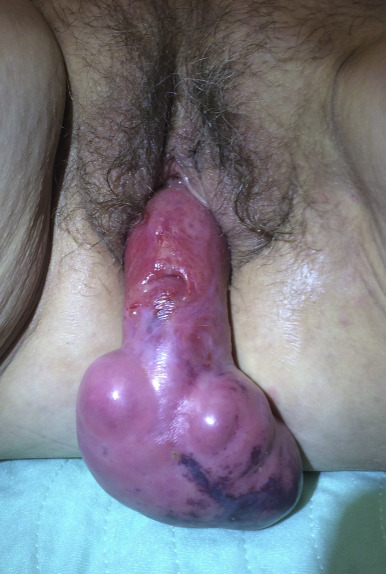
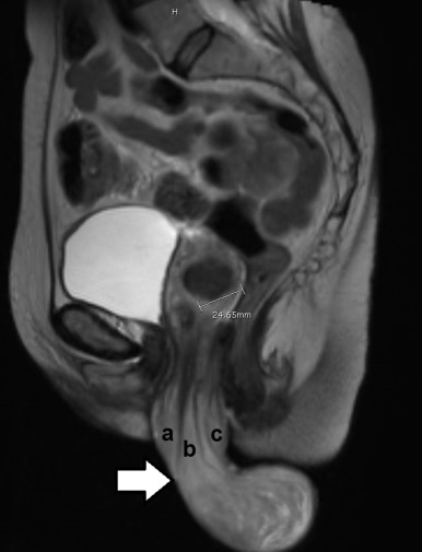
A 64-year-old female nurse, who was known to have a stable and reducible uterine prolapse, returned to her gynecologist’s office, after a few years of hiatus, with increasing vaginal discomfort. She reported no other acute symptoms and no significant change in her urinary, defecatory, or sexual function. Perplexed with the appearance of an irreducible, 7-cm, firm, ulcerated, hemorrhagic mass outside her introitus, she was referred to an urogynecologist ( Figure 1 ). Her cervical location was not clarified until magnetic resonance imaging (MRI), which revealed an 18-cm-long uterus, including a mass attached to the cervix. While the cervix was prolapsed and elongated to 7 cm, the mass accounted for 6 cm of this length. There was also a 2.5-cm leiomyoma in her 5-cm-long uterine corpus. The MRI suggested that this was a prolapsed pedunculated edematous uterine leiomyoma ( Figure 2 ).


Stay updated, free articles. Join our Telegram channel

Full access? Get Clinical Tree


