Ethnic or racial group
Number of patients reported
Korean
5
Japanese
2
Brazilian
1
Bolivian
1
Madagascan
1
Italian
5
German
11
Caucasian American
6
Hispanic American
2
African American
28
Jamaican
1
There are many etiologic factors implicated in periorificial dermatitis and CGPD (Table 39.2). The most common is systemic, topical, or inhaled corticosteroid use [4, 6]. About half of the patients with periorificial dermatitis present with a history of recent corticosteroid use [19]. The fluorinated corticosteroids appear to be the most problematic. Interestingly, in some patients, topical, inhaled, and systemic corticosteroids can temporarily improve periorificial dermatitis. However, a rebound effect is commonly observed after discontinuation of the corticosteroid medication [4]. A particular consideration in dark-skinned patients is the possible contribution of over-the-counter bleaching agents. These products are frequently used by patients with skin of color and may contain corticosteroids that could exacerbate periorificial dermatitis. Many of the bleaching agents that are available over the counter outside of the United States are known to contain potent corticosteroids, and these products may be imported by patients, their families, or even sold in ethnic stores within the United States.
Table 39.2
Etiologic factors associated with periorificial dermatitis
Etiologic factors | |
|---|---|
Corticosteroids | Inhaled |
Topical (particularly fluorinated) | |
Systemic | |
Cosmetics and skin care products | Moisturizers (petroleum or paraffin base) |
Bleaching creams | |
Essential oils | |
Lipstick | |
Greases | |
Physical sunscreens with high SPF | |
Antiseptic solutions | |
Medications/vaccines | Oral contraceptives |
Fluorinated toothpaste | |
Varicella vaccine | |
Microorganisms | Candida species |
Fusiform spirilla bacteria | |
Demodex folliculorum | |
Fusobacterium | |
Physical factors | Ultraviolet light |
Heat | |
Wind | |
Hot water | |
Drooling | |
Preexisting medical conditions | GI malabsorption |
Lip-licking cheilitis | |
Immunosuppression (esp. leukemia) | |
Emotional stress | |
Other | Latex gloves |
Black synthetic mesh | |
Musical instruments | |
Bubble gum | |
Formaldehyde |
Other factors which have been associated with the development of periorificial dermatitis include fluorinated toothpastes and overuse of skin-care ointments and creams, especially those with petrolatum or paraffin base or an isopropyl myristate vehicle [1, 20]. One of the proposed mechanisms is that persistent hydration of the horny layer from overuse of moisturizers may lead to impaired barrier function and proliferation of skin flora, resulting in the papular eruption. Physical sunscreens with high sun protection factors have also been implicated in periorificial dermatitis, particularly in patients with atopic dermatitis and poor skin barrier function [21]. In addition, physical factors such as ultraviolet light exposure, heat, such as from use of hot water, and wind have been associated with development of periorificial dermatitis [1].
There are also infectious agents including fusiform spirilla bacteria, Candida species, and Demodex folliculorum that have been implicated in the pathogenesis of this condition. Fusobacterium overgrowth is often found after use of fluorinated topical corticosteroids [1]. There is one reported case of CGPD that occurred after varicella vaccination [7]. A variety of other factors have also been associated with periorificial dermatitis including use of oral contraceptives, conditions of GI malabsorption, emotional stress, musical instruments, latex gloves, lipstick, essential oils in bubble gum, greases, formaldehyde preservatives, antiseptic solutions, black synthetic mesh, drooling, and lip-licking cheilitis [1, 7]. Finally, there appears to be an increased risk of periorificial dermatitis in immunocompromised children, especially in those patients with leukemia [1].
Clinical Presentation
Usually a perioral distribution, but perinasal and periocular are also seen
Mostly monomorphous small grouped papules with erythema and variable scale
Rarely symptomatic
Periorificial dermatitis usually presents as grouped very small erythematous to pink papules, occasionally with vesicles and pustules, sometimes with surrounding erythema and fine scale (Fig. 39.1) [4]. Papules can appear more flesh colored in darker skinned individuals. Furthermore, hyperpigmentation and hypopigmentation can be seen in patients with skin of color (Fig. 39.2). Papules, vesicles, and pustules are usually ~1–3 mm in size and in the same stage. The lesions are in a perioral distribution in 70–100 % of cases, perinasal in 43–57 %, and periocular in about 25 % (Fig. 39.3). Papules and pustules less commonly appear on the cheeks, chin, neck, and forehead. Lesions may recur over weeks to months [1]. There is usually a 3–5 mm border of normal skin separating the vermilion border of the lips from the affected area (Fig. 39.4) [1]. This can be a helpful clue in determining the diagnosis. In majority of the cases the eruption is asymptomatic, but a subset of patients experience pruritus and sometimes a burning sensation. The differential diagnosis may include acne vulgaris, seborrheic dermatitis, impetigo, acrodermatitis enteropathica, and tinea faciei.
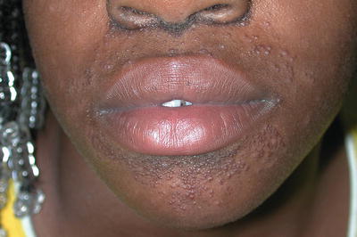
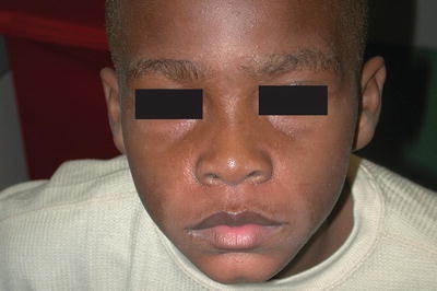
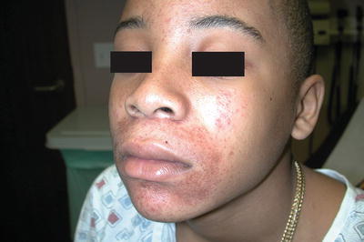
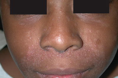
Fig. 39.1
An African-American girl with multiple erythematous monomorphic papules in a perioral distribution
Fig. 39.2
An African-American boy with multiple flesh-colored papules with surrounding hypopigmentation in a perioral distribution
Fig. 39.3
An African-American boy with erythematous monomorphous grouped papules in a perioral, perinasal, and periocular distribution
Fig. 39.4


Area of clearing between the vermilion border and the affected area
Stay updated, free articles. Join our Telegram channel
Full access? Get Clinical Tree
