Orthopedics


Orthopedics


Peter N. Fysh

Steven T. Tanaka


Orthopedic problems among children commonly are encountered in the busy chiropractic clinic. Because the doctor of chiropractic’s major area of expertise is the neuromusculoskeletal system, a child with symptoms of bone and/or joint pain might be brought to a chiropractor for evaluation. Although most adult orthopedic problems are straightforward in diagnosis, this is not always the case in the pediatric population. Pediatric spinal problems in particular require in-depth knowledge and experience for their accurate identification and management. Evaluation of pediatric problems requires a trained perception on the part of the doctor, in part because of communication difficulties, but also because of the young child’s inability or reticence to demonstrate and perform tests appropriately. Examination of the child’s movements during play and during relaxed walking or running is perhaps an ideal time to gather clinical data. Abnormalities of gait and posture are best evaluated without the child being made aware of the process, whereas ranges of joint motion can be estimated by visually examining the child’s body movements before the physical examination.

Some of the most common pediatric problems confronting the chiropractor include conditions such as otitis media, asthma, tonsillitis, headache, and enuresis (see Chapter 12), which, to the untrained, at first glance, may not appear to involve the neuromusculoskeletal system but often respond positively to improvement in neuromusculoskeletal function. For chiropractors, of course, that would involve correction of vertebral subluxation complexes. The orthopedic conditions appearing in the pediatric “top 10” include mainly spine-related conditions, such as neck pain, low back pain, thoracic pain, scoliosis, and growing pains, many cases of which respond well to chiropractic care.


DEVELOPMENT OF THE SPINE

An understanding of the growth and development of the vertebral column is essential to the ability of the doctor of chiropractic to develop and modify spinal evaluation and treatment plans to the needs of the pediatric patient. Here we present an overview of spinal development.

At birth, the length of the spinal column represents 40% of the total body height, the same percentage as in the adult. During embryonic development, the spine is shaped with a single primary anterior concave curve, which later changes in order to adapt to the human bipedal posture. At birth, the secondary lordotic curves in the cervical and lumbar spinal regions are not fully developed. The cervical and lumbar lordoses continue to develop after birth. The cervical curve develops in response to the upright posture of the head at around 3 months and should be established by the time the infant can sit upright, at about 9 months. The lumbar curve develops in response to the child adopting the standing position and appears by the age of 12 months. The lumbar lordosis is well developed by the age of 18 months in response to walking, an activity that requires a postural shift so that the center of gravity can be maintained over the legs (1).


Spinal Growth

A few weeks after conception, the primitive spine is visible as vertebrae and intervertebral discs through which the notochord passes. Cartilage fills in the vertebral bodies and pushes the notochordal material into the area that will be the disc spaces (2). When the vertebral body begins to ossify, the area that will develop into the endplates remains cartilaginous.

At birth, the length of the vertebral column is approximately 24 cm. By the end of the adolescent
growth phase, this length will have increased to 70 cm. During the first year of life, the spine will increase in length by 12 cm, and another 15 cm will occur between the ages of 1 and 5 years. The first 5 years is the time of greatest spinal growth, even greater than that of the adolescent growth period.
From age 5 to 10 years the annual rate of spinal growth slows to 10 cm but increases again once puberty is reached. From 10 to 18 years, the spinal length increases by 20 cm in boys and by 15 cm in girls.

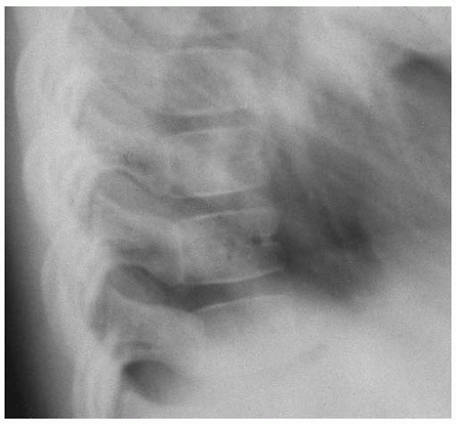
Around puberty, ossification centers appear for the vertebral secondary ring epiphyses and the tips of the spinous and transverse processes. Ossification of the spine is finally completed at approximately 22 to 25 years of age. The lateral spinal radiograph at birth shows an anterior notch in the center of each spinal vertebra, more prominent in the thoracic region, which is due to the persistence of the intersegmental artery (3) (Fig. 13-1).

Intervertebral Disc The adjacent surfaces of each vertebra are strongly connected to each other by the fibrocartilaginous intervertebral discs. In the newborn and young infant, the intervertebral space image appears to be greater than the actual height of the intervertebral disc because of the presence of the non-ossified and radiolucent elements of the adjacent vertebral bodies. Although this might give the spine the appearance of weakness; in fact, the connection of the vertebral body to the intervertebral disc is strong—in the case of vertebral fracture with displacement, there is no separation between the vertebral body and the intervertebral disc. As a result of this, the strength of the spine at birth is such that the bodies and the neural arches may be considered largely ossified.


FIGURE 13-1 Normal lateral radiograph of the thoracic spine demonstrating the radiolucency of the intersegmental artery.

At birth, the ratio of the height of the vertebra to the height of the disc is about 1:1. The vertebral body growth quickly outpaces that of the disc. Upon skeletal maturity, the ratio of vertebral height to disc height is from 3:1 to 5:1 (2).

Initially, there is primordial disc material that is composed of an outer and inner region and surrounds the notochordal material. The outer region develops into the anulus fibrosus. This outer region has fibers that anchor into the cartilaginous layer of the vertebral body and later develop into Sharpey’s fibers. The outer region is fibrous and has few cells. The inner region and the notochord remnants form the nucleus pulposus (2).

The cervical intervertebral discs at birth are composed largely of the nucleus pulposus (4), and they continue to have a proportionally larger nucleus pulposus than the lumbar spine in the mature spine. Few laminae form in the cervical anulus fibrosus. The posterior anulus laminae layers are nearly indistinguishable from the posterior longitudinal ligament because the fibers have a longitudinal orientation rather than the more typical oblique orientation (5,6). With maturation, a crevice is often found that can extend from the posterior aspect of the uncovertebral joints into the posterior disc. It may eventually bisect the disc into anterior and posterior halves. This is thought to facilitate cervical spine rotation (5,7,8).

Maturation of the Vertebrae Atlas The fetal proatlas is thought to be formed by the first cervical somite and fourth occipital somite. Unlike the rest of the cranial bones, which have a membranous origin, the occiput has a more endochondral bone form that probably is necessary for the ligamentous attachments of the skull to the odontoid and the formation of the eventual os terminale of the dens. The anterior arch of the atlas receives contributions from the first and second cervical somites (9). At birth, the anterior arch of the atlas consists primarily of fibrocartilage. The atlas vertebra has an anterior ossification center that usually appears by the end of the first year of life and is responsible for the development of the anterior arch and the greater portion of the lateral masses. Each neural arch has its own ossification center present at birth, which is usually ossified by 6 to 12 months of age. The posterior arch of the atlas is usually closed by the age of 3 years, but may occasionally remain ununited throughout life, as in spinal bifida occulta. The neural arches join with the anterior arch and fuse at about age 7. During the formative years, the anterior atlas would
seem to derive much of its strength from the attachment of the transverse ligament to the odontoid process and from the odontoid process attaching to the anterior arch by ligamentous structures.

Axis The odontoid process is probably derived from the first and second cervical somites. The C1 and C2 somites also contribute to the anterior arch of C1 and some supporting ligaments between the atlas and dens (9). The axis vertebra has four ossification centers at birth, which are already partially ossified. The odontoid begins ossifying sometime during the first and fifth prenatal months. The unossified sections are attached to each other by cartilaginous tissue. The synchondroses between the neural arches and the body fuse between 2 and 3 years of age. In early childhood, a horizontal cartilaginous translucency persists between the body and odontoid process and usually disappears when it ossifies around 6 years of age. Occasionally, the growth center may persist between the odontoid process and the axis body until about 11 years of age. This cartilaginous matrix is the homologue of the intervertebral disc. Traumatic disturbance of this growth center has been suggested as the cause of the development of an ossiculum odontoideum or ununited odontoid process (10). The mechanism is proposed as an unrecognized injury to the C2 vertebra early in life, which causes pseudoarthrosis of the dens, disrupts the blood supply, and results in progressive atrophy. The cephalad tip of the odontoid process at birth appears Y-shaped. Two secondary ossification centers at the tip of the odontoid appear at the age of 3 to 6 years and are called ossiculum terminale. Fusion to the rest of the odontoid occurs between ages 10 and 13 years (4). Failure of the apex to fuse to the body of the odontoid is called ossiculum terminale persistens of Bergman (11).

C3-C7 The bodies of the cervical vertebrae below the axis are almost fully ossified at birth. The neural arches close posteriorly around 2 to 3 years of age and anteriorly with the vertebral body at about age 3 to 6 years. The tips of the spinous processes and transverse processes appear by age 15 to 16 and fuse to the rest of the processes by age 25. Ossification of the peripheral portion of the vertebral bodies takes place at around age 20. Therefore, the uncovertebral joints of von Luschka, that is, lateral interbody joints, are not radiographically visible in the pediatric cervical spine.

Cervical Ribs The anterior portion of the transverse process of the seventh cervical vertebra develops from a separate ossification center. It usually fuses with the main ossification center around the age of 6. Aberrant ossification of this section may give rise to the development of a cervical rib. The shaft of the cervical rib grows laterally and forward into the posterior triangle of the neck. In this area, it may come into contact with the lower trunk of the brachial plexus and compress the C8 and T1 spinal nerves and the subclavian vessels against a hypertonic anterior scalene muscle, which may result in neurological and vascular symptoms (e.g., thoracic outlet syndrome).

Thoracic Spine The ossification of the thoracic spine is completed in the same pattern as that of the lower cervical spine, but closure of ossification centers generally occurs earlier than in the cervical spine. The ossification centers for the tips of the transverse and spinous processes appear at about 14 to 16 years of age and eventually fuse with more central structures around 25 years of age.

Lumbar Spine Ossification of the lumbar spine is completed in the same pattern as that of the lower cervical and thoracic spine, but closure of ossification centers generally occurs somewhat later. Similar to the cervical spine, the ossification centers for the tips of the transverse and spinous processes appear at about 15 to 16 years of age and finally fuse with the main structure by about age 25.

Sacrum At birth, the sacrum is mainly cartilaginous and represents typical vertebrae as each segment ossifies independently. Fusion of one sacral segment to another does not commence until around puberty and the bodies of the sacral segments do not unite with one another until after the age of 20. The sacral discs may remain unossified until mid-life (4,12). As an aside, decalcification of the fusion between the sacral segments may occur among the elderly, and the sacrum once again has separate, potentially moveable segments.

Coccyx At birth, the coccyx is made up of four rudimentary cartilaginous vertebrae. The ossification process starts after birth with the development of separate secondary ossification centers for each segment. Ossification centers develop at widely separated intervals from the first to the 20th year of life. The coccygeal segments fuse with each other progressively up until about the age of 20. Late in life, the sacrum and coccyx may eventually fuse together.

Growth of the Trunk Although the spine grows at a steady rate after the first year of life, the trunk undergoes a period of rapid increase in height during the adolescent growth spurt at about 12 years of age in girls and 14 years in boys. This increase is most significant in the lower extremities and accounts for this rapid height increase combined with a relatively steady lengthening spinal column (13).


DEVELOPMENTAL PATTERNS AND ANOMALIES


Crawling

Babies will usually be ready to crawl actively at about 9 to 10 months. Crawling is a natural protective reflex designed to prevent asphyxiation when the infant is lying face down. Delay in crawling may be an indication of orthopedic problems, such as congenital hip dislocation, or of neurological problems, such as cerebral palsy. However, the commencement of crawling is highly variable, and parents should not be overly concerned if the child is a late starter.

Crawling requires simultaneous use of opposite extremities, that is, right arm with left leg and vice versa, in a coordinated, reciprocating motion. Because of the decussation of the outflow tracts from the brainstem to the spinal cord, the nerve impulses for motor activity in the extremities originate on the opposite sides of the brain, that is, the left brain controls the right extremities and vice versa. Because crawling requires the simultaneous use of opposite extremities, each movement requires the use of both the right and left hemispheres of the brain. The act of crawling, therefore, is a complex action of neural coordination. Studies of children who were categorized as “early walkers,” that is, those who crawled for a comparatively short period before beginning to walk, demonstrated lower performance scores on pre-school assessment tests, thereby supporting the importance of early cross-crawl patterning movements in the development of sensory and motor systems of the body and general motor skills (14).


Baby Walkers

Baby walkers represent a cause of significant injury among the infant population. The use of walkers and other apparatus designed to prematurely assist infants to assume erect posture should be discouraged as a potential cause of injuries, including possible finger amputation. In a study of infants suffering trauma from the use of baby walkers, 88% of injuries were caused by a fall down stairs (15). A study using electromyography demonstrated that the use of infant walkers alters the mechanics of locomotion by inducing substantial mechanical errors in the walking process (16). Another study suggested that, for some infants, the excessive use of baby walkers alters the pathway of normal locomotor development (17). One 1991 US survey found that an estimated 27,804 injuries requiring treatment had resulted from the use of baby walkers, with most of the victims being younger than 2 years of age (16). A study of the mechanisms of walker-related injuries identified stairway falls (71%), tip-overs (21%), falls from a porch (3%), and burns (5%). Twenty-nine percent of the infants suffered significant injuries, including skull fractures, concussion, intra-cranial hemorrhage, cervical spine fracture, and death (17). Baby walker-related injuries represent the third most common cause of injury in infants from 7 to 14 months of age, and the use of such devices should be actively discouraged (18).

Sabir et al. reported on an 11-month-old girl who was in a walker and fell down a 15-step stairway, but initially she had no apparent neurological or imaging abnormalities. Rapidly, she became “apathetic.” Twenty-four hours after the injury and while under observation in the hospital, her limbs became paralyzed. She later developed respiratory failure and signs of kidney dysfunction. Over the course of a year, she regained full use of her limbs. More than 2 years later she died of cardiac arrest during an episode of respiratory distress (19). This case report illustrates the dangers of walkers (20,21); in this case, an infant fell down a stairway while using a walker did not show signs of spinal cord injury on radiographic studies. Needless to say, she did deteriorate. A chiropractor would wonder if the subsequent tragic ending could have been avoided with chiropractic evaluation and care. Of course, the most important question is why the parents allowed the infant to use a walker at the top of the stairs. It was not stated if there was fencing or another obstructive device at the top of the stairs that should have prevented the fall.


Walking

The age at which children commence walking can vary considerably. Typically, walking can begin at any time from 7 to 15 months of age. Parents of a “late walker” can be discouraged by the variability of the date that their child commences walking. However, in one British study of infants who had not yet begun to walk by the age of 18 months, 32% of the study group had an identifiable pathology to explain their late-walking (22). Be that as it may, the child’s body has an innate understanding of the appropriate stage at which the bones, ligaments, joints, muscles, and the nervous system are ready and coordinated to withstand the forces of erect stature. Encouraging children to walk prematurely may predispose them to increased stress on spinomusculoskeletal structures, as well as to possible delay in the development of neurological coordination.

Several studies have hypothesized the importance of early crawling experience—cross-crawl—in the development of sensory and motor systems of the body and general motor skills. One of the keystones of the Doman Institute’s work to help those with brain lesions or anomalies, such as non-development or loss of a cerebral hemisphere, is “cross-crawl exercise.” The intact hemisphere is often able to re-establish the motor function of the opposite missing hemisphere. As mentioned
above, one 1991 study that compared the performance of crawlers and non-crawlers on the Miller Assessment for Preschoolers showed that non-crawlers had lower average scores (23). It seems that the longer an infant crawls (to a point, of course), the better it is for the development of the nervous system.

Initially, the toddler walks on the forefoot or with a broad-based, flat-footed gait, with the hips held in slight flexion and no reciprocating arm swing. By the age of 2 years, most toddlers will have established an upright, heel-strike gait and will swing the arms. Abnormal walking patterns in young children are a source of much interest to clinicians.

A waddling gait may be indicative of infantile coxa vara or of an undetected congenital hip dislocation, whereas abnormalities in position and placement of the feet may be caused by congenital or acquired anomalies in the bones or joints of the lower extremities or pelvis. For example, medial or internally rotated (In) displacement of the ilium joint with fixation (using the posterior superior iliac spine [PSIS] as the point of reference) may produce lateral rotation of the ipsilateral leg and foot. The converse is also true: lateral or externally rotated (Ex) sacro-iliac displacement with fixation may cause medial displacement of the ipsilateral leg and foot. Because sacro-iliac joint subluxation is frequently accompanied by a physiologic short leg, frequent falls by a young infant while walking or running should be an indication to evaluate the position, alignment, and length of the lower extremities and pelvis.

Positional changes of the lower extremities should be evaluated to assess the likelihood of spontaneous correction or possible pathology. During the first year of life, rotational problems may present. At birth, the newborn infant’s feet will usually turn inward because of the typical position occupied in-utero. This internally rotated condition of the feet is called metatarsus adductus and usually resolves spontaneously by the end of the first year in 90% of infants (24). A most important step in examining an infant with metatarsus adductus is to check for congenital hip dysplasia (CHD) because this condition is more common among this group. Most cases of in-toeing resolve spontaneously by the end of the first year of life and require only observation on the part of the clinician. Many chiropractors have taken care of infants with toe-in and have seen them improve with adjustments, particularly but not isolated to the sacrum, specifically a posterior second sacral segment. Torsional deformities of the lower extremity are covered in more detail later in this chapter.

As the child begins to walk, the opposing sacro-iliac surfaces develop opposing and matching grooves and depressions. The importance of watching the walking pattern and the movement of the sacro-iliac joints and the symmetry of the gluteal muscles is of particular importance. If there is asymmetry of the gluteal muscles and minimal sacro-iliac movement with a corresponding “throwing forward” of the ipsilateral leg, there is likely fixation in the sacro-iliac joint (25). One may speculate, if the sacro-iliac joint dysfunction remains uncorrected, is it a significant risk factor for the development lower lumbar disc disorder because of the biomechanical relation between the pelvis and lumbar motion units?


Postural Abnormalities

Postural evaluation is an important component of the physical examination of the growing child. Evaluation of postural abnormalities should be made while searching for evidence of underlying musculoskeletal or neurological problems. The following postural distortions may indicate an underlying problem: lateral head tilt; head rotation; unleveled shoulders; prominent scapulae; thoracic kyphosis; unleveled iliac crests; lumbar hyperlordosis or hypolordosis; internal or external rotation of an ilium, leg, or foot; toe-in or toe-out; and measured discrepancy between the leg lengths.

Postural habits are problematic with the carrying of heavy school backpacks and long periods of time spent in front of media centers and computers. Many children and adolescents carry backpacks improperly. Backpacks should be worn high rather than with loose straps or being allowed to hang low or worn over one shoulder.

More than two-thirds of children who wear school backpacks either have loose straps, wherein the backpack sits at the lumbar or sacral level, or they strap the backpack over only one shoulder. It is common for the backpacks to weigh 10% to more than 20% of the child’s body weight. At 20% of body weight, ischemia of the skin under the strap can occur. It has been found that carrying the backpack low increases pressure over the shoulders and reduces contact with the back, thereby increasing the pressure on the shoulder. Wearing the backpack strapped over only one shoulder was found to alter shoulder and spinal angles to compensate for the asymmetrical load (26). Backpack use also significantly compressed the lumbar discs, particularly to the lower lumbar spine (27).

Many sit in front of computers and televisions hunched forward—flexed lower back, forward bend thoracic spine, and anterior shift of the cervical spine—for hours. Many continue to maintain that hunched position when standing and walking. Many computer stations that children use in school and at home have no ergonomic considerations. One obvious problem is that children come in many sizes, and most computer stations are “one size fits all.” Robbins et al. (28) did a 1-week test on the posture of children who sat at a computer. One group had onscreen warnings and was shown proper posture. The other group did not receive those instructions. Upon 1-week follow-up, the group that received instructions had less musculoskeletal pain than the control group (28).


Growth Rate

The velocity of growth among children varies with age, being particularly noticeable around the adolescent period. The average growth rate in children is around 4 to 6 cm each year, from 2 years of age until the commencement of the adolescent growth spurt. Once the adolescent growth spurt is reached, around age 11 to 12 in girls and 13 to 14 in boys, the rate of growth increases significantly to around 6 to 12 cm per year. The increase in growth during this period is almost entirely caused by the increase in the length of the lower extremities and is not because of increased growth rate of the spinal column, which remains stable at around 2 cm per year. The adolescent growth spurt normally lasts approximately 2 years, and completion of growth is normally achieved about 4 years after its start. The greater final height among men is reportedly because of a longer and more intense adolescent growth phase. The onset of the adolescent growth spurt can reliably be estimated by the stage of body development. Among girls, the adolescent growth spurt precedes sexual maturation and is nearly complete by menarche, whereas in boys it corresponds with the onset of testicular and penile enlargement.

Because the greatest risks of progressive spinal deformities have been identified as occurring during the rapid adolescent growth phase, it is important to have a reliable method of evaluating cessation of growth. Radiologic signs can be obtained from the wrist as an indication of growth completion. If the distal ulnar epiphysis is closed, growth can be considered to be almost complete. Risser’s sign is used by many to give some estimate of osseous maturity, typically to determine possible progression of scoliosis. It is one of the methods used to predict the possibility of future progression of scoliosis before skeletal maturity. The progressive ossification of the iliac crest is observed. When the crest is fully ossified, osseous maturity is pretty much complete (29). It is typically done with the AP x-ray film (PA partially obscures part of the iliac crest) and by some with the lateral film as well because the sagittally-oriented section of the iliac apophysis may be obscured by the PSIS and sacro-iliac joint shadows (29,30). The most reliable estimate of completion of spinal growth is closure of the vertebral secondary ring epiphyses, which usually occurs by the age of 17 to 18 years in girls and by 18 to 19 years of age in boys.

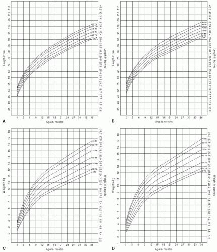
Normal rates of childhood growth have been extensively studied and are recorded in standard anthropometric charts. Length and weight percentiles for birth through 36 months are presented in Figure 13-2A-D. A slow rate of growth is of concern when the child’s height is less than the third percentile for age, that is, less than three standard deviations below the mean for that age. Growth rate should also be of concern when the child’s height drops below a previously established growth curve because this may be an indication of an underlying pathology or metabolic problem.


SPINAL PROBLEMS IN CHILDREN

The diagnosis and treatment of spinal problems in children is a specialized task and should be undertaken with great care to ensure that a serious problem is not masquerading as a relatively minor complaint. Spinal problems in children can be difficult to diagnose. In infants, spinal problems may present simply with irritability, poor feeding, and restlessness. Spinal pain can have many causes, such as birth trauma; trauma from falls, motor vehicle accidents, or child abuse; acquired torticollis; spondylolysis or spondylolisthesis; infections associated with meningitis; vertebral osteomyelitis or diskitis; and juvenile chronic arthritis (JCA) formerly known as “juvenile rheumatoid arthritis”. The causes of spinal pain, therefore, include not only the ubiquitous vertebral subluxation, but also disease processes that directly attack or refer symptoms to the vertebrae and joint structures.

Symptoms of back and neck pain in children can sometimes be difficult to interpret because of difficulty in communicating effectively with young patients and because such symptoms do not always appear to be associated with or to specifically relate to spinal problems. As a child’s age increases, so does their ability to communicate more effectively, thereby assisting the clinician in identifying the location and intensity of the symptoms and any associated activity that might have caused the problem. When evaluating the various spinal problems in children, one should be well aware that childhood back pain maybe caused by a wide range of disorders. Though studies have shown that adult back pain most often has a non-pathological cause, back pain in children is more frequently associated with identifiable pathology (31).

The spine in children is extremely flexible because of the elasticity of the muscles, ligaments, and intervertebral discs. Such increased elasticity provides for increased joint ranges of motion, which allow for the extreme spinal positions associated with normal childhood activity.

Increased elasticity of spinal capsular ligaments, coupled with the falls and trauma of childhood, can predispose young children to vertebral and pelvic subluxation (see Chapter 3). Around the time of puberty, the spine gradually loses some of its elasticity and continues to lose flexibility as the teenage years progress.

During the adolescent phase, the developing spine is rapidly gaining strength through increasing bone density and progressive ossification, which results in the closure of more epiphyseal growth centers. The adolescent patient will go through phases of rapid
growth wherein height might increase by 6 in. in as many months. This rapid growth phase, known as the “adolescent growth spurt,” is associated more with growth of the lower extremities than it is with growth of the spine, as noted above. Growth of the spine remains fairly constant throughout the adolescent period, growing at about the same rate from the age of 5 years onward. The adolescent phase is also a time when body structures are subjected to increasing trauma through sporting activities, increasing body weight, and increased velocity of movement. The effect of these changes can amplify any existing congenital or acquired spinal weaknesses, such as facet tropism, spondylolysis, spondylolisthesis, or the results of previous spinal trauma. Spinal problems in children can be due to many causes and can be categorized by age group (Table 13.1). Efficient proprioception from all joints is needed to reduce the risk of injury.


FIGURE 13-2 A: Length by age percentiles for girls aged birth to 36 months. B: Length by age percentiles for boys aged birth to 36 months. C: Weight by age percentiles for girls aged birth to 36 months. D: Weight by age percentiles for boys aged birth to 36 months. (Reprinted from Hamill PW, Drizd TA, Johnson CL, et al. Physical growth: National Center for Health Statistics percentiles. Am J Clin Nutr 1979;32:609-612, with permission.)


TABLE 13.1


Causes of Back and Neck Pain by Age Group


Infants (0-2 y)


Trauma/subluxation; falls, motor vehicle accidents, child abuse; congenital deformities


Spinal cord malformation; tethering of the cord


Spina bifida


Klippel-Feil syndrome


Malignancy; spinal cord tumor


Birth trauma: rotation, lateral flexion or traction of neck


Infection; vertebral osteomyelitis, diskitis


Torticollis


Pre-school and grade-school years (3-10 y)


Trauma/subluxation; falls, motor vehicle accidents, child abuse


Infection; vertebral osteomyelitis, diskitis


Spondylolysis, spondylolisthesis


Tumor: leukemia


Juvenile chronic arthritis


Torticollis


Adolescent years (11-19 y)


Trauma/subluxation: sports injuries, overuse syndrome, motor vehicle accidents


Poor posture


Spondylolysis, spondylolisthesis


Facet tropism (overuse syndrome)


Scoliosis


Ankylosing spondylitis


Scheuermann’s disease


Infection; vertebral osteomyelitis, diskitis


Tumors (benign): osteoid osteoma


Tumors (malignant): osteoblastic sarcoma, Ewing’s sarcoma


Birth Trauma

The trauma of the typical medical birth process, with its commonly applied rotation, lateral flexion, and traction of the cervical spine, has been identified as a possible cause of neurological trauma as well as vertebral misalignment with subluxation/fixation of the upper cervical spinal segments. Spinal problems associated with birth trauma have been associated with poor feeding, regurgitation, fussiness, tremors, and/or sleeping difficulties in the newborn infant. Atlas (C1) or occipital vertebral subluxation may be an etiological factor for a newborn infant who has difficulty feeding, who spits up consistently during and after each feeding, and who sleeps for only short periods of time. The problem with an infant who will only nurse from one breast and refuses or is difficult to feed on the opposite side can sometimes be corrected by restoring normal function to the involved dysfunctional cervical spinal segment(s).


Falls

It is not uncommon for infants to suffer trauma from falls encountered while negotiating their way through the developmental processes of the first 2 years of life. Such traumas may be caused by rolling off a bed or changing table, crawling onto a stairway, or the monumental task of learning to walk. All of these examples can be common situations encountered during the infant’s life that predispose the child to spinal problems and warrant regular spinal examinations. With increased mobility, growing children will encounter an even greater number of traumatic situations that may affect the spine, possibly resulting in back or neck pain or asymptomatic joint subluxation. At this stage of life, the spine is still undergoing secondary ossification; that is, the vertebral structures are still largely made up of cartilage, which is progressively ossifying. Because of the presence of such an abundant mass of cartilage and its relative flexibility, young children are less disposed to vertebral fractures. A further characteristic of the young spine is the comparative elasticity of the intervertebral ligament structures. Though such increased elasticity provides a child with increased joint ranges of motion, it can also be the cause of vertebral subluxation, hypermobility, or dislocation.


Motor Vehicle Injuries

Motor vehicle accidents (see Chapter 20) frequently involve the entire family. However, it is not uncommon for infants and children involved in such incidents to go unchecked, either medically or by a chiropractor, simply because they have no overt symptoms. This is in contrast to the parents, who will receive an appropriate
clinical evaluation. Because of the increased flexibility of an infant’s spinal column, spinal trauma such as that which might be encountered during a motor vehicle accident is more likely to result in spinal cord injury with associated neurological symptoms rather than result in severe damage to the motion segment soft tissues, although this is extremely variable. It is important that every occupant involved in a motor vehicle accident, whether old or young, have a physical examination to check for the presence of such potential injuries. The potential for a relatively minor stiff neck after an auto accident to be the precursor to long-term spinal degeneration, which may eventually destroy the alignment, biomechanics, and integrity of joint structures, particularly in the cervical spine, must be recognized.

Accident death rates for pediatric motor vehicle occupants are extremely high. The highest death rate for infants occurs when an unrestrained infant is riding on the lap of an adult. The reason for such a high mortality rate is because the child’s effective weight, which is markedly increased during the deceleration phase of a collision, far exceeds the ability of the adult to hold onto the child. Furthermore, the weight transfer of the adult during the collision may crush the infant against the interior of the vehicle. In many areas, laws require all passengers in automobiles to be restrained by seat belts, and those children who are too small for seat belts are required to be properly secured in government-approved car seats. The highest death rate for motor vehicle injuries is among the adolescent population. Although teenagers travel fewer overall miles than adults, their lack of driving experience, coupled with excessive driving speed and poor judgment, has been identified as the likely cause of this increased mortality.

Infants and young children who are involved in auto accidents may suffer symptoms such as irritability, lethargy, poor feeding, repeated squinting of the eyes, and restlessness. Symptoms such as these may be the only evidence that a young infant has suffered injuries. In older children who are better able to communicate, symptoms may include fatigue, irritability, headache, and/or neck pain with or without restricted range of motion. Injuries suffered by a child in an auto accident can be the cause of spinal problems that may not be evident for months or, in some cases, years after the incident. But, worse than this: the children involved may have suffered significant spinal injuries that require immediate chiropractic attention but remain quite hidden because of the inability of children to communicate their symptoms.

The need for a detailed and thorough examination of the spine of all children involved in auto accidents is obviously of great importance because of an immature spine. Because children have greater flexibility in their neck, they are more likely to suffer spinal cord damage rather than fractures of the spinal vertebrae. This is because of the different rates of stretch between the spinal column—the vertebrae, muscles, and ligaments— and the spinal cord, which carries all of the important nerve impulses to and from the brain. The spinal column has been shown experimentally to have up to 5 cm (2 in.) of stretch before vertebral or ligament damage occurs, whereas the spinal cord residing inside the spinal column has only 1.25 cm (0.5 in.) of stretch before rupture and hemorrhage can occur (31). As a result of this difference in the rate of stretch between these two structures, young children are more likely to suffer spinal cord and nerve damage rather than fractured vertebrae or spinal ligament injuries. As a result of this potential for children to suffer spinal injuries without any apparent symptoms, it is important that all children involved in auto accidents, no matter how minor, receive a detailed and thorough spinal examination to ensure that a proper differential diagnosis has been made. It should include a detailed and thorough examination of the nervous system to rule out any possible spinal cord damage. The neurologic component of this examination requires evaluation of cranial and peripheral nerves for both motor and sensory deficits. In very young infants (younger than 6 months of age), evaluation of the primitive reflexes will also provide additional information about the status of the nervous system (see Chapter 6). Parents sometimes ignore the possibility of pediatric injury that can lead either to short-term effects on the child’s health, long-term problems such as premature spinal degeneration, or both. The long-term costs of ignoring the child’s need for a spinal examination can be significant and may destine the child to a future of spinal pain, stiffness, spinal degeneration and instability, headaches, and other associated problems.


Child Abuse

Child abuse should be suspected in any infant presenting with injuries that do not correlate with the case history data (see Chapter 16). Injuries caused by abuse may sometimes be the result of the frustration of parents to control an infant’s constant crying or other persistent symptom. Some infant habits may have a physical cause that can include early spinal trauma. All cases of non-accidental trauma must be recognized and reported by the attending primary care physician in a manner that is consistent with the laws of the physician’s particular jurisdiction. Child abuse incidents may be the cause of complaints of back and neck pain in children who are brought in for treatment by a parent or guardian. In its simplest form, child abuse may be the cause of joint injuries or of recurrent headaches that have no other clinical explanation. More severe problems, such as fractures and joint dislocations, can be the result of
increasingly violent trauma inflicted upon a child by a parent or guardian. Fractures are the most common result of child abuse in the toddler, and skeletal x-rays are indicated in any case where suspicion of child abuse is present (32). Occasionally, an x-ray finding of an old, healed fracture site that does not correlate with the history and for which detailed questioning cannot uncover the cause may be evidence of previous or ongoing child abuse. Skeletal injuries can be dated so that the date of injury can be accurately estimated. The radiographic appearance of a healing bone after acute trauma follows a predictable pattern: 0 to 5 days, radiolucent fracture line is apparent but there is no evidence of healing; 5 to 10 days, periosteal new bone formation is evident; 10 to 14 days, soft callous formation; 14 to 21 days, hard callous formation (33).


PROBLEMS OF THE NECK AND CERVICAL SPINE

Children can present with an array of problems associated with the cervical spine. This section includes a discussion of problems such as torticollis, Klippel-Feil syndrome, and developmental anomalies such as spina bifida, hemivertebra, and odontoid anomalies. In addition to descriptions of these classic conditions, this chapter also includes discussion of other significant problems of the newborn cervical spine, as well as complaints of neck pain in children after involvement in an automobile accident, because this can be a significant cause of cervical spinal trauma (34).

Newborn infants can have subluxated spinal vertebrae. The upper cervical complex is an area of the spine that is frequently subjected to significant trauma during the birth process. Studies have identified the cervical spine and the atlanto-occipital junction as the locations of multiple clinical findings affecting the newborn infant. Published studies have identified signs and symptoms of suboccipital strain/sprain in newborn infants and have highlighted the apparent clinical effectiveness of spinal adjustments in correcting the problem (35).

The prevalence of neck pain and headaches in pre-adolescents is quite high. Hellstenius (36) found a prevalence of 40%, with girls predominating in the 12-to 13-year-old age range. Below that age, the prevalence was equal between boys and girls. In many preadolescents the pain was severe or frequent enough that it affected their daily activities. This study also found that 60% of the children with headaches and/or neck pain had a history of head trauma. An interesting finding by Hellstenius (36) was that the children had self-reported considerably more frequent neck pain and headaches than their patients reported.


KISS Syndrome

Kinematic imbalances caused by suboccipital strain, otherwise known as KISS syndrome, identify the pathogenic potential of the craniovertebral junction to produce a constellation of symptoms and clinical findings. The most common clinical findings associated with KISS syndrome include restricted motion of the cervical spine, torticollis, scoliosis of the cervical spine, asymmetric paraspinal muscle development, delayed development of the femoral heads and acetabulae, and asymmetrical or slow development of motor skills. Other findings include facial scoliosis, opisthotonus (retraction of the head and arching of the back, with the infant unable to hold the head erect), deformities of the feet, restless sleep, and poor feeding (35).

The most common etiological factors causing suboccipital strain include intrauterine malposition of the fetus, the use of forceps or vacuum extraction during the birth process, prolonged labor, and multiple fetuses. The incidence of these risk factors in affected infants varies significantly from the established normal birth statistics (35). If one child suffers from KISS, there is an increased risk that other children of the same sex in the family will have KISS syndrome as well (37).

KISS has been divided into two groups. KISS I, or fixed lateral flexion, is characterized by torticollis, unilateral microsomia, asymmetry of the skull, C-scoliosis of the neck and trunk, asymmetry of the motion of the limbs, asymmetry of the gluteal area, and retardation of motor development on one side. KISS II, or fixed retroflexion, is characterized by hyperextension while asleep, asymmetrical occipital flattening, shoulders pulled up, fixed supination of the arms, inability to lift trunk from the prone position, orofacial muscular hypotonia, and difficulty with breastfeeding on one side. Sometimes the two types may be combined. The most common combination is a significant scoliosis combined with retroflexion (37).

Correcting subluxation/fixations of the occipitoatlantal and the atlantoaxial joints of the cervical spine simplifies and shortens the course of the infant’s problems and significantly reduces the need for physiotherapy. Indications for spinal adjustment in affected infants depend on first recognizing the clinical symptoms and then confirming them with the physical and radiologic findings. Radiographic findings can be used to accurately evaluate the alignment of the atlanto-axial and occipitoatlantal joints as an aid in determining the most appropriate direction of adjustment, as well as in ruling out the presence of any spinal deformities. Reducing asymmetry of the cervical spine in an infant is important for proper development of proprioceptors in this area (37).

Identification of suboccipital strain requires delicate digital palpation. Initial indications may be increased pain sensitivity of the suboccipital and upper cervical
spinal regions and/or restricted movements of the head and neck. The atlas vertebra fixed on the right side relative to the occiput has been reported in several studies to be the more frequent finding (35,38). Suboccipital strain can be managed effectively with specific adjustments of the cervical spine, usually only with a few treatments (35).

Biedermann (37) states that the neural imprint of the memory of the postural asymmetries may continue throughout life even if the problem seems to have been treated successfully. During times of stress or rapid growth, the asymmetry may return temporarily (37).


Torticollis

Torticollis is characterized most commonly by unilateral spasm or contracture of the sternocleidomastoid (SCM) muscle. This condition is recognizable in a patient by the tilting of the head to one side while the chin is rotated away to the opposite side. Torticollis is generally classified as either congenital or acquired. The etiological factors include birth trauma, especially with breech birth and forceps delivery; spinal cord tumor; and congenital spinal anomalies such as hemivertebra in the cervical spine and subluxation of a cervical segment. Atlantoaxial rotatory subluxation is now a wellrecognized cause of childhood torticollis. Such rotatory displacements may occur spontaneously in-utero or be associated with trauma (39). Subluxations in other locations of the spine can also result in torticollis or lateral head tilt presentation.

Congenital Torticollis Congenital torticollis, also known as congenital wryneck, may be present in the newborn infant or may appear within the first few weeks of life. Forty percent are associated with difficult deliveries (40). The characteristic positional changes, caused by spasms of the SCM and adjacent muscles, may be noted at birth or, in some cases, may not be evident until the infant is 2 to 4 weeks of age. Congenital torticollis commonly is associated with hematoma and unilateral fibrotic contractures in the SCM, possibly caused by stretching trauma during the birth process or by the position of the head in-utero. If fibrotic contracture or a hematoma is the cause, then a mass, usually 1 to 3 cm in diameter, should be palpable in the involved SCM muscle. If no mass can be palpated in the SCM, then x-rays of the cervical spine should be obtained to evaluate other possible causes of congenital torticollis. Congenital torticollis is frequently found in conjunction with CHD, and all newborns with congenital torticollis should be examined for this associated hip condition. Other causes of torticollis may include subluxation of the cervical or thoracic spine. Less commonly, vertebral dislocation, spinal cord tumor, or osseous developmental anomalies associated with hemivertebra, Klippel-Feil syndrome, and Sprengel’s deformity may be the cause of congenital torticollis. Newborn infants with torticollis accompanied by a mass in the contracted SCM should undergo a treatment of chiropractic adjustments where indicated and passive stretching of the involved SCM. This protocol nearly always is successful if started before the age of 1 year. Newborns without a detectable mass in the SCM and without x-ray evidence of congenital anomalies of the cervical spine should be evaluated for subluxation in the cervical spine or mid to upper thoracic regions. If subluxation is present, the newborn should be treated with specific adjustment of the involved spinal motion segment(s).

Medically, treatments of congenital torticollis can range from neurodevelopment stretching to surgery. Surgery can include cervical muscle denervation or fusion (40).

Stone-McCoy et al. (41) describe a 4-month-old girl who had presented to the chiropractic office with congenital torticollis. Previous care had included physical therapy, cranio-sacral therapy, and myofascial release therapy, all of which provided limited improvement. She had also been fitted with a tubular orthosis for torticollis collar. The infant had a left head tilt with severely limited left head rotation and resistance when passively flexing the left upper extremity. The birth of this infant had been difficult and prolonged. Based upon the spinal analysis, a toggle adjustment of the left atlas was performed. During the second visit, both the atlas (toggle) and C5 (activator) were adjusted. Gradually, the left shoulder motion improved, as did cervical rotation and head tilt. Over the course of nine visits within a few weeks, improvements continued, with occasional exacerbations and various segments adjusted throughout the spine. At the time the report was written, which was 4 months after the initiation of care, the infant received care on an as-needed basis (41).

Colin (40) writes of a 7-month-old boy who was diagnosed with muscular congenital torticollis. He was premature by 3 weeks, jaundiced, and had no sucking reflex. During chiropractic examination, his head was tilted laterally to the left and flexed to the chest. He could not sit up by himself. Passive head movement did not elicit distress. A vertebral subluxation was found at C3-C4 and various asymmetries were found throughout the spine, torso, and extremities. After he was analyzed, a light adjustment was given at C3-C4 and light pressure was applied to the pelvis, hips, temporomandibular joint (TMJ), and the cervical, thoracic, and lumbar spine. Myofascial release was done to the spinal musculature. After the initial care he was able to sit and was more relaxed. On the third visit he had regressed
slightly. Adjustments began in the pelvis, which seemed to aggravate the torticollis. TMJ and cranial adjustments improved it. Cervico-thoracic adjustment allowed him to sit and the torticollis to improve. Over a period of 6 weeks, with periods of slight regression, he improved: his head was more centered, he was able to sit, although he was somewhat unstable, and he began to roll over and pull himself up (40).

Pederick (42) presented a case of a 7-month-old boy who had a difficult birth and could not turn his head to the right or extend it. He also had a misshapen and large head. There was tension in the right SCM and scalene muscles, which elicited an avoidance reaction when lightly palpated. Cranial adjusting and fascial release was employed. Quite rapidly, movement of the head improved and head shape became more symmetrical. Unfortunately, a few months later, he died of sudden infant death syndrome (42).

Toto (43) described a 7-month-old boy who had a significant left head tilt since birth. He regurgitated several times a day and had weekly projectile vomiting. He also had ear infections since birth. Myospasms were noted on the left SCM and trapezius muscles. Mild facial asymmetries were also found. He could not rotate nor laterally bend his head to the right. The left side of the atlas and the C3-C4 vertebrae were adjusted. Trigger point and passive stretching were done on the aforementioned muscles. He was seen initially three times per week over a 3-month period. The head tilt and muscle spasms decreased. After 2 weeks of care he cried less and began laughing more frequently. After 5 months of care, head tilt and muscle spasms had resolved (43).

Guttman (44) found atlantal “blockage” in a 10-monthold child with congenital torticollis who was developing facial and skull asymmetries as well. After manual correction of the blockage, the infant gained improvements in posture, motor responses, mental and linguistic development, and facial and skull asymmetries (44).

Acquired Torticollis Acquired torticollis is a condition not present at birth but has its onset at a later time, months or years after birth. Most cases of acquired torticollis among older children have sudden onset and may follow strenuous activity, mild trauma, or sudden change in neck position. Significant spasm of the SCM can be identified and tenderness can be elicited in the belly of the muscle and in the area of the synovial articulations on the uninvolved side. X-rays will indicate a significant scoliosis in the cervical region, which may account for the tenderness and irritation of the ligamentous structures on the convex side of the spinal curve. Most medical pediatric textbooks now list the most common cause of acquired torticollis as rotational subluxation of the atlanto-axial complex.

Although the upper cervical spine can be involved in a sizable number of torticollis cases, it is important to examine the full spine carefully. Subluxations in the thoracic and lower cervical spine can also cause torticollis or lateral tilt presentations. If the patient’s posture appears to be more of an antalgic lean caused by a lower cervical/upper thoracic subluxation, the pain will be provoked on the side the patient leans away from when the doctor attempts to passively straighten the patient’s neck and head. Where the torticollis is caused by upper cervical subluxation, the spasming SCM is painful on the ipsilateral side of tilt when the doctor attempts to passively straighten the patient’s head and neck.

Other less common causes of acquired torticollis include intervertebral disc calcification, retropharyngeal infections, osteomyelitis of the cervical vertebrae, drug reactions, spinal tumors, and atlanto-axial instability as seen in Morquio’s and Down syndromes. The use of radiographic studies and bone scans usually will be sufficient to identify any of the above-mentioned causes of acquired torticollis.

Older children with acute torticollis may have vertebral subluxation(s), typically in the lower cervical and upper to mid-thoracic spine, which usually respond quickly to care. A positive response is usually seen after the first adjustment unless it is chronic. If needed, gentle manual traction can be applied to the contracted myofascial elements. In mild cases, torticollis and postural signs should substantially be resolved during the first week of care. The nature of the injury will determine the need for follow-up care to decrease the findings of subluxation. Particularly in colder climates, a soft collar worn during the initial few days of care may help to provide support and keep the neck muscles warm, although most cases can be managed without such appliances.

Moore and Pfiffner (45) describe a case of a 4-year-old boy who presented with acute traumatic torticollis. The child had fallen off of the bed and hit the floor head-first. He had his head in left lateral flexion and slight right rotation and had a moderate level of pain. Turning the head was painful. Muscle spasms were noted in left trapezius and cervical paravertebral muscles. X-rays showed a left lateral tilt of the head, left lateral mass of the atlas overhanging the C2 facet, and asymmetry of the paraodontoid space. C3-C4 was adjusted using a diversified cervical spine adjustment. Follow-up 2 weeks after the adjustment showed complete resolution of signs and symptoms of torticollis (45).

Bolton and Bolton presented three cases of acute torticollis in adults. All three patients presented with sustained lateral head tilt and severe neck pain. In all cases, the problem was resolved with a toggle-recoil adjustment to the atlas. The direction of adjustment was based on examination and x-ray findings (46).


Klippel-Feil Syndrome

The majority of cervical spine anomalies occur between the occiput and C2. Below the C2 level, developmental changes usually occur as multiple segmentation anomalies, of which, Klippel-Feil syndrome is an example (47). Klippel-Feil syndrome is a congenital spinal condition that is characterized by a short neck and limitation of head and neck movement. This rather characteristic picture is caused by failure of segmentation of the developing cervical vertebrae with the presence of multiple block vertebrae. Clinically, the neck appears short and the hairline may appear low. Cervical ranges of motion are restricted, and the neck may also be webbed. This webbing phenomenon is known as pterygium colli. Radiographs will reveal the underlying bony abnormality of block vertebrae at multiple levels (see Chapter 4). The presence of multiple block vertebrae may lead to hypermobility at the remaining intervertebral motion segments. Other anomalies frequently associated with Klippel-Feil syndrome include Sprengel’s deformity (described later in the chapter), hearing impairment, and genitourinary, cardiopulmonary, and/or nervous system anomalies.


Odontoid Anomalies

Anomalies of the odontoid process of the C2 vertebra are well-recognized in the radiologic literature. The true incidence of anomalous defects in the C2 vertebra is unknown, but certainly the prevalence is quite low. Disorders such as Klippel-Feil syndrome, Down syndrome, and Morquio’s syndrome have a higher incidence of odontoid anomalies than can be identified in the general population (48). Anomalies of the odontoid process may include congenital agenesis or hypoplasia, ossiculum odontoideum, and ossiculum terminale.

Agenesis or Hypoplasia The odontoid process is formed from the first and second cervical somites. Failure of separation between the anterior arch of C1 and the odontoid process can lead to failure of formation or hypoplasia of the odontoid process. This condition can be associated with instability of the atlanto-axial region. Radiographic flexion/extension views should be obtained to evaluate for possible instability, and orthopedic consultation and opinion should be sought. Spinal manipulation to this area in the presence of instability is contraindicated.

Ossiculum Odontoideum Ossiculum odontoideum is considered to have either of two causes. Traditionally, os odontoideum was considered to be a congenital non-union of the dens at the neurocentral synchondrosis; however, more recent explanations suggest that the presence of an identifiable instability at the base of the dens is caused by traumatic disturbance of the growth plate and possibly osteonecrosis, giving the appearance of an ununited fracture (49,50). The atlas transverse ligament is usually intact in this condition. However, symptoms caused by cord compression or vertebral artery compromise may be present. Radiographic evidence of an os odontoideum in a young child is based on the finding of an odontoid process that is unstable relative to the base of the C2 vertebra. This is best identified on cervical flexion-extension studies. Surgical stabilization of an unstable odontoid process should be provided as an option for the patient because further trauma may compromise the spinal cord and/or vascular structures in this region.

Ossiculum Terminale Ossiculum terminale is a condition in which the tip of the odontoid fails to unite with the rest of the dens. Secondary ossification in this region is usually complete around the age of 12 years. Before this time, the odontoid tip may not be seen on plain film radiographs. In its place a V- or Y-shaped odontoid groove may be seen. Little clinical significance is attached to this anomaly, and no instability is associated with its presence.


Skull Asymmetry

Chiropractic care has been of benefit in many instances of skull asymmetries or deformational plagiocephaly. Many cases seem to be associated with cervical spine dysfunction (51). A retrospective study of 25 patients with non-stenotic, deformational plagiocephaly who were receiving chiropractic care found complete resolution of the condition in 2 to 5 months with an average of 1.8 adjustments. Vertebral subluxation complexes were found in the cervical spine and pelvis. No description of the adjustments given was discussed (51).


PROBLEMS OF THE THORACIC SPINE AND TRUNK


Scoliosis

Few problems affect the thoracic spine and trunk of children, but those that do can represent serious disabilities that may significantly affect the child’s future well-being. Possibly the most serious long-term disability of the thoracic spine and trunk that can afflict a child is scoliosis. Because scoliosis is a physical finding and does not represent a diagnosis, its etiology should be investigated in all cases and its classification established before commencing treatment (see Chapter 14). Scoliosis can be readily detected by postural evaluation,
and many cases of scoliosis are found during routine chiropractic physical examinations. Scoliosis screening is such an effective process for locating previously unidentified cases of scoliosis that screenings have become a regular occurrence in most schools.

Adolescent idiopathic scoliosis is the most common form of scoliosis and is a classification that is reserved for those scolioses that cannot be classified into any other appropriate category. The label idiopathic scoliosis may therefore be considered to be a diagnosis of exclusion. Adolescent idiopathic scoliosis has no associated spinal pain; therefore, any patient who present with scoliosis and also complains of back pain should be carefully evaluated for an alternative cause. This type of scoliosis is more common among girls and tends to progress more rapidly during an adolescent growth spurt.

Congenital scoliosis is associated with failure of the spine to form appropriately during embryological development. It may be because of specific vertebral anomalies, such as hemivertebrae or improper segmentation of the vertebral structures. Congenital scoliosis frequently presents concurrently with other developmental anomalies, such as genitourinary anomalies, cardiac anomalies, and spinal cord tethering. The goal of any management program is to prevent the progression of the scoliosis. Classically, bracing has been the method of choice to prevent further progression of spinal curvatures associated with congenital scoliosis. Initial evaluation and progressive monitoring of congenital curvatures, especially small ones, is the appropriate course of action. Some congenital spinal curvatures can be non-progressive, but this can only be determined by evaluation over a period of 6 to 12 months. The detailed evaluation and management protocols for the child with scoliosis are delineated elsewhere in this text (see Chapter 14).

At this point, it is sufficient to point out that not all cases of scoliosis destine the child to a life of physical deformity. For each case of scoliosis, a careful and thorough evaluation must always be carried out to identify the type of scoliosis, and regular follow-up evaluations must be made to assess the appropriateness and success of the selected program of scoliosis management.


Ankylosing Spondylitis

Ankylosing spondylitis (AS) is a chronic inflammatory condition of unknown etiology that primarily affects the joints of the spine and pelvis. AS is classified as a sero-negative spondyloarthropathy. The sero-negative classification is defined as the absence of antinuclear antibodies and rheumatoid factor in the blood; however, these patients frequently are positive for the human leukocyte antigen (HLA) B27. The pathophysiology of AS involves chronic inflammation of the cartilage, ligaments, and synovial linings of the joints. AS is a painful, progressive arthritic condition that occurs mainly in men between the ages of 18 and 30 years. AS is four times more common among men, with the overall incidence being reported at about 30 cases per 100,000 (52). Frequently the diagnosis of AS is difficult because back pain is not always present early in the course of the disease. Characteristic clinical findings include night pain and morning stiffness in the hips and spine, which is relieved by physical activity. Exquisite tenderness and inflammation of the tendons, especially at the insertion of the Achilles and patella tendons, has also been described as a common early clinical finding with AS (53).

Presenting symptoms may be low-back pain and stiffness, particularly around the sacro-iliac joints, or lower extremity joint pain, characteristic of the arthritides. After initially involving the sacro-iliac joints, this disorder progresses upward along the vertebral column to involve the lumbar, thoracic, and, in some cases, the cervical spine. This disorder eventually results in bony ankylosis of the spinal joints, with the inflammatory process eventually subsiding after about 10 to 15 years.

The best clinical diagnostic indicator has been suggested to be low-back pain and stiffness lasting for 3 months. Thoracolumbar pain has been shown to be a poor indicator for the diagnosis of AS (54). During examination, marked limitation of motion is usually encountered in the involved areas of the spine. Sacroiliac joint involvement can be identified by palpation and provocative stress testing. Restricted breathing capacity may be caused by involvement of the costo-vertebral joints. Permanent stiffness of the affected joints usually results. The earliest radiographic changes show a fuzziness of the sacro-iliac joints caused by loss of cortical bone at the iliac margins of the sacro-iliac joint. Destruction of the hyaline cartilage joint margins produces a joint outline that can no longer be clearly depicted, thus producing a pseudo-widening of the joint space. As joint destruction progresses, discrete areas of destruction and sclerosis occur along the iliac margins or the joint, leading to the classic “rosary bead” appearance. This is the stage in which the diagnosis of AS is usually made. Later x-ray changes may show complete obliteration of the sacro-iliac joint spaces with bony ankylosis of the lumbar, thoracic, and occasionally the cervical intervertebral joints.

Management of the patient with AS should involve activity rather than rest. Spinal adjustments may be used to maintain flexibility at the involved intervertebral motion segments. Spinal exercises have been shown to provide significant short-term improvements in spine and hip range of motion and should be instituted early in the course of treatment. In the longer term, spinal
exercises have been shown to reduce the rate at which the loss of joint mobility occurs (55). Spinal exercises and adjustments should be instituted for the purpose of increasing joint range of motion, or at least to preserve what movement remains. The patient should be advised to sleep flat on the back, on a firm mattress, with a low pillow under the head and neck to prevent exacerbating the flexion deformity of the spine. The following five assessment criteria for accurately measuring progress in the treatment of AS have been identified: cervical rotation, tragus to wall distance, lateral flexion, modified Schober’s test, and intermalleolar distance (56). These dimensions should be regularly monitored as a measure of the patient’s progress.


Postural Kyphosis

The normal thoracic kyphotic curve should be present from birth. When evaluating the thoracic spine, palpation of the alignment of this curve should reveal a smooth kyphotic curve devoid of flat spots, excessive ridges, or lateral deviation. Individual vertebrae should be assessed for symmetry and conformity with the adjacent vertebral structures and for the presence of a slight springing motion. Tenderness and edema during palpation can be indicators for a more detailed examination of the involved area, the most common cause being vertebral subluxation at that level.

Postural kyphosis is an exaggerated kyphosis seen in the thoracic spine of adolescents that is essentially caused by faulty posture. Occasionally, this posture may be habitual and be also seen in the patient’s parents or role models. This posture can be helped with range of motion exercises and usually resolves with time and without excessive treatment. In today’s era of home entertainment systems, computers, smart phones, the Internet, and videogames, postural and ergonomic recommendations are important, particularly for those with still-developing spines. A lot of young people have spines that appear to have kyphotic lumbar and have thoracic and cervical spines that appear hyperlordotic or kyphotic and anteriorly shifted.


Scheuermann’s Disease

Scheuermann’s disease is a relatively common disorder of unknown etiology that primarily affects the adolescent thoracic spine and produces pain and cosmetic deformity. It is usually associated with increased thoracic kyphosis (57). Some cases are thought to be hereditary. Some opine that vitamin A and D deficiencies might be involved (58

Only gold members can continue reading. Log In or Register to continue

Stay updated, free articles. Join our Telegram channel

May 24, 2016 | Posted by in PEDIATRICS | Comments Off on Orthopedics

Full access? Get Clinical Tree

Get Clinical Tree app for offline access