Race/ethnicity
Fitzpatrick types
Skin reactions
Asian/East Asian
II–IV
Risk of sun damage, dyspigmentation/melasma, facial sensitivity, and keloids
Black/African American/Afro-Caribbean
IV–VI
Risk of dyspigmentation, especially hyperpigmentation
Obfuscation of diagnosis due to pigmentary alterations
Xerosis
Keloids and traction/styling related hair damage
hair damage
Caucasian
I, II (rarely III)
Risk of phototoxic reactions, damage, and vascular reactivity in response to trauma and ultraviolet light exposure
South east Asian/Indian
III–V
Risk of pigmentary disturbance, especially hyperpigmentation and hypopigmentation
Xerosis
Hispanic/Latino
II–V
Risk of dyspigmentation, both hyperpigmentation and hypopigmentation, obfuscation of diagnosis due to pigmentary alterations, xerosis, traction-related hair damage, keloids, and photodamage
Native American
II–IV
Risk of dyspigmentation, both hyperpigmentation and hypopigmentation, xerosis, obfuscation of diagnosis due to pigmentary alterations, and photodamage
Middle Eastern
III–VI
Risk of dyspigmentation, both hyperpigmentation and hypopigmentation, xerosis, obfuscation of diagnosis due to pigmentary alterations, and photodamage
Normal Skin in Children of Color
Newborns of color often have hyperpigmentation of the genitalia.
The Mongolian spot is frequently noted in newborns of Black, Asian, and Hispanic descent.
Patterned pigmentation including pigmentary mosaicism, café au lait macules, and nevus depigmentosus typically develop in the first 2 years of life.
Newborn Caucasian children are lightly pigmented with light colored vellus hairs and indeterminate eye color; children of color will have only partial pigmentation at birth with darker lanugo hairs (especially Hispanic and Latino) and eye color, which may not further change with development. Cutis marmorata, a vascular pattern in the extremities which disappears with warming, may be more notable in lightly pigmented children. When very extensive at birth in children of color, this can represent a form of neonatal lupus erythematosus. Milia, sebaceous hyperplasia, and neonatal acne are common in all skin tones, races, and ethnicities. Transient neonatal pustular melanosis, which can leave extensive spotty pigmentary alteration in black infants, is more likely to be noted at birth in children of color. The scales of infantile seborrheic dermatitis will likely be pigmented in pigmented children (Fig. 4.1 ). Congenital hairy pinnae may be noted in newborns of diabetic moms or children with hyperinsulinism, but is also seen more frequently in the South Pacific, India, Sri Lanka, and Africa [2].
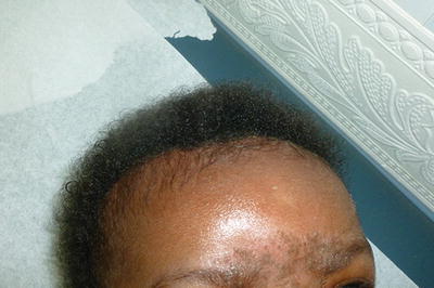
Fig. 4.1
Dark scales in a dark infant with seborrheic dermatitis
Hyperpigmentation is noted at birth or within the first few days of life in black/African-American and Afro-Caribbean children including pigmentation of the lips, fingertips, genitalia (especially in the midline), nipples, umbilicus, axillae, and anal orifice. Pigmentation persists in most areas, except the axillae, and the rest of the body will eventually develop further pigmentation to match these sites. Only axillary hyperpigmentation disappears by the end of the first year [3]. Scrotal to perineal hyperpigmentation including peri-anal is notable in most male Black infants at birth, becoming more prominent as pigmentation develops in the first 6–12 months of life.
Pigmentary development is brisk in the first 2 years of life, obviating any underlying pigmentary mosaicism, café au lait spots, and nevus depigmentosus. During this stage of rapid pigmentary development, children of Black and Hispanic/Latino descent have more pigmentary lability with extensive hypopigmentation developing in response to insults, such as seborrheic dermatitis and/or atopic dermatitis. Eventually, with age, hyperpigmentation in response to atopic dermatitis will become the predominant pigmentation response to irritation for most children who are Black or darker Hispanic/Latino.
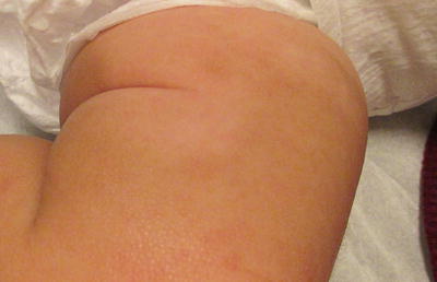
The Mongolian spot is a benign self-resolving pigmentation of newborns. The infants affected are generally over 2,500 g and usually 35 weeks of gestation or greater. The Mongolian spot is a bluish-gray pigmentation which develops due to the presence of melanocytes in the dermis. Whether these melanocytes are in the process of migration or local factors stimulate their accumulation (e.g., flexural positioning in utero) remains unknown. The dermal melanocytes create a Tyndall effect, which is a blue coloration due to pigmentary filtration. Mongolian spots are noted primarily at birth, but can appear up to a month later. The leading location is the sacrum and gluteal region and these lesions are rarely paired with port-wine stains in the phakomatosis pigmentovascularis type II. The Mongolian spot is a normal variant in children of color, found in 9.5–18.9 % of Caucasian infants, 46 % of Hispanic /Latino, 62.2 % of Indian, 83.6 % of Asian , and 96 % of Black newborns [4–6]. Eccentric lesions outside of the sacrum can be noted. Rarely, extensive lesions can be seen in the setting of a mucopolysaccharidosis such as Hunter’s or Hurler’s disease.
One can note eccentric mongolian spots/lesions over the chest, abdomen, and extremities, notably sparing the umbilicus and breast tissue. On the extremities, lesions will be noted over the extensor surfaces (especially proximally). Resolution over the first few years of life is typical, but occasional persistence into adolescence will be noted [7, 8].
Fig. 4.2
Mongolian spot over the sacrum
Café au lait macules (CALM) are localized post-zygotic gene mutations affecting pigment production. Darker ovoid pigmentation is a result of giant pigment granules and the presence around keratinocyte nuclei of melanosome complexes [9]. The presence of a CALM is statistically more common in black children under the age of 5 years than in Caucasian children of the same age range; the prevalence of café au lait in black vs. Caucasian children is for one lesion (22 % vs. 11 %) or two lesions (5 % vs. 2 %), respectively [10]. One of the diagnostic criteria of neurofibromatosis type I is 6 or more CALM of 5 mm+ in children or 15 mm+ in adults; however, neurofibromatosis is not more common in Black children.
Six or more CALM is the leading cutaneous marker of Neurofibromatosis type I (Fig. 4.3) [11]. McCune Albright syndrome can also be associated with a large segmental CALM, distributed over a large cutaneous surface area of the central chest or abdomen. CALM are usually one to two Fitzpatrick types darker than the skin tone of the observed individual. While there may be delayed appearance, most uncomplicated CALM are noted within the first 5 years of life, with slight delay in appearance for Black children due to lack of early pigmentary maturation. Fitzpatrick type VI patients may have such dark café au lait lesions as to require dermoscopy to rule out a pigmentary network of a congenital melanocytic nevus.
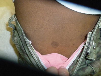
Fig. 4.3
Multiple café au lait over the back. A child with these many lesions should have a thorough family history, physical, neurological and developmental examinations as well as ophthalmologic evaluation to identify a second criterion to establish the diagnosis of neurofibromatosis type I
Ovoid hypopigmentation is more common in children of Hispanic/Latino, Indian, and Asian descent (Fig. 4.4). The nevus depigmentosus is a misnomer, as there is only a reduction of pigmentation, not an absence of pigmentation in these lesions. Sun protection may aid in avoidance of further exacerbation of the pigmentary differences. In these lesions localized increased pheomelanin to eumelanin ratio and reduced melanosome number with clumping occur. The appearance of the nevus depigmentosus when present, will be noted at birth for 50 % and almost all have formed by 1 year of life, primarily over the chest, abdomen, and extremities [