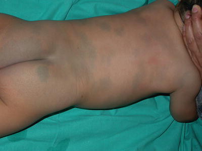
Fig. 6.1
Mongolian spot in the most common location: lumbosacral and buttock area

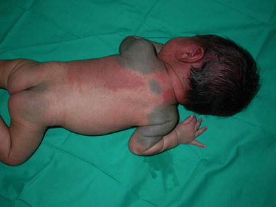
Fig. 6.2
Extensive Mongolian spot

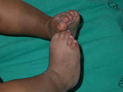
Fig. 6.3
Mongolian spot on the extremity. These Mongolian spots in locations other than the lumbosacral area have been called aberrant Mongolian spots
Mongolian spots may be seen in association with capillary malformation (port-wine stains) in the so-called phakomatosis cesioflammea (Fig. 6.4) or with segmental cafe au lait macules (phakomatosis pigmentopigmentalis) [4, 5].


Fig. 6.4
Phakomatosis pigmentovascularis (cesioflammea). The coexistence of Mongolian spot and capillary malformation (nevus flammeus)
Extensive Mongolian spots may be a manifestation of GM1 gangliosidosis and mucopolysaccharidosis type I (Hurler’s disease), and less commonly mucopolysaccharidosis type II (Hunter’s syndrome), mucolipidosis, Niemann–Pick disease, and mannosidosis [6, 7]. In these cases of associated lysosomal storage diseases Mongolian spots usually involve both the dorsal and ventral trunk, in addition to the skin of the sacrum and extremities, and may be progressive in nature [7].
Biopsy of Mongolian spots shows elongated and stellate melanocytes in the reticular dermis.
Pathogenesis
Mongolian spots probably represent abnormal migration of melanocytes from the neural crest to the epidermis. The cause of Mongolian spots is unknown.
Treatment
No treatment is usually needed
Therapy is usually unnecessary because Mongolian spots are benign lesions and tend to fade with age. There is a report on the use of Q-switched alexandrite laser and also of intense pulsed light treatment with some success [8–11]. Treatment of lesions in children of color should be undertaken carefully as the risk of hyperpigmentation and hypopigmentation with laser therapies can be quite high, especially with Fitzpatrick type V and VI patients.
Prognosis
Mongolian spots are benign. When associated with lysosomal storage diseases the prognosis is that of the associated disorder.
Stay updated, free articles. Join our Telegram channel

Full access? Get Clinical Tree


