Fig. 20.1
Pearly papules of molluscum contagiosum on the face and upper lip
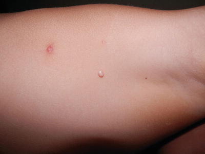
Fig. 20.2
Giant molluscum contagiosum in the axilla
Children often develop hypersensitivity reactions to the MCV that manifest as eczematous patches surrounding the molluscum papules (Fig. 20.3). These eruptions are often referred to as molluscum dermatitis. In addition, the hypersensitivity reaction can sometimes occur at distant sites (i.e., the id reaction). These eczematous reactions are often asymptomatic or slightly pruritic. Some authors believe that these id reactions represent an immunologically mediated host response to the virus and herald the onset of regression [6]. These reactions do not require any treatment other than emollients. However, in symptomatic patients, a short course of topical corticosteroids is helpful.
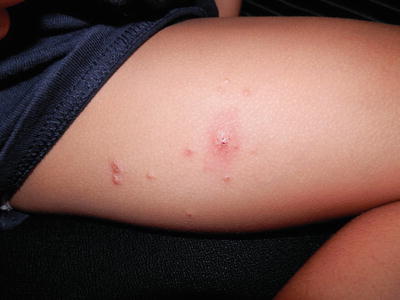
Fig. 20.3
Molluscum dermatitis on the thigh
Secondary bacterial infection is an uncommon complication.
Management
Molluscum contagiosum resolves spontaneously over several months to years
In younger children, relatively painless treatments like topical tretinoin, topical salicylic acid, and imiquimod cream are good options
In older children, curettage, cryotherapy, and cantharidin are effective options
The diagnosis of molluscum is usually made clinically. Differential diagnoses include plane warts, milia, and papular eczema. Rarely coccidioidomycosis or other fungal infections of the immunocompromised host can mimic molluscum. In uncertain cases, the presence of intracytoplasmic eosinophilic inclusion bodies, also known as molluscum bodies, on histologic examination of a skin biopsy will clinch the diagnosis.
In healthy children, molluscum contagiosum heals spontaneously within months to several years. However, parents often request for treatment as it may help to reduce autoinoculation and transmission to close contacts and improve clinical appearance.
A recent Cochrane review found that no single intervention had been shown to be convincingly effective in the treatment of molluscum contagiosum [7].
A prospective randomized study compared curettage, cantharidin, combination of salicylic acid and lactic acid, and imiquimod for molluscum contagiosum in children [8]. Curettage was found to be the most efficacious treatment and had the lowest rate of side effects. Cantharidin was a useful alternative office procedure, but had moderate complications due to blisters and necessitated more visits. The salicylic acid/lactic acid combination was effective but too irritating for children. Topical imiquimod was promising, but expensive, and the optimum treatment schedule had not yet been determined.
The infected areas should be covered with clothing or bandage and good hand hygiene should be practiced to prevent spreading to others, especially during contact sports. Personal items like towels and clothing should not be shared. Bath sponges should be avoided due to risk of autoinoculation.
Office Treatment
Cantharidin 0.7 % in flexible collodion is a vesicant that should be carefully applied to the molluscum contagiosum papules with a wooden stick and allowed to dry thoroughly. It is a quick, painless, and effective treatment, but causes blisters on the sites of application. It should be applied with great caution to flexural areas, like the axillae, cubital, and popliteal fossae, because of the risk of extensive blistering. The extent of blistering and discomfort varies in different individuals, and hence the initial treatment should be done conservatively. The blistering is limited to the epidermis, and does not scar unless there is secondary infection [9–12]. Several treatment sessions are usually required for complete clearance. Unfortunately, cantharidin is not readily available in some countries.
Curettage is very effective in removing molluscum contagiosum. However, it is painful, time-consuming, and best done in a cooperative child who has received adequate topical anesthesia, e.g., with eutectic mixture of lidocaine and prilocaine 2.5 % cream. There is a risk of methemoglobinemia with usage over large surface areas, and the prescribing guide should be consulted for maximum volume that can be used in one session based on the child’s weight. For children with multiple lesions, several treatment sessions are usually required for complete clearance [13, 14].
Cryotherapy is an effective procedure. Light freezing of the lesion and minimal icing of the surrounding skin usually suffices. However, it is painful and often induces hypopigmentation in children with skin of color that may persist for weeks. Topical anesthesia is often required in younger children.
Trichloroacetic acid 12 % solution has been reported to be an effective and safe therapy for facial molluscum contagiosum in children [15].
Home Treatment
Topical keratolytic agents like tretinoin (up to 0.1 %) and salicylic acid (up to 17 %) can be applied to the papules on alternate days until an inflammatory response is produced [16].
Imiquimod 5 % cream, a toll-like receptor 7 agonist, induces alpha-interferon and cytokines upon topical application. It is applied 3–5 days a week to molluscum lesions until they become inflamed. It is slow acting and expensive but effective, painless, and a good option in young children with numerous small lesions or lesions on the face [17, 18].
Prognosis
The prognosis is very good. Molluscum contagiosum is a self-limiting infection in healthy children. Spontaneous resolution may take up to several years. Most of the lesions resolve without scarring, although some may leave slightly depressed scars.
Ongoing Research
Further research is required to better characterize MCV viability in swimming pools.
Conclusion
Molluscum contagiosum is a common viral infection in children. It usually resolves spontaneously, but treatment is often sought to reduce cutaneous dissemination and social stigma. For younger children, relatively painless treatments like cantharidin, topical keratolytics, and imiquimod are good options. For older children and adolescents, curettage, cryotherapy, and cantharidin are effective options.
Viral Warts
Introduction
Warts are caused by the human papillomavirus (HPV), which is a double-stranded DNA virus. More than 100 types of HPV have been identified [22]. HPV 1, 2, 27, and 57 are the most prevalent HPV types found in cutaneous warts in the general population. Warts caused by HPV 1 have a distinct clinical profile, being related to children aged less than 12 years, plantar location, duration less than 6 months, and to patients with less than 4 warts [23].
Warts can affect any area on the skin and mucosa. The HPV virus infects the epithelium only, and systemic dissemination does not occur.
HPV is spread by direct skin contact or indirectly by contact with contaminated surfaces. Autoinoculation may occur, causing cutaneous dissemination. The incubation period for HPV usually ranges from 1 to 6 months, but may be as long as 2 years.
Epidemiology
Prevalence of warts among children is between 22 and 33 %.
An increased risk of warts is found in children with a family history of warts.
Warts spontaneously regress in up to half of children within 2 years.
Warts are uncommon in infancy and early childhood, increase in incidence among school-aged children, and peak at 12–16 years [24]. The prevalence of warts among primary schoolchildren is reported to be 22–33 % [25, 26]. In the USA, warts are more common in white than black patients [27].
An increased risk of the presence of warts was found in children with a family member with warts, and in children where there was a high prevalence of warts in the school class [25]. The use of public swimming pools has been associated with a higher risk of infection [28].
Clinical Features
Clinical subtypes include common warts, filiform warts, plantar warts, plane warts, and anogenital warts.
Filiform warts are usually seen on the face.
Plane warts are flat-topped and flesh-colored papules that may koebnerize.
Common warts (verruca vulgaris) appear as hyperkeratotic papules with a rough, irregular surface. They can occur on any part of the body but are seen most commonly on the hands and knees (Fig. 20.4).
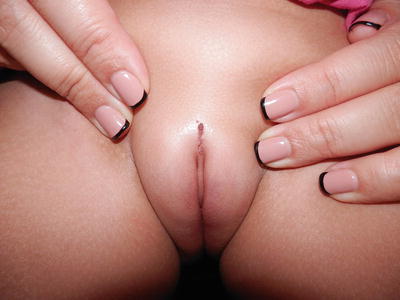
Fig. 20.4
Viral warts in genital area
Filiform warts are long and narrow growths, usually seen on the face around the lips, eyelids, or nares.
Plantar warts (myrmecia) begin as papules and progress to well-demarcated round lesions with a rough keratotic surface (Fig. 20.5). They grow deep and may be painful. Occasionally, they are covered by calloused skin; paring this away will reveal the thrombosed capillaries appearing as black dots.
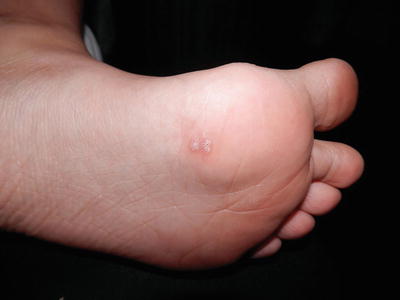
Fig. 20.5
Plantar warts
Plane warts (verruca plana) are slightly elevated, flat-topped, smooth, and flesh-colored papules. The face, dorsum of hands, and shins are the most common areas affected. These warts often exhibit the Köebner phenomenon.
Anogenital warts rarely occur in childhood. They appear as clusters of papillomatous lesions. Sexual abuse is a concern, but it is uncommon. The infection more often results from virus inoculation through perinatal transmission, autoinoculation from another body site, and infection from other family members and caregivers [31, 32].
Epidermodysplasia verruciformis is a rare inherited autosomal recessive disorder characterized by chronic HPV infection. Mutations in the EVER1 and EVER2 genes result in defective cell-mediated immunity. Warts usually appear in childhood on the sun-exposed areas. The lesions may appear like plane warts and resemble seborrheic keratoses or tinea versicolor. They tend to persist and may develop into squamous cell carcinoma in early to mid adulthood.
Management
Forty to fifty percent of warts in children resolve spontaneously within 2 years
Salicylic acid is a cost-effective treatment for warts
Regardless of the treatment used, warts may recur due to normal-appearing perilesional skin harboring HPV
The diagnosis of warts is often made on clinical findings. Paring of warts may reveal minute black dots, which represent thrombosed capillaries. A skin biopsy can be considered if the diagnosis is in doubt.
Multiple modalities are available for the treatment of warts, but none is uniformly effective [33]. For children, it is appropriate to begin treatment with the least painful methods. The more invasive procedures should be reserved for refractory warts.
Home Treatment
Salicylic acid has a proven therapeutic effect in the treatment of nongenital cutaneous warts. Compared to placebo, it significantly increased the chance of clearance of warts at all sites [34]. It is cheap, readily available without a prescription and can be applied at home. It is probably the most cost-effective treatment for warts to date. Salicylic acid should be avoided on the face and intertriginous and anogenital areas as it may cause severe irritation and erosions in these areas.
Imiquimod is an immune response modifier that has been successfully used for the treatment of common warts [35, 36]. It can be applied to nongenital cutaneous warts once or twice daily and to anogenital warts once every other day. Once the warts or the surrounding skin become inflamed, imiquimod can be temporarily discontinued, and then resumed when the inflammation settles.
Tretinoin cream has been used for the treatment of viral warts. It is probably the most effective for facial and plane warts. Sixteen out of 20 children with extensive warts who were given oral etretinate 1 mg/kg daily up to 3 months showed complete regression of the disease without relapse [37]. The use of systemic retinoids may be considered in children with widespread warts, e.g., epidermodysplasia verruciformis.
5-Fluorouracil (5-FU) is a chemotherapeutic agent that has been reported to be safe and effective in treating warts in children [38]. It is thought to work by interfering with DNA and RNA synthesis. The 5 % 5-FU cream can be applied once daily. Adverse reactions include erythema, ulceration, and hypopigmentation.
Contact sensitizers like squaric acid and dinitrochlorobenzene have been shown to be effective and safe topical immunotherapies for warts in children. They offer an alternative therapeutic option for patients with multiple recalcitrant warts. Their main side effect is allergic contact dermatitis, which may cause blistering and secondary spread in the more severe cases [39–41].
Zinc is an immune modulator that has been shown to be effective in the treatment of children with recalcitrant warts in two randomized placebo-controlled trials done in the Middle East [42, 43]. The majority of the children in the trials had low zinc levels. They were given oral zinc sulfate at a dose of 10 mg/kg daily up to 600 mg/day. The most common side effects were nausea and mild epigastric pain.
Office Treatment
Cantharidin can be applied topically to warts in the clinic and washed off after several hours. It causes localized blistering within 1–2 days. For plantar warts, a combination compound of cantharidin 1 %, podophyllotoxin 5 %, and salicylic acid 30 % is effective and safe [44, 45]. However, patients should be warned that the compound can cause significant blistering and severe pain, and the first treatment should be done conservatively.
Liquid nitrogen (−196 °C) can be applied using a cotton bud applicator or cryospray to the wart and a 1–2 mm rim of normal skin tissue around the wart. It is repeated every 2 weeks for approximately 3 months, as required [46]. It is painful and may cause blistering, hypopigmentation, or scarring after treatment. There is a risk of keloid induction with deep cryotherapy over the chest. Pigmentary alteration, especially hypopigmentation, may occur in dark patients, including blacks, dark Asians, and Hispanics. Cryotherapy must be used with caution on the sides of fingers to avoid injury to underlying structures and nerves. Cryotherapy can rarely result in a central clearing with an annular recurrence of the wart surrounding the treated area (“doughnut wart”). Cure rates ranging from 43 to 83 % have been reported. Wart clearance may be achieved through necrotic destruction of viral infected keratinocytes or by inducing local inflammation that triggers a cell-mediated immune response. Paring the wart, in addition to two freeze–thaw cycles, has been a valuable adjunct to cryosurgery for plantar warts [47].
The carbon dioxide laser produces nonselective tissue destruction. Wounds heal by secondary intention in several weeks. It is a therapeutic option for recalcitrant warts in children [48]. Local anesthesia is required and potential side effects include pain, infection, and scarring.
The pulsed dye laser targets the wart microvasculature. It appears to be an effective and safe alternative therapy of recalcitrant warts in children [49, 50]. Compared to the carbon dioxide laser, it is less painful and has a lower risk of scarring, but requires more treatment sessions.
Surgical treatment is very effective in treating older children with a few viral warts, especially filiform and common warts. These warts can be snipped, curettaged, or shaved away, followed by electrocautery of the bases. These surgical procedures carry risks of scarring, keloid formation, and dyspigmentation.
Prognosis
Forty to fifty percent of warts in children resolve spontaneously within 2 years. When warts resolve spontaneously, no scarring is seen. However, scarring can occur as a result of the more invasive treatment methods, e.g., carbon dioxide laser and surgical treatment. Periungual or subungual warts may cause permanent nail dystrophy.
Treatment failures and wart recurrences are common, more so among immunocompromised patients. Normal-appearing perilesional skin may harbor HPV, which helps to explain recurrences.
Ongoing Research
Further research is required to determine whether any therapeutic modality is more cost-effective than topical salicylic acid.
Conclusion
The viral wart is a common viral infection in children. It usually resolves spontaneously, but treatment is often sought to reduce cutaneous dissemination, and to remove painful or disfiguring warts. In the author’s experience, a combination of therapeutic modalities is often required to treat viral warts effectively and quickly. For younger children, relatively painless treatments like topical keratolytics and imiquimod are good options. For older children and adolescents, cryotherapy, surgery, or the combined cantharidin, podophyllotoxin, and salicylic acid lotion are effective options.
Tinea Versicolor
Introduction
Tinea versicolor, also called pityriasis versicolor, is a common benign superficial fungal infection localized to the stratum corneum. It is caused by dimorphic lipophilic fungus in the genus Malassezia, a component of the normal skin flora.
As Malassezia is a component of the normal flora, tinea versicolor is not regarded as a contagious disease. Predisposing factors include genetic predisposition, warm humid environments, immunosuppression, and malnutrition.
The predominant Malassezia species causing tinea versicolor varies in different populations, and most epidemiological studies were carried out in adults. In temperate climates, M. globosa appears to be the predominant species found in the lesions of tinea versicolor [51]. In a Japanese population, M. restricta overwhelmingly predominated at ages over 16–18 years in males and 23–29 years in females. M. globosa and M. restricta together accounted for more than 70 % of Malassezia species recovered [52]. In an Argentinian population, M. sympodialis and M. globosa were the most commonly isolated species [53].
Stay updated, free articles. Join our Telegram channel

Full access? Get Clinical Tree


