Menopause and the Perimenopausal Transition


Menopause and the Perimenopausal Transition


image

Throughout recorded history, multiple physical and mental conditions have been attributed to the menopause. Although medical writers often wrote colorfully in the past, unfortunately they were also less than accurate, unencumbered by scientific information and data. A good example of the stereotypical, inaccurate thinking promulgated over the years is the following written in 18872:


The ovaries, after long years of service, have not the ability of retiring in graceful old age, but become irritated, transmit their irritation to the abdominal ganglia, which in turn transmit the irritation to the brain, producing disturbances in the cerebral tissue exhibiting themselves in extreme nervousness or in an outburst of actual insanity.

The belief that behavioral disturbances are related to manifestations of the female reproductive system is an ancient one that has persisted to contemporary times. This belief regarding the menopause is not totally illogical; there is reason to associate the middle years of life with negative experiences. The events that come to mind are impressive: onset of a major illness or disability (and even death) in a spouse, relative, or friend; retirement from employment; financial insecurity; the need to provide care for very old parents and relatives; and separation from children. Thus, it is not surprising that a middle-age event, the menopause, shares in this negative outlook.

The scientific study of all aspects of menstruation has been hampered by the overpowering influence of social and cultural beliefs and traditions. Problems arising from life events have often been erroneously attributed to the menopause. But data, especially more reliable community-based longitudinal data, now establish that the increase in most symptoms and problems in middle-aged women reflects social and personal circumstances, not the endocrine events of the menopause.3,4,5,6,7,8,9,10,11 and 12 The variability in menopausal reactions makes
the cross-sectional study design particularly unsuitable. Longitudinal studies are better for documenting what is normal and the variations around normal.

The Massachusetts Women’s Health Study, a large and comprehensive prospective, longitudinal study of middle-aged women, provides a powerful argument that the menopause is not and should not be viewed as a negative experience by the vast majority of women.4,13 The cessation of menses was perceived by these women (as have the women in other longitudinal studies) as having almost no impact on subsequent physical and mental health. This was reflected by women expressing either positive or neutral feelings about menopause. An exception was the group of women who experienced surgical menopause, but there is good reason to believe that the reasons for the surgical procedure were more important than the cessation of menses.

Changes in menstrual function are not symbols of some ominous “change.” There are good physiologic reasons for changing menstrual function, and understanding the physiology will do much to reinforce a healthy, normal attitude. Attitude and expectations about the menopause are very important. Women who have been frequent users of health services and who expect to have difficulty do experience greater symptoms and higher levels of depression.5,9,10 The symptoms that women report are related to many variables within their lives, and the hormonal change at menopause cannot be held responsible for the common psychosocial and lifestyle problems we all experience. It is important to stress the normalcy of this physiologic event. Menopausal women do not suffer from a disease (specifically a hormone deficiency disease), and postmenopausal hormone therapy should be viewed as specific treatment for symptoms in the short term and preventive pharmacology in the long term.

It can be further argued that physicians have had a biased (negative) point of view, because the majority of women, being healthy and happy, do not seek contact with physicians.14,15 It is vital, therefore, that clinicians not only are familiar with the facts relative to the menopause but also have an appropriate attitude and philosophy regarding this period of life. Medical intervention at this point of life should be regarded as an opportunity to provide and reinforce a program of preventive health care. The issues of preventive health care for women are familiar ones. They include family planning, cessation of smoking, control of body weight and alcohol consumption, prevention of cardiovascular disease and osteoporosis, maintenance of mental well-being (including sexuality), cancer screening, and treatment of urologic problems.


Growth of the Older Population

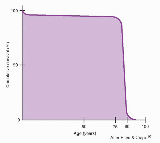
We are experiencing a relatively new phenomenon: we can expect to become old. We are on the verge of becoming a rectangular society. This is a society in which nearly all individuals survive to advanced age and then succumb rather abruptly over a narrow age range centering around the age of 85.

In 1000 B.C., life expectancy was only 18 years. By 100 B.C., the time of Julius Caesar, it had reached 25 years. In 1900, in the U.S., life expectancy still had reached only 49 years. In 2005, the average life expectancy was 80.7 years for women and 75.4 for men.16 Today, once you reach 65, if you are a man you can expect to reach 82.2, if you are a woman, age 85.17 We can anticipate that eventually about two-thirds of the population will survive to 85 or more, and more than 90% will live past age 65—this would be the nearly perfect rectangular society.18,19 Currently, Sweden and Switzerland are closest to this demographic composition.


image

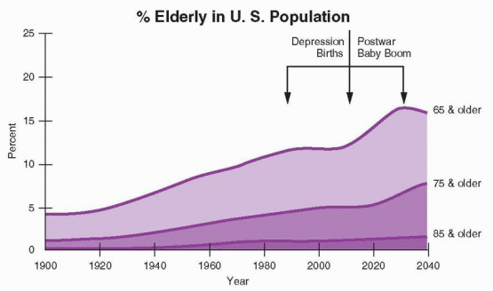
A good general definition of elderly is 65 and older, although it is not until age 75 that a significant proportion of older people show the characteristic decline and problems. Today the elderly population is the largest contributor to illness and human need in the U.S. There are more old people (with their greater needs) than ever before.21 In 1900, there were approximately 3 million Americans 65 and older (about 4% of the total population), and in 2000, there were 35 million (about 12% of the total population). By 2030, the elderly population in the U.S. will reach about 70 million, and about one in five Americans will be elderly.21 The world’s elderly population will more than double from 1998 to 2025, rising from 264 million in 2009 to 416 million in 2050.22 Population aging must be added to population growth as very important social problems.

Two modern phenomena have influenced the rate of change. The first was the post-World War II baby boom (1946-1964) that temporarily postponed the aging of the population, but now is causing a faster aging of the general population. The second major influence
has been the modern decrease in old-age mortality. Our success in postponing death has increased the upper segment of the demographic contour. By 2050, the current developed nations will be rectangular societies. China, by 2050, will contain more people over age 65 than the number of people of all ages currently living in the U.S.


image


Current World Population Changes23


Births


Deaths


Growth


Year


140,773,000


51,315,000


89,458,000


Month


11,731,080


4,276,250


7,454,834


Week


2,707,173


140,589


245,090


Hour


16,070


5,858


10,212


Minute


268


96


170


Second


4.5


1.6


2.8


This is a worldwide development, not limited to affluent societies.23 The population of the earth will continue to grow until the year 2100 or 2150, when it is expected to stabilize at approximately 11 billion. After 2020, all of this growth will occur in developing countries.22 In 2000, the poorest countries (located in Africa and Asia) accounted for 87% of the world’s population. In most developing countries, the complications associated with pregnancy, abortion, and childbirth are either the first or second most common cause of death, and almost half of all deaths occur in children under age 5. Limiting family size to two children would cut the annual number of maternal deaths by 50% and infant and child mortality also by 50%.24 Thus, it is essential to focus attention on population control, but declining fertility will increase population aging. In 1950, only 40% of people 60 and older lived in developing countries. By 2050, about 80% will live in those countries, as fertility in the developing regions is expected to drop from 2.73 children per woman in 2005-2010 to 2.05 by 2050.22
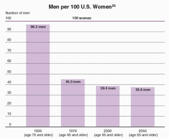
In 1900, men over age 65 in the U.S. outnumbered women 102 to 100. Now, there are only 70 men for every 100 women over age 65.25 By age 85, only 39 men are alive for every 100 women. Nearly 90% of white American women can expect to live to age 70. Vital statistics data indicate that this gender difference is similar in both the black and white populations in the U.S.26 Approximately 55% of girls, but only 35% of boys, live long enough to celebrate their 85th birthday.27 One in 5,600 individuals can expect to live to be 100.25


image


The 15 leading causes of death in the U.S. in 2006 were as follows:16


1.Diseases of the heart


— 26%


2.Malignant neoplasms


— 23%


3.Cerebrovascular diseases


— 5.7%


4.Chronic lower respiratory diseases


5.Accidents


6.Alzheimer’s disease


7.Diabetes mellitus


8.Influenza and pneumonia


9.Renal diseases


10.Septicemia


11.Suicides


12.Liver diseases


13.Hypertensive diseases


14.Parkinson’s disease


15.Homicides


Men and women reach old age with different prospects for older age, a sex differential that (it can be argued) is due in significant part to the sex hormone-induced differences in the cholesterol-lipoprotein profile and other cardiovascular factors, and thus the greater incidence of atherosclerosis and earlier death in men. From a public health point of view, the greatest impact on the sex differential in mortality would be gained by concentrating on lifestyle changes designed to diminish atherosclerosis: low-cholesterol diet, no smoking, optimal body weight, and active exercise. The death rate is higher for men at all ages, and coronary heart disease accounts for 40% of the mortality difference between men and women. Another one-third is from lung cancer, emphysema, cirrhosis, accidents, and suicides. It is interesting to note that in our society the mortality difference between men and women is largely a difference in lifestyle. Smoking, drinking, coronary-prone behavior, and accidents account for most of the higher male mortality rate over age 65. It has been estimated that perhaps two-thirds of the difference has been due to cigarettes alone. But we should emphasize that this is due to a greater prevalence of smoking in men. Women whose smoking patterns are similar to those of men have a similar increased risk of morbidity and mortality.28


The Older U.S. Female Population25


Age


1990


2000


2010


2020


55-64


10.8 mill.


(8.6%)


12.1 mill.


(9.0%)


17.1 mill.


(12.1%)


19.3 mill.


(12.9%)


65-74


10.1


(8.1%)


9.8


(7.3%)


11.0


(7.8%)


15.6


(10.4%)


> 75


7.8


(6.2%)


9.3


(7.0%)


9.8


(6.9%)


11.0


(7.3%)


Total


28.7


31.2


37.9


45.9


The mortality sex difference has been decreasing since 1979. The U.S. Census Bureau projects that the difference in life expectancy between men and women will increase until the year 2050, and then level off. In 2050, life expectancy for women will be 82 years and for men, 76.7 years.29 There will be 33.4 million women 65 and older, compared with 22.1 million men.

In addition to the growing numbers of elderly people, the older population itself is getting older. For example, in 1984, the 65-74 age group in the U.S. was over 7 times larger than in 1900, but the 75-84 group was 11 times larger and the 85 and older group was 21 times larger. In the 1990s, the population 85 years and older increased by 38%.25 The most rapid increase is expected between 2010 and 2030 when the post World War II baby boom generation will be age 65 and over. In the next century, the only age groups in the U.S. expected to experience significant growth will be those past age 55. In this older age group, women will outnumber men by 2.6 to 1. By the year 2040 in the U.S., there will be 8 million to 13 million people 85 years of age or older; the estimate varies according to pessimistic to optimistic projections regarding disease prevention and treatment.

Unmarried women will be an increasing proportion of the elderly. Elderly women are more likely to be widowed (59%) than elderly men (22%).30 Half of men 85 and older live with their wives, but only 10% of elderly women live with their husbands.31 Because the unmarried tend to be more disadvantaged, there will be a need for more services for this segment of the elderly population. Older unmarried people are more vulnerable, demonstrating higher mortality rates and lower life satisfaction.


The Rectangularization of Life

The lifespan is the biologic limit to life, the maximal obtainable age by a member of a species. The general impression is that the human lifespan is increasing. Actually lifespan is fixed, and it is a biologic constant for each species.20 In fact, differences in species’ lifespans argue in favor of a species-specific genetic basis for longevity. If lifespan were not fixed, it would mean an unlimited increase of our elderly. But a correct analysis of survival reveals that death converges at the same maximal age; what has changed is life expectancy—the number of years of life expected from birth. Life expectancy cannot exceed the lifespan, but it can closely approximate it. Thus the number of old people will eventually hit a fixed limit, but the percentage of a typical life spent in the older years will increase.

Our society has almost eliminated premature death. Diseases of the heart and the circulation, and cancers are now the leading causes of death. The reason for this is not an increase or an epidemic; it is a result of our success in virtually eliminating infectious diseases. Now the major determinant is chronic disease, affected by genetics, lifestyle, the environment, and aging itself. The major achievement left to be accomplished is in cardiovascular diseases. But even if cancer, diabetes, and all circulatory diseases were totally eliminated, life expectancy would not exceed 90 years.18


J.F. Fries described three eras in health and disease.32 The first era existed until sometime in the early 1900s, and was characterized by acute infectious diseases. The second era, highlighted by cardiovascular diseases and cancer, is now beginning to fade into the third era, marked by problems of frailty (fading eyesight and hearing, impaired memory and cognitive function, decreased strength and reserve). Much of our medical approach is still based on the first era (find the disease and cure it), and now we have conditions that require a combination of medical, psychological, and social approaches. Our focus has been on age-dependent, fatal chronic diseases. The new challenge is with the nonfatal, agedependent conditions, such as Alzheimer’s disease, osteoarthritis, osteoporosis, obesity, and incontinence. It can be argued that health programs in the future should be evaluated by their impact on years free of disability, rather than on mortality.


The Concept of the Compression of Morbidity

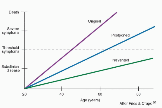
Chronic illnesses are incremental in nature. The best health strategy is to change the slope, the rate at which illness develops, thus postponing the clinical illness, and if it is postponed long enough, effectively preventing it. There has been a profound change in public consciousness toward disease. Disease is increasingly seen as something not necessarily best treated by medication or surgery, but by prevention, or more accurately, by postponement.

Postponing illness was expressed by J.F. Fries as the compression of morbidity.20,33 We would live relatively healthy lives and compress our illnesses into a short period of time just before death. Is this change really possible? A good affirmative example is the decrease in atherosclerosis in the U.S. Reasons include changes in the use of saturated fat, more effective detection and treatment of hypertension, increased exercise, and decreased smoking.


image

Physician smokers have declined from a high of 79% to a small minority.34 It is interesting, and amusing, to note that the greatest decrease has been among pulmonary surgeons, not surprising, while the least decrease has been among proctologists. From the mid 1970s to the early 1990s, smoking among physicians in the U.S. declined from 18.8% to 3.3%. Unfortunately, that still amounted to approximately 18,000 physicians who smoke. Approximately 35% of people in the U.S. who have not obtained a high school diploma are smokers, but only 12% of those with higher education are smoking, only 5.7% of those with
graduate degrees. Currently, approximately 23% of men and 18% of women are smokers.17 Cigarette smoking among high school students peaked in 1997, then declined to the current level of 20%.17 In addition, 14% of high school students smoke cigars and 8% use chewing tobacco. The use of chewing tobacco, pipe smoking, and cigars contributes significantly to morbidity and mortality. Tobacco, therefore, continues to be the single most preventable cause of premature illness and death in the U.S. It is important to note that smoking has a greater adverse effect on women compared with men.35 Women who smoke only 1 to 4 cigarettes per day have a 2.5-fold increased risk of fatal coronary heart disease.36

Physicians and older patients may be skeptical that quitting smoking after decades of smoking could be beneficial, but the effects are at least partly reversible within 1 to 5 years after quitting. In the Nurses’ Health Study, 61% of the excess risk of coronary heart disease mortality and 42% of stroke mortality was eliminated within 5 years after quitting smoking.37 The improvement in respiratory disease mortality is slower, and a small increased risk of lung cancer mortality persists even after 30 years. However, by 20 years after cessation, all the excess risk of vascular mortality and death due to respiratory diseases other than lung cancer reached the level of a never smoker. Even older patients who already have coronary artery disease have improved survival if they quit smoking.38 No matter how old you are, if you continue to smoke, you have an increased relative risk of death. But no matter how old you are, if you quit smoking, your risk of death decreases. Nevertheless, the risk of lung cancer remains elevated even in long-term ex-smokers.39

Since 1970, the death rate from coronary heart disease has declined approximately 50% in the U.S. Between 1973 and 1987 in the U.S., cardiovascular mortality declined in nearly every age group. In the combined age groups up to 54 years, cardiovascular mortality decreased 42%, and in people 55 to 84 years old, 33%.35 Despite our progress, we must continue to exert preventive efforts on the risk factors associated with cardiovascular disease, especially obesity, hypertension, and lack of physical activity.

The effort to improve the quality of life has an important value to society; it will decrease the average number of years that people are disabled and a liability. Frailty and disability are now major health and social problems of society. Most significantly, this is a major financial challenge for health care systems and social programs. With evolution toward a rectangular society, the ratio of beneficiaries to taxpayers grows rapidly, jeopardizing the financial support for health and social programs. Compression of morbidity is at least one attractive solution to this problem.


Menopause as an Opportunity

Clinicians who interact with women at the time of the menopause have a wonderful opportunity and, therefore, a significant obligation. Medical intervention at this point of life offers women years of benefit from preventive health care. This represents an opportunity that should be seized.

It is logical to argue that health programs should be directed to the young. It makes sense to create good lifelong health behavior. While not underrating the importance of good health habits among the young, we would argue that the impact of teaching preventive care is more observable and more tangible at middle age. The prospects of limited mortality and the morbidity of chronic diseases are viewed with belief, understanding, and appreciation during these older years. The chance of illness is higher, but the impact of changes in lifestyle is greater.


The Perimenopausal Transition


Definition of the Perimenopausal Transition

There is only one marker, menstrual irregularity, that can be used to objectively define and establish what is called the perimenopausal transition. This irregularity will be perceived by patients as skipped menstrual periods or longer durations (about 40 to 60 days) between periods.40 There is no universal pattern; each woman will perceive a change that is her own individual characteristic alteration.

The menopause is that point in time when permanent cessation of menstruation occurs following the loss of ovarian activity. Menopause is derived from the Greek words men (month) and pausis (cessation). The years prior to menopause that encompass the change from normal ovulatory cycles to cessation of menses are known as the perimenopausal transitional years, marked by irregularity of menstrual cycles. Climacteric, an older, more general, and less precise term, indicates the period of time when a woman passes from the reproductive stage of life through the perimenopausal transition and the menopause to the postmenopausal years. Climacteric is from the Greek word for ladder.

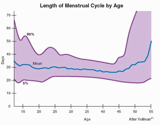
Menstrual cycle length is determined by the rate and quality of follicular growth and development, and it is normal for the cycle to vary in individual women. Informative data come from two seminal longitudinal studies (with very similar results): the study of Vollman of more than 30,000 cycles recorded by 650 women and the study of Treloar of more that 25,000 woman-years in a little over 2,700 women.41,42 The observations of Vollman and Treloar documented a normal evolution in length and variation in menstrual cycles.


image

Menarche is followed by approximately 5-7 years of relatively long cycles at first, and then there is increasing regularity as cycles shorten to reach the usual reproductive age pattern. In the 40s, cycles begin to lengthen again. The highest incidence of anovulatory cycles is under age 20 and over age 40.43,44 At age 25, over 40% of cycles are between 25 and 28 days in length; from 25 to 35, over 60% are between 25 and 28 days. The perfect 28-day cycle is indeed the most common mode, but it totaled only 12.4% of Vollman’s cycles.
Overall, approximately 15% of reproductive-age cycles are 28 days in length. Only 0.5% of women experience a cycle less than 21 days long, and only 0.9% a cycle greater than 35 days.45 Most women have cycles that last from 24 to 35 days, but at least 20% of women experience irregular cycles.46

When women are in their 40s, anovulation becomes more prevalent, and prior to anovulation, menstrual cycle length increases, beginning 2 to 8 years before menopause.42 Cycles greater than 40 days in length are prevalent in the year before menopause.47 In an Australian longitudinal study, when cycle length exceeded 42 days, menopause predictably followed within 1 or 2 years.48 This period of longer cycles uniformly precedes menopause no matter the age when menses cease, whether menopause is early or late.49 The duration of the follicular phase is the major determinant of cycle length.50,51 This menstrual cycle change prior to menopause is marked by elevated follicle-stimulating hormone (FSH) levels and decreased levels of inhibin, but normal levels of luteinizing hormone (LH) and slightly elevated levels of estradiol.52,53,54,55,56,57 and 58 Most importantly, even irregular cycles with long intervals (greater than 50-60 days) can be ovulatory, as many as 25%, meaning that late perimenopausal women can be at risk for pregnancy.59

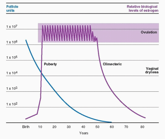
In the average woman, continuing follicular depletion and declining fertility begin at age 37-38, and menopause follows approximately 13 years later (average age 51). However, in epidemiologic studies approximately 10% of women in the general population become menopausal by the age of 45,60,61 probably because they were born with a smaller than normal ovarian follicular pool that is functionally depleted at an earlier age. Menopause occurs when the number of remaining follicles falls below a critical threshold, about 1,000, regardless of age.

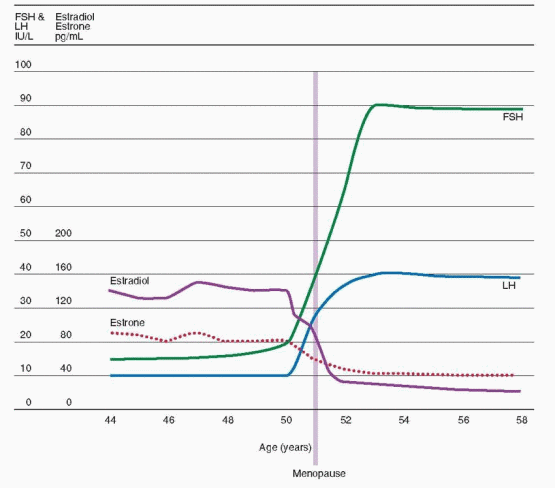
Contrary to older belief (based on the report by Sherman et al., in 197650), estradiol levels do not gradually wane in the years before the menopause, but remain in the normal range, although slightly elevated, until about 1 year before follicular growth and development cease.

The Sherman et al. data were from a small cross-sectional study of one cycle collected from only 8 women, ages 46-56. More recent longitudinal studies of women as they pass through the perimenopausal transition reveal that estrogen levels do not begin a major decline until about a year before menopause.56,62,63 Indeed, women experiencing the perimenopausal transition actually have higher overall estrogen levels, a response that is logically explained by an increased ovarian follicular reaction to the increase in FSH secretion during these years.64 Variability in estrogen levels is characteristic of the perimenopausal transition, with greater variability observed in menstrual cycles that display greater irregularity.65

As noted, most women experience a 2- to 8-year period of time prior to menopause when anovulation becomes common.42 During this period of time ovarian follicles continue their rate of loss until eventually the supply of follicles is finally depleted.66,67 In a study of human ovaries, the loss that began when the total number of follicles reached approximately 25,000, usually at age 37-38, correlated with a subtle but real increase in FSH and decrease in inhibin.68 These changes, including the increase in FSH, reflect the reduced quantity of aging follicles, and their reduced secretion of inhibin, the granulosa cell product that exerts an important negative feedback influence over FSH secretion by the pituitary gland. It is possible that both inhibin-A and inhibin-B may be involved, because luteal-phase levels of inhibin-A and follicular-phase levels of inhibin-B decrease with aging and antedate the rise in FSH.69,70 and 71 A careful study in Australia, however, indicated that the increase in FSH was correlated only with a decrease in inhibin-B, and in response, estradiol concentrations increased slightly.62

Decreasing inhibin production could reflect either a shrinking number of follicles, or a reduced functional capacity of older follicles, or both.72 The observation that preovulatory follicular fluid inhibin concentrations are similar in young and older cycling women suggests that the number of remaining follicles is the most important factor.73


image

As FSH levels increase and the follicular phase becomes shorter, estradiol levels rise earlier, suggesting that higher FSH levels stimulate more rapid follicular development.74 Careful studies indicated that the earlier acute rise in estradiol levels results from advanced follicular development at the beginning of the cycle and earlier selection of the dominant follicle.75,76 Follicular phase and overall cycle length reach their nadir at approximately age 42. Over the subsequent 8-10 years preceding the menopause, average cycle length and variability steadily increase as ovulations become less regular and less frequent.41 The age-related changes in the endocrine characteristics of the menstrual cycle that result from progressive follicular depletion correlate with a measurable decrease in ovarian volume and in the number of antral follicles observed by transvaginal ultrasonography during the early follicular phase.77,78,79,80,81,82 and 83

The inverse and tight relationship between FSH and inhibin indicates that inhibin is a sensitive marker of ovarian follicular competence and, in turn, that FSH measurement is a clinical assessment of inhibin.53,54 The decrease in inhibin secretion by the ovarian follicles begins early (around age 35), but accelerates after 40 years of age. This is reflected in the decrease in fecundity that occurs with aging (as discussed in Chapter 27). Furthermore, the ineffective ability to suppress gonadotropins with postmenopausal hormone therapy is a consequence of the loss of inhibin, and for this reason FSH cannot be used clinically to titer estrogen dosage in postmenopausal hormone therapy.

The Michigan Bone Health and Metabolism Study is a longitudinal, assessment of the perimenopausal transition in a cohort of 629 women initiated in 1992-1993. The initial rise in FSH in these women was modest until 7 years prior to menopause, then accelerated with
an even greater increase in the 2 years before menopause, finally reaching a plateau about a year after menopause.84 The major decrease in estradiol levels began about 2 years before menopause.85 Declining levels of inhibin B and anti-müllerian hormone (AMH) reached a low to nondetectable point about 5 years before menopause.86 Although the inhibin B and AMH results are in general agreement with other reports, the exactness of the timing is limited by the fact that the blood samples were obtained from only 50 women in the study. Nevertheless, the Michigan study confirms the validity of AMH as a marker for the ovarian reserve of follicles. Unlike inhibin B, AMH is not a participant in the feedback relationship between the ovary and the pituitary gonadotropins, rather AMH, a product of granulosa cells, reflects the number of follicles present in the ovaries awaiting FSH stimulation.87 The variability in these measurements from individual to individual, however, precludes the practical use of these tests to predict with accuracy the future date of menopause.

The perimenopausal years are a time period during which postmenopausal levels of FSH (greater than 20 IU/L) can be seen despite continued menstrual bleeding, while LH levels still remain in the normal range. Occasionally, corpus luteum formation and function occur, and the perimenopausal woman is not safely beyond the risk of an unplanned and unexpected pregnancy until elevated levels of both FSH (>20 IU/L) and LH (>30 IU/L) can be demonstrated.55 However, even under these circumstances, fluctuations can occur, with a period of ovarian failure followed by resumption of ovarian function.54 Because variability is the rule, it would be wise to recommend the use of contraception until the postmenopausal state is definitely established. According to the Guinness Book of World Records, a woman from Portland, Oregon, holds the modern record for the oldest spontaneous pregnancy, conceiving when 57 years and 120 days old. Several months of amenorrhea together with an FSH level of 40 IU/L or more are reliable signals that menopause is either near or already passed.88

In the longitudinal Massachusetts Women’s Health Study, women who reported the onset of menstrual irregularity were considered to be in the perimenopausal period of life.89 The median age for the onset of this transition was 47.5 years. Only 10% of women ceased menstruating abruptly with no period of prolonged irregularity. The perimenopausal transition from reproductive to post-reproductive status was, for most women, approximately 4 years in duration. In the study by Treloar, the average age for entry into the perimenopausal transition was 45.1, and the age range that included 95% of the women was 39-51.60 The mean duration of the perimenopausal transition was 5.0 years, with a range of 2 to 8 years.


The Perimenopausal Transition42,60,89


Average age of onset — 46


Age of onset for 95% of women — 39 to 51


Average duration — 5 years


Duration for 95% of women — 2 to 8 years


Preventive Health Screening of Healthy Perimenopausal Women

The most important contribution a clinician can provide to the perimenopausal woman is the education she needs and desires to make therapeutic choices. This early educational process will help to build a solid relationship with patients, a relationship they will want to
continue as they age. The following recommendations are derived from our own clinical experience:


  • Provide guidance and education to facilitate a patient’s decision making.


  • Provide time and an appropriate location for sensitive and uninterrupted discussions.


  • Use educational materials, especially handouts, but also explain them using your own words.


  • Involve family members during counseling and educational visits.


  • Be accessible. Consider designating a member of your staff as the menopause resource person. Encourage phone calls and emails.


  • Be involved in community and hospital educational programs for the public.


  • Use an effective, well-trained counselor for patients who need in-depth help in coping with life’s trials and tribulations.

Preventive intervention during the perimenopausal years has three major goals. The overall objective is to prolong the period of maximal physical energy and optimal mental and social activity. A specific goal is to detect as early as possible any of the major chronic diseases, including hypertension, heart disease, diabetes mellitus, and cancer, as well as impairments of vision, hearing, and teeth. Finally, the clinician should help perimenopausal women to smoothly traverse the menopausal period of life. Preventive health care and management of the later reproductive years give clinicians an excellent opportunity to function as a woman’s primary care provider.


The Age of Menopause

Designating the average age of menopause has been somewhat difficult. Based on crosssectional studies, the median age was estimated to be somewhere between 50 and 52.90 These studies relied on retrospective memories and the subjective vagaries of the individual being interviewed. Until recently, studies with longitudinal follow-up to observe women and record their experiences as they pass through menopause were hampered by relatively small numbers. The Massachusetts Women’s Health Study provides us with data from 2,570 women.89

The median age for menopause in the Massachusetts Study was 51.3 years. Only current smoking could be identified as a cause of earlier menopause, a shift of approximately 1.5 years. Those factors that did not affect the age of menopause included the use of oral contraception, socioeconomic status, and marital status. Keep in mind that a median age of menopause means that only half the women have reached menopause at this age. In the classic longitudinal study by Treloar, the average age of menopause was 50.7, and the range that included 95% of the women was 44 to 56.91 In a survey in the Netherlands, the average age of menopause was 50.2, and in an Italian longitudinal study, 50.9.61,92

The Study of Women’s Health Across the Nation (SWAN) is an ongoing, national study, recording the health of American women as they pass through the perimenopausal transition (http://www.edc.gsph.pitt.edu/swan/). The study began in 1994 in seven research centers and enrolled 3,302 participants with five racial/ethnic groups and a variety of backgrounds for an initial cross-sectional survey. In 1996, these women began a longitudinal, follow-up study with extensive data collection occurring annually.

In the SWAN study, the median age of menopause was 51.4, with an earlier onset associated with current smoking, lower education, and lower socio-economic status, whereas a later age was associated with parity and prior use of oral contraceptives.93 In contrast, a Dutch study concluded that prior use of oral contraceptives was associated with an earlier (less than 1 year) menopause.94 About 1% of women have been reported to experience menopause before the age of 40.95 The SWAN study reported a similar percentage of 1.1%, with a slightly higher rate in black and Hispanic women and a lower rate of 0.5% in Chinese women and 0.1% in Japanese women.96 Hispanic women experienced menopause about 6 months earlier compared with other ethnic groups, whereas Japanese women were about 3 months later.

Two large cohorts of European women reported average ages of menopause in various countries that centered around age 51, slightly higher in Northern Europe and slightly lower in Southern Europe.97 Some countries, like India, report an average age of menopause as much as 5 years earlier.98 In epidemiologic studies, approximately 10% of women in the general population become menopausal by the age of 45.60,61 Pedigree analysis has revealed that the genetic features of early menopause (age 40-45) and premature ovarian failure are similar and suggest a dominant pattern of inheritance through maternal or paternal relatives.99,100 There are two studies indicating that daughters of mothers with an early menopause (before age 46) also have an early menopause.101,102 and 103

There is sufficient evidence to believe that undernourished women and vegetarians experience an earlier menopause.101,104 Because of the contribution of body fat to estrogen production, thinner women experience a slightly earlier menopause.105 Frequent consumption of alcohol is associated with a later menopause.102 This is consistent with the reports that women who consume alcohol have higher blood and urinary levels of estrogen, and greater bone density.106,107,108,109 and 110

In multiple studies, there has been no correlation between age of menarche and age of menopause, with the exception of one Swedish study concluding that an earlier menarche
and earlier menopause go together.61,91,101,111,112 In most studies, race, parity, and height have no influence on the age of menopause; however, three cross-sectional studies found later menopause to be associated with increasing parity.61,89,93,101,105 Two studies found that irregular menses among women in their early 40s predicts an earlier menopause.113,114 A French survey detected no influence of heavy physical work on early menopause (before age 45).115 An earlier menopause has been reported to be associated with living at high altitudes.116,117 And most intriguing, an earlier age of menopause has been reported in lefthanded women compared with right-handed women.118,119 Finally, earlier menopause is associated with growth retardation in late gestation.120

It has been argued that premature ovarian failure can occur in women who have previously undergone abdominal hysterectomy or endometrial ablation, presumably because ovarian vascular flow has been compromised, but the only prospective study could find no elevations of FSH within the first 2 years after surgery.121,122 and 123

Multiple studies have consistently documented that an earlier menopause (an average of 1.5 years earlier) is a consequence of smoking. There is a dose-response relationship with the number of cigarettes smoked and the duration of smoking.124,125 Even former smokers show evidence of an impact.97

Unlike the decline in age of menarche that occurred with an improvement in health and living conditions, most historical investigation indicates that the age of menopause has changed little since early Greek times.126,127 Others (a minority) have disagreed, concluding that the age of menopause did undergo a change, starting with an average age of about 40 years in ancient times, and in Sweden an increase of about 1 year over the last 80 years.112,128 If there has been a change, however, history indicates it has been minimal. Even in ancient writings, an age of 50 is usually cited as the age of menopause.


Sexuality and Menopause

Sexuality is a lifelong behavior with evolving change and development. It begins with birth (maybe before) and ends with death. The notion that it ends with aging is inherently illogical. The need for closeness, caring, and companionship is lifelong. Old people today live longer, are healthier, have more education and leisure time, and have had their consciousness raised in regard to sexuality.

Younger people, especially physicians, underrate the extent of sexual interest in older people. In a random sample of women aged 50 to 82 in Madison, Wisconsin, nearly one-half of the women reported an ongoing sexual relationship.129 In the Duke longitudinal study on aging, 70% of men in the 67 to 77 age group were sexually active, and 80% reported continuing sexual interest, while 50% of all older women were still interested in sex.130 In the Postmenopausal Estrogen-Progestin Interventions (PEPI) trial, 60% of women 55-64 years old were sexually active.131 In a national sample of American men and women, the prevalence of sexual behavior declined with aging; however, 26% of individuals age 75 to 85 years were still sexually active.132 Therefore a significant number of postmenopausal women are sexually active, and only a relatively small percentage complain of sexual problems. The prevalence of self-reported sexual problems peaks in middle-aged women, sufficient to cause distress in about 22% of U.S. women, and about 12% of women aged 45 to 64.133

The decline in sexual activity with aging is influenced more by culture and attitudes than by nature and physiology (or hormones). The two most important influences on older sexual interaction are the strength of a relationship and the physical condition of each
partner.131,132,134 The single most significant determinant of sexual activity for older women, therefore, is the unavailability of partners due to divorce and the fact that women are outliving men. Given the availability of a partner, the same general high or low rate of sexual activity can be maintained throughout life.5,135 Longitudinal studies indicate that the level of sexual activity is more stable over time than previously suggested.136,137 and 138 Individuals who are sexually active earlier in life continue to be sexually active into old age. However, aging is associated with a decline in sexual function in many women, and this has been documented in the menopausal transition.139,140 A significant component of this decline can be attributed to menopausal symptoms associated with decreasing estrogen levels, a problem that is easily ameliorated by estrogen treatment.

There are two main sexual changes in the aging woman. There is a reduction in the rate of production and volume of vaginal lubricating fluid, and there is some loss of vaginal elasticity and thickness of the epithelium. Less vaginal atrophy is noted in sexually active women than in inactive women; presumably the activity maintains vaginal vasculature and circulation. The dyspareunia associated with postmenopausal urogenital atrophy includes a feeling of dryness and tightness, vaginal irritation and burning with coitus, and postcoital spotting and soreness. Of course, these changes are effectively prevented by estrogen treatment. Indeed, estrogen therapy has a positive impact on sexuality beyond its effects on vaginal tissue.131 In an Australian study assessing changes in sexual functioning during the perimenopausal and menopausal transition, a correlation with a decline in sexuality was demonstrated with estradiol levels, but not with testosterone levels.141 However, the prior level of sexual activity and the partner status and relationship were more important factors than hormone levels in determining midlife sexual function during the perimenopausal and menopausal transition.142


Illness and Sex

It is not uncommon to encounter women who have had surgery that affects sexuality. The list includes vulvectomy and surgery of the breast. Sexual problems are not limited, however, to surgical procedures and illnesses of the genitalia. Altered self-image can occur with diseases of any site; however, studies have not found hysterectomy to have a detrimental impact on sexuality.131,143

Sexual counseling, to be effective, must be provided to couples both before and after surgery. It is not unexpected that the surgeon may not be fully capable of providing this counseling. A major contribution from an older woman’s primary clinician is to arrange for competent and experienced sexual counseling. Unfortunately, most physicians operate on the principle that if no questions are raised there is no problem. The expert surgeon should be grateful for the help of experts in psychosexual therapy. Seek out the potential for post treatment sexual morbidity before the surgery. Assess the patient’s abilities for coping and her sense of body image. Consider the quality of the patient’s relationship, and be sensitive to the absence of a relationship. This entire effort may take some time. The normal state of presurgical anxiety, fear, and denial hampers good communication.

Antihypertensive agents are frequently responsible for male sexual dysfunction, but little information is available regarding female sexual function. However, remember that vaginal lubrication is the female counterpart to the male erection, and, therefore, vaginal dryness is a likely consequence. Adrenergic blocking agents are especially noted to affect libido and potency in men. Similarly, psychotropic drugs of all categories have been associated with inhibition of sexual function. Finally, one should always suspect alcoholism when patients complain of sexual dysfunction. Androgen treatment for decreased sexuality is discussed in Chapter 18.


Hormone Production After Menopause

Shortly after the menopause, one can safely say that there are no remaining ovarian follicles.63,144 Eventually there is a 10-20-fold increase in FSH and approximately a 3-fold increase in LH, reaching a maximal level 1-3 years after menopause, after which there is a gradual, but slight, decline in both gonadotropins.145,146 Elevated levels of both FSH and LH at this time in life are conclusive evidence of ovarian failure. FSH levels are higher than LH because LH is cleared from the blood so much faster (initial half-lives are about 20 min for LH and 3-4 h for FSH), and perhaps because there is no specific negative feedback peptide for LH like inhibin. The age-related decline in gonadotropin levels in the latter years of postmenopausal life is believed to reflect aging of the pituitary gonadotropin-secreting cells, specifically a decrease in the ability to respond to gonadotropin releasing-hormone (GnRH).

After menopause, the ovary secretes primarily androstenedione and testosterone, but the circulating level of androstenedione after menopause is about one-half that seen prior to menopause.147 Most of this postmenopausal androstenedione is derived from the adrenal gland, with only a small amount secreted from the ovary, even though androstenedione is the principal steroid secreted by the postmenopausal ovary.148,149 Dehydroepiandrosterone (DHEA) and its sulfate (DHEAS), originating in the adrenal gland, decline markedly with aging; in the decade after menopause the circulating levels of DHEA are approximately 70% less and levels of DHEAS are about 74% less than the levels in young adult life.150, 151

Testosterone production decreases by approximately 25% after menopause, but the postmenopausal ovary in most, but not all women, secretes more testosterone than the premenopausal ovary, at least in the first years of the postmenopausal period. With the disappearance of follicles and estrogen, the elevated gonadotropins drive the remaining tissue in the ovary to a level of increased testosterone secretion. The ovarian cells of origin are uncertain; presumably the steroidogenic tissue is that which has accumulated from ovarian follicles undergoing atresia because stromal cells believed to be of mesenchymal origin lack steroidogenic capability.152 Suppression of gonadotropins with gonadotropin-releasing hormone (GnRH) agonist or antagonist treatment of postmenopausal women results in a significant decrease in circulating levels of testosterone, indicating the gonadotropindependent postmenopausal ovarian origin.153,154 and 155

The total amount of testosterone produced after menopause, however, is decreased because the amount of the primary source, peripheral conversion of androstenedione, is reduced. The early postmenopausal circulating level of androstenedione decreases approximately 62% from young adult life.150 The menopausal decline in the circulating levels of testosterone is not great, from no change in many women to as much as 15% in others.56,146,150,156,157 In an excellent longitudinal Australian study from 5 years before menopause to 7 years after menopause, the circulating levels of testosterone did not change.151 Indeed, because of a decrease in sex hormone-binding globulin, this Australian study calculated an increase in free androgens.

Later in the postmenopausal years, the circulating androgen levels are nearly all, if not all, derived from the adrenal gland. A careful study could detect no circulating androgens in postmenopausal women (averaging 12 years distant from menopause) with complete adrenal insufficiency, and no intraovarian testosterone or androstenedione.158

The circulating estradiol level after menopause is approximately 10-20 pg/mL, most of which is derived from peripheral conversion of estrone, which in turn is mainly derived from the peripheral conversion of androstenedione.147,160,161 The circulating level of estrone
in postmenopausal women is higher than that of estradiol, approximately 30-70 pg/mL. The average postmenopausal production rate of estrogen is approximately 45 μg/24 h, almost all, if not all, being estrogen derived from the peripheral conversion of androgens. The androgen/estrogen ratio changes drastically after menopause because of the more marked decline in estrogen, and an onset of mild hirsutism is common, reflecting this marked shift in the sex hormone ratio. With increasing postmenopausal age, a decrease can be measured in the circulating levels of dehydroepiandrosterone sulfate (DHEAS) and dehydroepiandrosterone (DHEA), whereas the circulating postmenopausal levels of androstenedione, testosterone, and estrogen remain relatively constant.146,147


Blood Production Rates of Steroids159


Reproductive Age


Postmenopausal


Oophorectomized


Androstenedione


2-3 mg/day


0.5-1.5 mg/day


0.4-1.2 mg/day


Dehydroepiandrosterone


6-8


1.5-4.0


1.5-4.0


Dehydroepiandrosterone sulfate


8-16


4-9


4-9


Testosterone


0.2-0.25


0.05-0.18


0.02-0.12


Estrogen


0.350


0.045


0.045


Changes in Circulating Hormone Levels at Menopause56, 147, 160


Premenopause


Postmenopause


Estradiol


40-400 pg/mL


10-20 pg/mL


Estrone


30-200 pg/mL


30-70 pg/mL


Testosterone


20-80 ng/dL


15-70 ng/dL


Androstenedione


60-300 ng/dL


30-150 ng/dL


Estrogen production by the ovaries does not continue beyond the menopause; however, estrogen levels in postmenopausal women can be significant, principally due to the extraglandular conversion of androstenedione and testosterone to estrogen. The clinical impact of this estrogen varies from one postmenopausal woman to another, depending on the degree of extraglandular production, modified by a variety of factors.

The percent conversion of androstenedione to estrogen correlates with body weight. Increased production of estrogen from androstenedione with increasing body weight is probably due to the ability of fat to aromatize androgens. This fact and a decrease in the levels of sex hormone-binding globulin (which results in increased free estrogen concentrations) contribute to the well-known association between obesity and the development of endometrial cancer. Body weight, therefore, has a positive correlation with the circulating levels of estrone and estradiol.147 Aromatization of androgens to estrogens is not limited to adipose tissue, however, because almost every tissue tested has this activity.

Eventually, the ovarian steroidogenic tissue is exhausted and, despite huge reactive increments in FSH and LH, no further steroidogenesis of importance results from gonadal activity. The postmenopausal ovary weighs less than 10 g, but it can be visualized by ultrasonography.162 With increasing age, the adrenal contribution of precursors for estrogen production proves inadequate. In this final stage of estrogen availability, levels are insufficient to sustain secondary sex tissues.


image

In summary, the symptoms frequently seen and related to decreasing ovarian follicular competence and then estrogen loss in this protracted climacteric are:


  • Disturbances in menstrual pattern, including anovulation and reduced fertility, decreased flow or hypermenorrhea, irregular frequency of menses, and then, ultimately, amenorrhea.


  • Vasomotor instability (hot flushes and sweats).


  • Atrophic conditions: atrophy of vaginal epithelium; formation of urethral caruncles; dyspareunia and pruritus due to vulvar, introital, and vaginal atrophy; general skin atrophy; urinary difficulties such as urgency and abacterial urethritis and cystitis.


  • Health problems secondary to long-term deprivation of estrogen: the consequences of osteoporosis and cardiovascular disease.

A precise understanding of the symptom complex the individual patient may display is often difficult to achieve. Some patients experience severe multiple reactions that may be disabling. Others show no reactions, or minimal reactions that go unnoticed until careful medical evaluation.

It is helpful to classify the hormonal problems in three categories:


  • Those associated with relative estrogen excess such as dysfunctional uterine bleeding, endometrial hyperplasia, and endometrial cancer.


  • Those associated with estrogen deprivation such as flushes, atrophic vaginitis, urethritis, and osteoporosis.


  • Those associated with hormone therapy (Chapter 18).


Problems of Estrogen Excess


Exposure to Unopposed Estrogen

Throughout the perimenopausal period, there is a significant incidence of dysfunctional uterine bleeding. In the SWAN study, about 20% of cycles even early in the perimenopausal transition were anovulatory, associated with shorter intervals at the beginning of the transition and longer intervals later.163 Irregular bleeding was most often due to anovulation, whereas heavy menstrual bleeding was associated with obesity and uterine abnormalities.

Although the greatest concern provoked by this symptom is endometrial neoplasia, the usual finding is non-neoplastic tissue displaying estrogen effects unopposed by progesterone. This results from anovulation in premenopausal women and from extragonadal endogenous estrogen production or estrogen administration in postmenopausal women. There are 4 mechanisms that could result in increased endogenous estrogen levels:


  • Increased precursor androgen (functional endocrine tumors, liver disease, stress).


  • Increased aromatization (obesity, hyperthyroidism, and liver disease).


  • Increased direct secretion of estrogen (ovarian tumors).


  • Decreased levels of SHBG (sex hormone-binding globulin) leading to increased levels of free estrogen.

In all women, whether premenopausal or postmenopausal, whether on or off hormone therapy, specific organic causes (neoplasia, complications of unexpected pregnancy, or bleeding from extrauterine sites) must be ruled out. In addition to careful history and physical examination, dysfunctional uterine bleeding requires endometrial evaluation. Transvaginal ultrasonographic measurement of endometrial thickness can be utilized in postmenopausal women to avoid unnecessary biopsies.164 In perimenopausal and postmenopausal women with abnormal bleeding, endometrial biopsy is considered unnecessary when the endometrial thickness is less than 5 mm because the risk of endometrial hyperplasia or cancer is remote.165,166 and 167 Substantial evidence is lacking to support the application of this criterion to premenopausal women. We believe that biopsy is unnecessary in perimenopausal women when the endometrial thickness is less than 5 mm, that biopsy is indicated when the clinical history suggests long-term unopposed estrogen exposure even when the endometrial thickness is “normal” (5-12 mm), and that biopsy should be performed when endometrial thickness is greater than 12 mm even when clinical suspicion of disease is low.

If the uterus is normal on examination, for reasons of both accuracy and cost-effectiveness, the method of biopsy should be an office aspiration curettage, NOT the older, more costly and risky, in-hospital dilation and curettage (D&C). We recommend the use of a plastic endometrial suction device. It is easy to use, requires no cervical dilation, and is frequently painless. This device is as efficacious as older more painful techniques. Insertion should first be attempted without the use of a tenaculum. In many patients, this is feasible and avoids the sensation of the tenaculum grasping the cervix. Once the suction is applied, the endometrial cavity should be thoroughly curetted in all directions, just as one would with a sharp curette during a D&C. If the cannula fills up with tissue, a second and even a third cannula should be inserted until tissue is no longer obtained. Although most patients report no problems with cramps or pain, the application of suction in some patients stimulates cramping that usually passes within 5-10 minutes. Because cramping occurs in such a small minority of patients, it is not our practice to routinely give an inhibitor of prostaglandin synthesis. For repeat biopsies, in patients known to cramp, it is helpful to use such an agent at least 20 min before the procedure.


Less than 10% of postmenopausal women cannot be adequately evaluated by office biopsy. Most commonly, the reason is the inability to enter the uterine cavity. In such instances, dilation and curettage (D&C) are in order; however pretreatment with misoprostol or cervical laminaria may avoid a D&C. Furthermore, if the uterus is not normal on pelvic examination (enlarged and irregular), the office endometrial biopsy must yield to D and C with hysteroscopy in order to achieve accuracy of diagnosis.

If the vulva, vagina, and cervix appear normal on inspection, perimenopausal bleeding can be assumed to be intrauterine in origin. Confirmation requires the absence of abnormal cytology on the Pap smear. The principal symptom of endometrial cancer is abnormal vaginal bleeding, but carcinoma will be encountered in patients with bleeding in less than 3% of postmenopausal endometrial biopsies.168,169 and 170 Normal endometrium is found over half the time, polyps in approximately 3%, endometrial hyperplasia about 15% of the time, and atrophic endometrium in the rest of patients with postmenopausal bleeding. Postmenopausal bleeding should always be taken seriously. Approximately 10% of patients who have benign findings at the initial evaluation subsequently develop significant pathology within 2 years.169 The persistence of abnormal bleeding demands repeated evaluation.

Additional procedures include the following:


Colposcopy and cervical biopsy for abnormal cytology or obvious lesions.

Endocervical assessment by curettage for abnormal cytology (the endocervix must always be kept in mind as a source for abnormal cytology).

Hysterogram, hysteroscopy, or sonohysterography with endometrial biopsy if bleeding persists to determine the presence of endometrial polyps or submucosal fibroids, and to rule out the presence of endometrial cancer.171

Keep in mind that the pathologic reading, “tissue insufficient for diagnosis,” when a patient is on estrogen-progestin treatment, often represents atrophic, decidualized endometrium that yields little to the exploring curet. If the clinician is confident in his or her technique, knowing that a full investigation of the intrauterine cavity has been accomplished, then as long as the patient does not persist in bleeding, this reading can be interpreted as comforting and benign, the absence of pathology.

In the absence of organic disease, appropriate management of uterine bleeding is dependent on the age of the woman and endometrial tissue findings. In the perimenopausal woman with dysfunctional uterine bleeding associated with proliferative or hyperplastic endometrium (uncomplicated by atypia or dysplastic constituents), periodic oral progestin therapy is mandatory, such as 5-10 mg medroxyprogesterone acetate or 200 mg micronized progesterone given daily for the first 14 days of each month. If hyperplasia is present, follow-up aspiration curettage after 3-4 months is required, and if progestin is ineffective and histologic regression is not observed, formal curettage is an essential preliminary to alternate therapeutic surgical choices. Progestins can mask abnormal tissue, and, therefore, follow-up biopsy is best scheduled 3 months after progestin treatment. Because hyperplasia with atypia carries with it a risk of cancer (even invasive), hysterectomy is the treatment of choice. Persistence or progression of abnormal endometrium was observed in 28.4% of women with complex hyperplasia and in 26.9% of women with atypical hyperplasia despite treatment with a progestational agent.172 However, progestational response was better with higher dosage and longer duration treatment. If treatment with a progestational agent is elected, we recommend a minimum duration of 3 to 6 months, with 20 mg medroxyprogesterone or 40 mg megesterol acetate daily.

When monthly progestin therapy reverses simple hyperplastic changes (which it does in 95-98% of cases) and controls irregular bleeding, treatment should be continued until withdrawal bleeding ceases. This is a reliable sign (in effect, a bioassay) indicating the onset of estrogen deprivation and the need for the addition of estrogen. If vasomotor disturbances
begin before the cessation of menstrual bleeding, a combined estrogen-progestin program can be initiated as needed to control the flushes.

If contraception is required, the healthy, nonsmoking patient with normal blood pressure should seriously consider the use of estrogen-progestin contraception. The anovulatory woman cannot be guaranteed that spontaneous ovulation and pregnancy will not occur. The use of a low-dose estrogen-progestin contraceptive will at the same time provide contraception and prophylaxis against irregular, heavy anovulatory bleeding and the risk of endometrial hyperplasia and neoplasia.

Clinicians have often utilized a traditional postmenopausal hormone regimen to treat a woman with the kind of irregular cycles usually experienced in the perimenopausal years. This addition of exogenous estrogen without a contraceptive dose of progestin when a woman is not amenorrheic or experiencing menopausal symptoms is inappropriate and even risky (exposing the endometrium to excessively high levels of estrogen). And most importantly, a postmenopausal hormonal regimen does not inhibit ovulation and provide contraception.173 The appropriate response is to regulate anovulatory cycles with monthly progestational treatment along with an appropriate contraceptive method or to utilize lowdose estrogen-progestin contraception. An oral contraceptive that contains 20 mg estrogen provides effective contraception, improves menstrual cycle regularity, diminishes bleeding, and relieves menopausal symptoms.174 Treatment with the transdermal or vaginal method of estrogen-progestin contraception (Chapter 23) would also be appropriate.

A common clinical dilemma is when to change from estrogen-progestin contraception to postmenopausal hormone therapy. It is important to change because even with the lowest estrogen dose contraceptive available, the estrogen dose is 4-fold greater than the standard postmenopausal dose, and with increasing age, the dose-related risks with estrogen become significant. One approach to establish the onset of the postmenopausal years is to measure the FSH level, beginning at age 50, on an annual basis, being careful to obtain the blood sample on day 6 or 7 of the estrogen-progestin-free week in a standard 3-week regimen (when steroid levels have declined sufficiently to allow FSH to rise). Friday afternoon works well for patients who start new estrogen-progestin treatment on Sunday. When FSH is greater than 20 IU/L, it is time to change to a postmenopausal hormone program. Because of the variability in FSH levels experienced by women around the menopause, this method is not always accurate.175,176 Indeed, in some women, FSH will not rise until 2 weeks after the last steroid contraception exposure. A 2-week wait is not very practical and places the patient at risk for an unwanted pregnancy. The treatment-free week method is practical and works for most women. Women who are dependent on contraceptives to prevent pregnancy can be allowed to enter their mid fifties on low-dose estrogen- progestin contraception, and then empirically switched to a postmenopausal hormone regimen. The empirical approach is necessary with patients using the newer extended-day or continuous dosing regimens of estrogen-progestin contraception.

Because of the favorable impact of locally released progestin on the endometrium, the levonorgestrel IUS (intrauterine system) is very effective for the treatment of menorrhagia, as effective as the administration of oral progestins (with less side effects), and compares favorably with endometrial resection or ablation.177,178,179,180 and 181 In addition, this IUD can be used to treat endometrial hyperplasia.182,183,184,185,186 and 187 Comparison studies of endometrial hyperplasia indicate that the levonorgestrel IUS is as effective, and probably better than standard treatment with an oral progestin.183,188,189 The levonorgestrel IUS may be associated with a slight increase in the formation of ovarian cysts, but they are asymptomatic and resolve spontaneously.190

In postmenopausal women, one must view any adnexal mass as cancer until proven otherwise. Surgical intervention is usually necessary, and appropriate consultation must be obtained not only for the surgical procedure but also for suitable preoperative evaluation
and preparation. Nonpalpable, asymptomatic ovarian cysts are commonly detected by ultrasonography. Cysts that are less than 10 cm in diameter and without septations or solid components (unilocular) have a very low potential for malignant disease and can be managed with serial ultrasound surveillance (at 3 months, 6 months, 12 months, and then annually), especially if the serum CA 125 is normal.191,192 Surgery is recommended for symptomatic cases, if growth occurs, if internal echoes are obtained, if fluid develops in the pelvis, or if there is a family history of breast or ovarian cancer.


The Impact of Postmenopausal Estrogen Deprivation

The menopause should serve to remind patients and clinicians that this is a time for education. Certainly preventive health care education is important throughout life, but at the time of the menopause, a review of the major health issues can be especially rewarding. Besides the general issues of good health, attention is appropriately focused on cardiovascular disease and osteoporosis.

During the menopausal years, some women experience severe multiple symptoms, whereas others show no reactions or minimal reactions that can go unnoticed. The differences in menopausal reactions in symptoms across different cultures is poorly documented, and indeed, it is difficult to do so. Individual reporting is so conditioned by sociocultural factors that it is hard to determine what is due to biologic versus cultural variability.193,194 For example, there is no word to describe a hot flush in Japanese, Chinese, and Mayan.195 Nevertheless, there is reason to believe that the nature and prevalence of menopausal symptoms are common to most women, and that variations among cultures and within cultures reflect not physiology, but differences in attitudes, societies, lifestyles, socioeconomic status, and individual perceptions.196,197,198,199,200 and 201 Hormone levels during the perimenopausal years vary little among different ethnic groups; differences are mainly because of varying body sizes.202


Vasomotor Symptoms

The vasomotor flush is viewed as the hallmark of the female climacteric, experienced to some degree by most postmenopausal women. The term “hot flush” or “hot flash” is descriptive of a sudden onset of reddening of the skin over the head, neck, and chest, accompanied by an increase in heart rate and a feeling of intense body heat. The flush is sometimes concluded by profuse perspiration. The duration varies from a few seconds to several minutes and, rarely, for an hour. The frequency may be rare to recurrent every few minutes. Flushes are more frequent and severe at night (when a woman is often awakened from sleep) or during times of stress. In a cool environment, hot flushes are fewer, less intense, and shorter in duration compared with a warm environment.203 Most importantly, hot flushing can affect a woman’s quality of life and interfere with work or recreational activities.

In the longitudinal follow-up of a large number of women, fully 10% of the women experienced hot flushes before menopause, while in other studies as many as 15-25% of premenopausal women reported hot flushes.9,89,204,205 The frequency has been reported to be even higher in premenopausal women diagnosed with premenstrual syndrome.206 In the Massachusetts Women’s Health Study, the incidence of hot flushes increased from 10% during the premenopausal period to about 50% just after cessation of menses.89 By approximately 4 years after menopause, the rate of hot flushes declined to 20%. In a community-based
Australian survey, 6% of premenopausal women, 26% of perimenopausal women, and 59% of postmenopausal women complained of hot flushing.207 A large American cross-sectional survey reported that 57% of perimenopausal women and 49% of early postmenopausal women experienced significant hot flushing.200 Another national survey in the U.S. reported hot flushing in 79% of perimenopausal women and 65% of postmenopausal women.208

In cross-sectional surveys, up to 40% of premenopausal women and 85% of menopausal women report some vasomotor complaints.205 A longitudinal study in Gothenburg, Sweden, recorded a maximal prevalence of 60% at age 52-54, with a decline to 30% at age 60 and 9% at age 72.209 In the SWAN study, 57% of perimenopausal women experienced hot flushing, and about 50% after menopause up to age 55.210 There is no difference in the prevalence of vasomotor complaints in U.S. surveys of black and white women.211,212 Overweight women report more hot flushing, perhaps reflecting the effect of body fat causing a higher core body temperature.200,202,213 Exact estimates on prevalence are hampered by inconsistencies and differences in methodologies, cultures, and definitions.214 The prevalence in different societies is influenced by personal and social attitudes, individual psychological and physical health, familiarity with the portrayal of menopausal issues in the literature and media, ethnic variation, different diets, and dissimilar living conditions; however, accounting for cultural differences, the overall prevalence and experience are similar throughout the world.215,216

Although the flush can occur in the premenopause, it is a major feature of postmenopause, peaking in the first year after the last menses, lasting in 50% of women for 4 to 5 years, but in some (as many as 25%) for longer than 5 years, and up to 15 years in 10%.217 In an excellent Australian longitudinal cohort study, the average duration of vasomotor symptoms was 5.2 years (with a range of 2 to 10 years) in nonusers of hormone thrapy, and slightly longer, 5.5 years, in hormone users.218

The physiology of the hot flush is still not understood. Studies suggest that women with hot flushes have a more narrow zone of temperature regulation, and therefore, smaller changes in core body temperature produce compensatory responses, such as shivering or flushing.219 MRI scanning of the brain during hot flushing indicates widely distributed cortical activation rather than a precise location.220 Hot flushes are definitely brought about by a decline in estrogen; however, not all hot flushes are due to estrogen deficiency. Flushes and sweating can be secondary to diseases, including pheochromocytoma, carcinoid, leukemias, pancreatic tumors, and thyroid abnormalities.221 Unfortunately, the hot flush is a relatively common psychosomatic symptom, and women often are unnecessarily treated with estrogen. When the clinical situation is not clear and obvious, estrogen deficiency as the cause of hot flushes should be documented by elevated levels of FSH.

The correlation between the onset of flushes and estrogen reduction is clinically supported by the effectiveness of estrogen therapy and the absence of flushes in hypoestrogen states, such as gonadal dysgenesis. Only after estrogen is administered and withdrawn do hypogonadal women experience the hot flush. Although the clinical impression that premenopausal surgical castrates suffer more severe vasomotor reactions is widely held, this was not borne out in the only objective study ever performed.222

Although the hot flush is the most common problem of the postmenopause, it presents no inherent health hazard. The flush is accompanied by a discrete and reliable pattern of physiologic changes.219,223 The flush coincides with a surge of LH (not FSH) and is preceded by a subjective prodromal awareness that a flush is beginning. This aura is followed by measurable increased heat over the entire body surface. A flush is triggered by a small elevation in core body temperature. The body surface experiences an increase in temperature, accompanied by changes in skin conductance, and then the flush is followed by a fall in core temperature—all of which can be objectively measured. In short, the flush is not a
release of accumulated body heat but is a sudden inappropriate excitation of heat release mechanisms. Its relationship to the LH surge and temperature change within the brain is not understood. The observation that flushes occur after hypophysectomy indicates that the mechanism is not dependent on or due directly to LH release. In other words, the same brain event that causes flushes also stimulates gonadotropin-releasing hormone (GnRH) secretion and elevates LH. This is probably secondary to hypothalamic changes in neurotransmitters that increase neuronal and autonomic activity.224

Premenopausal women experiencing hot flushes should be screened for thyroid disease and other illnesses. A comprehensive review of all possible causes is available.225 Clinicians should be sensitive to the possibility of an underlying emotional problem. Looking beyond the presenting symptoms into the patient’s life is an important service to the patient and her family that eventually will be appreciated. This is far more difficult than simply prescribing estrogen, but confronting problems is the only way of reaching some resolution. Prescribing estrogen inappropriately (in the presence of normal levels of gonadotropins) only temporarily postpones, by a placebo response, dealing with the underlying issues.

A striking and consistent finding in most studies dealing with menopause and hormonal therapy is a marked placebo response (at least 51% in the first weeks of treatment)226 in a variety of symptoms, including flushing. In an English randomized, placebo-controlled study of women being treated with estrogen implants and requesting repeat implants, there was no difference in outcome in terms of psychological and physical symptoms comparing the women who received an active implant to those receiving a placebo.227

A significant clinical problem encountered in our referral practice is the following scenario: a woman will occasionally undergo an apparently beneficial response to estrogen, only to have the response wear off in several months. This leads to a sequence of periodic visits to the clinician and ever-increasing doses of estrogen. When a patient reaches a point of requiring large doses of estrogen, a careful inquiry must be undertaken to search for a basic psychoneurotic or psychosocial problem. To help persuade a patient that her symptoms are not due to low levels of estrogen, we find it very helpful and convincing to measure the patient’s blood level of estradiol and share the result with her.


The Hot Flush


Premenopausal


10-25% of women


Perimenopausal


60%


Postmenopausal:


No flushes


15-25%


Daily flushing


15-20%


Duration


1-2 years average


5 or more years: 25%


Other Causes


Psychosomatic


Stress


Thyroid disease


Subacute, chronic infections


Pheochromocytoma


Carcinoid


Leukemia


Cancer


Atrophic Changes

With extremely low estrogen production in the late postmenopausal age, or many years after castration, atrophy of vaginal mucosal surfaces takes place, accompanied by vaginitis, pruritus, dyspareunia, and stenosis. Genitourinary atrophy leads to a variety of symptoms that affect the ease and quality of living. Urethritis with dysuria, urgency incontinence, and urinary frequency are further results of mucosal thinning, in this instance, of the urethra and bladder. Recurrent urinary tract infections are effectively prevented by postmenopausal intravaginal estrogen treatment.228 Vaginal relaxation with cystocele, rectocele, and uterine prolapse, and vulvar dystrophies are not a consequence of estrogen deprivation.

Deprived of estrogen, the vagina loses collagen, adipose tissue, and the ability to retain water. As the vaginal walls shrink, the rugae flatten and disappear. The surface epithelium loses its outer fibrous layer and thins to a few layers of cells, markedly reducing the ratio of superficial to basal cells. As a result, the vaginal surface is left friable, prone to bleeding with minimal trauma. While these changes are occurring, the blood vessels in the vaginal walls narrow, and secretions from sebaceous glands diminish. Over time the vagina itself contracts and loses flexibility, while the labia minora become paler and smaller. In addition, pH becomes more alkaline, making the vaginal environment less hospitable to lactobacilli and more susceptible to infection by urogenital and fecal pathogens. Infecting organisms can ascend into the urinary system to cause urethritis, urinary tract infections, and cystitis.

Dyspareunia, sometimes with postcoital bleeding, is the inevitable consequence of a severely atrophied vagina and scanty lubrication. Even for women who are not sexually active, atrophic vaginitis can cause itching, irritation, and burning. These symptoms often go unmentioned, and it is important to inspect for signs of vaginal atrophy even in the absence of complaints. Measuring pH is a simple way to determine estrogen’s influence or absence. A pH greater than 4.5 is almost always observed with estrogen deficiency.229,230

Dyspareunia seldom brings older women to our offices. A basic reluctance to discuss sexual behavior still permeates our society, especially among older patients and physicians. Gentle questioning may lead to estrogen treatment of atrophy and enhancement of sexual enjoyment. Objective measurements have demonstrated that vaginal factors that influence the enjoyment of sexual intercourse can be maintained by appropriate doses of estrogen.231 Both patient and clinician should be aware that a significant response can be expected by 1 month, but it takes a long time to fully restore the genitourinary tract (6-12 months), and clinicians and patients should not be discouraged by an apparent lack of immediate response. Raloxifene and tamoxifen have little impact on the vaginal epithelium, and vaginal dryness is worse with aromatase inhibitors. Sexual activity by itself supports the circulatory response of the vaginal tissues and enhances the therapeutic effects of estrogen. Therefore, sexually active older women have less atrophy of the vagina even without estrogen.

Although it is argued that genuine stress incontinence is not affected by treatment with estrogen, others contend that estrogen treatment improves or cures stress incontinence in over 50% of patients due to a direct effect on the urethral mucosa.232,233 and 234 A meta-analysis concluded that improvement was reported only in nonrandomized studies.235 Two randomized trials dedicated to this clinical problem failed to demonstrate a beneficial effect of estrogen treatment.236,237 Most cases of urinary incontinence in elderly women are a mixed problem with a significant component of urge incontinence that is believed to be improved by estrogen therapy. However, the Heart and Estrogen-progestin Replacement Study (HERS) randomized trial indicated a worsening of incontinence with hormone therapy for both urge and stress incontinence, and the Nurses’ Health Study reported a small increase of incontinence in hormone users.238,239 There is no convincing support for a
beneficial impact of estrogen treatment on incontinence. In the SWAN study, only 15% of incontinent women reported a worsening of urinary incontinence during the perimenopausal transition, largely because of weight gain.240 The majority of incontinent women experienced either no change or an improvement. The SWAN study strongly documents that urinary incontinence is not a major symptom of menopause and the perimenopausal transition.240,241 Incontinence at midlife is not a consequence of hormonal changes, but largely the effect of excess body weight or diabetes mellitus.

A decline in skin collagen content, elasticity, and skin thickness that occurs with aging can be considerably avoided by postmenopausal estrogen therapy.242,243,244,245 and 246 The effect of estrogen on collagen is evident in both bone and skin; bone mass and collagen decline in parallel after menopause, and estrogen treatment reduces collagen turnover and improves collagen quality.247,248 One study demonstrated not only an increase in facial skin thickness, but an improvement in wrinkles with topical estrogen.249 A randomized trial demonstrated improvements in skin elasticity, skin hydration, and skin thickness comparing hormone treatment with placebo.250 More impressively, data from the U.S. First National Health and Nutrition Examination Survey indicated that estrogen use was associated with a lower prevalence of skin wrinkling and dry skin.251 Smoking is a major risk factor for facial skin wrinkling, and hormone therapy cannot diminish this impact of smoking.252 In a 1-year clinical trial, hormone therapy did not improve skin wrinkling already present.253

One of the features of aging in men and women is a steady reduction in muscular strength. Many factors affect this decline, including height, weight, and level of physical activity. Women currently using estrogen have been reported to demonstrate a lesser decline in muscular strength, although at least one study could detect no impact of estrogen.254,255,256,257,258 and 259 This is an important issue because of the potential protective consequences against fractures, as well as a benefit due to the ability to maintain vigorous physical exercise.


Psychophysiologic Effects

The view that menopause has a deleterious effect on mental health is not supported in the psychiatric literature, or in surveys of the general population.204,205,260,261 The concept of a specific menopause-induced psychiatric disorder (involutional melancholia) has been abandoned. Indeed, depression is less common, not more common, among middle-aged women, and the menopause cannot be linked to psychological distress.3,4,5,6,7,8 and 9,262 The longitudinal study of premenopausal women indicates that hysterectomy with or without oophorectomy is not associated with a negative psychological impact among middle-aged women.263 Longitudinal data from the Massachusetts Women’s Health Study document that menopause is not associated with an increased risk of depression.264 Although women are more likely to experience depression than men, this sex difference begins in early adolescence, not at menopause.265

The U.S. National Health Examination Follow-up Study includes both longitudinal and cross-sectional assessments of a nationally representative sample of women. This study has found no evidence linking either natural or surgical menopause to psychologic distress.266 Indeed, the only longitudinal change was a slight decline in the prevalence of depression as women aged through the menopausal transition. Results in this study were the same in estrogen users and nonusers.

A negative view of mental health at the time of the menopause is not justified; many of the problems reported at the menopause are due to life events.11,12,267,268 Thus, there are problems encountered in the perimenopausal transition and the early postmenopause that are seen frequently, but their causal relation with estrogen is unlikely. These problems
include fatigue, nervousness, headaches, insomnia, depression, irritability, and palpitations. Indeed, at this stage of life both men and women express a multitude of complaints that do not reveal a gender difference that could be explained by a hormonal cause.269,270 Nevertheless, midlife women report complaints more often than men,270 perhaps reflecting the generally negative perceptions and connotations our cultures and societies have attributed to the menopause.

Two longitudinal cohort studies assessed the new onset of depressive symptoms and disorders during the perimenopausal transition. The Penn Ovarian Aging Study followed over 8 years 436 women with no history of depression and correlated hormonal changes with the onset of depressed mood.271 Fifty percent of the women developed an increase in measures of depression and 26% met the criteria for a clinical diagnosis of depressive disorder. Using the women as their own controls, the depression group was 2.5 times more likely to develop clinical depression comparing status during the perimenopausal transition to the premenopausal state. These symptoms during the perimenopausal transition were associated with greater variability (but no average differences) in estradiol levels, suggesting that fluctuations of estradiol can be an important destabilizing factor.

The Harvard Study of Moods and Cycles is a prospective cohort of women with and without histories of depression.272 In the women who entered the perimenopausal transition, the risk of new depression was almost doubled compared with premenopausal women, from 9.5% to 16.6%, and this risk was linked to the presence of vasomotor symptoms. Importantly, a statistically significant increase in risk of new depressive symptoms was present only in women with a history of adverse life events (the events are not defined or specified in the report). Also of note, 83% of the women experienced no mood changes.

The SWAN study reported similar results. A first episode of depression in perimenopausal women was linked to poor physical health, anxiety disorders, stressful life events, and hot flushing.273

This area of concern has been very difficult to study. Inconsistent results can reflect variations in study designs, selection of subjects, methods used to measure mood, and the definition of menopausal status. However, the best reports provide reliable evidence of a vulnerable population of women. Depressive mood changes are influenced by other factors, including body weight, smoking, premenstrual syndrome (PMS, defined in Chapter 14), employment, and marital status. Premenopausal PMS is a strong predictor of depressive symptoms arising in the menopausal transition.

The most important questions are whether truly normal women experience an increase in depression during the menopausal transition, and are there subtle or even clinically apparent psychological problems that identify a susceptible subgroup? The cohort studies support the argument that there is a vulnerable group of perimenopausal women who are responsible for the increase of new depression observed during the perimenopausal transition. The data are consistent with the idea that fluctuations in hormone levels are related to mood symptoms, but it is impossible to know if this is a true cause and effect relationship.

In summary, most women (about 85%) experience the perimenopausal transition without mood difficulties. Some women are at greater risk of new onset depressive symptoms, and this is probably enhanced by hormonal variations and vasomotor symptoms. These vulnerable women are likely derived from a group of premenopausal women with underlying psychological problems (although “problem” may be too strong of a word). It is also possible that perimenopausal hormone changes create a state that makes an individual less able to deal with adverse events in life.

Attempts to study the effects of estrogen on these problems have been hampered by the subjectivity of the complaints (high placebo responses) and the “domino effect” of what
a reduction of hot flushes does to the frequency of the symptoms. Using a double-blind crossover prospective study format, Campbell and Whitehead concluded many years ago that many symptomatic “improvements” ascribed to estrogen therapy result from relief of hot flushes—a “domino” effect.274 Studies that have controlled for menopausal symptoms conclude that mood is very affected by vasomotor symptoms and sleep disturbances, besides reflecting life problems.139,275

A study of 2,001 Australian women aged 45-55 focused on the utilization of the health care system by women in the perimenopausal period of life.14 Users of the health care system in this age group were frequent previous users of health care, less healthy, and had more psychosomatic symptoms and vasomotor reactions. These women were more likely to have had a significant previous adverse health history, including a past history of premenstrual complaints. This study emphasized that perimenopausal women who seek health care help are different from those who do not seek help, and they often embrace hormone therapy in the hope it will solve their problems. Similar findings have been reported in a cohort of British women.276 It is this population that is seen most often, producing biased opinions among clinicians regarding the menopause. We must be careful not to generalize to the entire female population the behavior experienced by this relatively small group of women. Most importantly, perimenopausal women who present to clinicians often end up being treated with estrogen inappropriately and unnecessarily. Nevertheless, it is well established that a woman’s quality of life is disrupted by vasomotor symptoms, and estrogen therapy provides impressive improvement.277,278 and 279 Patients are grateful to be the recipients of this “domino” effect.

The Women’s Health Initiative (discussed in Chapter 18) concluded that estrogen-progestin therapy had no beneficial impact on health-related quality of life.280 However, only 12.7% of the participants had moderate to severe vasomotor symptoms at entry to the study, and the severity can be questioned because the participants were willing to take placebo medication. The overall baseline quality of life in this study was relatively high, and the study participants were older (the average number of years distant from menopause was 12+). This randomized, clinical trial did not study the appropriate population of women in order to assess the effect of hormone therapy on measures of quality of life.

The Women’s International Study of Long Duration Oestrogen after the Menopause (WISDOM) trial was a randomized, controlled trial in the U.K., Australia, and New Zealand, of 3,721 women aged 50-69 treated with either combined 0.625 mg conjugated estrogens-2.5/5.0 mg medroxyprogesterone or placebo.281 The original plan was to randomize 22,300 women to the study that would last 10 years. The study was canceled in October 2002 in reaction to the initial reports from the WHI. Unfortunately, the premature cancellation precludes the possibility of any long-term data from WISDOM. In 2,130 women who completed one year, there were statistically significant improvements in the treated women in the categories of vasomotor, sexual, and sleep symptoms. Treated women reported a reduction in aching joints and muscles, night sweats, insomnia, and vaginal dryness. The treated group reported more breast tenderness, but the percentages were notably low (16% in the treated group and 7% in the placebo group).

The WISDOM trial investigators argued that the small effects on quality of life reported by the WHI and HERS can be attributed to the insensitive measurement tools used in those clinical trials. The WISDOM trial used a survey tool specifically designed to assess postmenopausal physical and emotional wellbeing, plus a validated, generic questionnaire, the European quality of life instrument. Only the specific questionnaire detected significant changes; the European generic tool did not. This emphasizes the importance of using the appropriate study tool to investigate this area of postmenopausal health. Similar results with vasomotor symptoms, sleep, and joint complaints were actually reported by the WHI, but with a smaller difference between treated and placebo groups. The WHI survey had only one question devoted to sexuality.


The results of the WISDOM trial are not surprising; they reflect what all clinicians have observed in their own practices. The most important point to be made is this: the WISDOM, WHI, and HERS trials were all similar in that they enrolled postmenopausal women heavily tilted towards the oldest age group without symptoms. It is a simple and logical conclusion that hormone therapy in a younger, symptomatic group of postmenopausal women would produce greater quality of life benefits than that quantified in the clinical trials. All three clinical trials, therefore, underestimated the beneficial impact because of age and symptom status of their participants. However, in the WISDOM trial, even older postmenopausal women who were symptomatic benefited from hormone therapy. Age should not be the sole guiding factor in decision-making.

Emotional stability during the perimenopausal period can be disrupted by poor sleep patterns. Hot flushing does have an adverse impact on the quality of sleep.282,283 and 284 Estrogen therapy improves the quality of sleep, decreasing the time to onset of sleep and increasing the rapid eye movement (REM) sleep time.277,285,286 In the SWAN study, one-third of the women reported sleep problems, even without hot flushes or night sweats, and the prevalence of vasomotor symptoms was associated with an increased risk of sleep disturbances; hormone therapy improved sleep quality.287,288 Perhaps flushing may be insufficient to awaken a woman but sufficient to affect the quality of sleep, thereby diminishing the ability to handle the next day’s problems and stresses. An improvement in sleeping with estrogen treatment can even be documented in postmenopausal women who are reportedly asymptomatic.286

Thus, the overall “quality of life” reported by women can be improved by better sleep and alleviation of hot flushing. However, it is still uncertain whether estrogen treatment has an additional direct pharmacologic antidepressant effect or whether the mood response is totally an indirect benefit of relief from physical symptoms and, consequently, improved sleep. Utilizing various assessment tools for measuring depression, improvements with estrogen treatment were recorded in oophorectomized women.289,290 In the large prospective cohort study of the Rancho Bernardo retirement community, no benefit could be detected in measures of depression in current users of postmenopausal estrogen compared with untreated women.291 Indeed, treated women had higher depressive symptom scores, presumably reflecting treatment selection bias; symptomatic and depressed women seek hormone therapy. Others report that estrogen therapy has a more powerful impact on women’s well-being beyond the relief of symptoms such as hot flushes.277,292,293 In elderly depressed women, improvements in response to fluoxetine were enhanced by the addition of estrogen therapy.294 In a 12-week, randomized, placebo-controlled trial of 55 perimenopausal women with clinically significant major depression, estradiol treatment with the 100 mg transdermal method significantly improved mood.295 A similar American short-term study of 34 perimenopausal women with both major and minor depressions treated with 50 mg estradiol transdermally demonstrated improvements independently of an effect on vasomotor symptoms.296 These small clinical trials argue that estrogen treatment is beneficial for the treatment of clinical depression. This conclusion is supported by the successful treatment of postpartum depression with estradiol treatment.297,298

The most common cause of perimenopausal mood problems is already-existing depression,10,299 but there does exist a small population of women whose moods are sensitive to hormonal changes. In the American SWAN study, the prevalence of mood changes increased from the premenopause to the early perimenopause, from about 10% to about 16.5%.299 There are three possible explanations: (1) the decline in estrogen at menopause affects neurotransmitters that regulate mood; (2) mood is adversely affected by vasomotor symptoms (the domino theory); (3) mood is affected by the vicissitudes of life that are commonly prevalent around menopause. Some would argue that these mood swings are in response to the hormonal fluctuations that occur during the perimenopausal years. These fluctuations do indeed occur,64 but whether they cause any symptoms remains to be determined. It seems logical that individuals with mood problems can reflect all of these mechanisms.


Cognition and Alzheimer’s Disease

Depending on the method of assessment, evidence for beneficial effects of estrogen on cognition can be found in the literature, especially in verbal memory.300,301 However, the effects in healthy women are not impressive, and perhaps of little clinical value. A short-term study failed to document an objective improvement in memory, although a slight improvement in mood was recorded.302 Another short-term (3 months) randomized, double-blind study could detect no improvement in cognitive performance compared with placebo treatment.303 The Melbourne Women’s Midlife Health Project could not document an effect on verbal memory during the menopausal transition.304 A longitudinal study in Chicago could not detect a cognitive decline through the menopause, as assessed by working memory and perceptual speed.305 On the other hand, estrogen treatment of women immediately after bilateral oophorectomy was associated with improvement in certain, but not all, specific tests of memory, and healthy postmenopausal women taking estrogen scored higher on tests of immediate and delayed recall.306,307 and 308 In a case-control study of women aged 55-93 years, estrogen users had better recall of proper names, but no improvement in word recall.309 Women in the Baltimore Longitudinal Study of Aging who were using estrogen performed better in tests of visual learning and memory.310,311 In a New York City cohort of women, the use of estrogen was associated with better performance in tests of cognition, and better performance in verbal memory, but the cohort in the Study of Osteoporotic Fractures demonstrated no effect of estrogen use on the age-related decline in cognition.312,313 In Connecticut, a randomized, placebo-controlled trial demonstrated better reading ability and verbal memory in the estrogen-treated group of postmenopausal women.314 Perhaps a lack of agreement is due to the variability in test vehicles and the specific aspects of memory function studied. Furthermore, there is impressive individual variability, and when differences have been observed they have not been large, and perhaps of little clinical importance. In addition, any beneficial effects may be attenuated by progestational agents.301

Another possibility for the variable effects of estrogen treatment on cognition is the variability among women in endogenous estrogen levels. Using sensitive assays for free, non-protein-bound estradiol and bioavailable (loosely bound) estradiol, cognitive decline occurred at a greater rate in women with low estradiol levels.315 Studies of cognition may have to differentiate between low- and high-risk women according to endogenous, biologically active estradiol levels. Similarly, a beneficial effect on cognitive decline has been observed only in women negative for the gene associated with Alzheimer’s disease, APOE-e4, which encodes the e4 allele of the glycoprotein known as apolipoprotein E, which has as one of its functions, the shuttling of lipids during neuronal repair.316

Up to three times as many women as men develop Alzheimer’s disease. Estrogen is capable of protecting central nervous system function by means of multiple mechanisms. For example, estrogen protects against neuronal cytotoxicity induced by oxidation; estrogen reduces the serum concentration of amyloid P component (the glycoprotein found in Alzheimer’s neurofibrillary tangles); and estrogen increases synapses and neuronal growth, especially dendritic spine density.317,318 and 319 Estrogen protects against the cerebrovascular toxicity exerted by amyloid peptides, and promotes synaptic formation and neuronal growth and survival.320,321 and 322 Progestational agents do not exert similar actions.

Case-control and cohort findings indicated that Alzheimer’s disease and related dementia occurred less frequently (perhaps as much as 60% less) in estrogen users, and the effect was greater with increasing dose and duration of use.323,324 and 325 In the Baltimore Longitudinal Study of Aging (a prospective cohort), the risk of Alzheimer’s disease was 54% reduced; in a cohort in New York City, the risk was reduced 60%; and in the Italian Longitudinal Study of Aging, the risk was 72% reduced in estrogen users.326,327 and 328 The findings are not uniformly positive; a case-control study with accurate information on clinical diagnoses and estrogen use from the U.K. General Practice Research Database could detect no impact of estrogen
treatment on the risk of developing Alzheimer’s disease, but the number of estrogen users was very small.329

The short-term administration of unopposed estrogen to patients with Alzheimer’s disease (secondary prevention) has been reported to improve cognitive performance, but mostly to have no effect.330,331,332,333 and 334 The administration of combinations of estrogen and progestin has also failed to demonstrate a beneficial impact in Alzheimer’s disease.335 The presence of estrogen therapy has been reported to enhance the beneficial response to tacrine in women with Alzheimer’s disease,336 but overall, the evidence is consistent with a failure of estrogen to influence already-existing Alzheimer’s disease or other forms of dementia.337

The data do support, however, a primary preventive effect. Most revealing is a prospective cohort study of women living in Cache County, Utah.338 Hormone therapy provided about a 41% reduced risk of developing Alzheimer’s with any use and an 83% reduction with 10 or more years of use. This cohort also demonstrated improved cognition in estrogen users.339 Most importantly, if women had initiated hormone therapy within a period of time that encompassed 10 years before the development of clinical symptoms, there was no effect. The Utah study strongly suggested that hormone therapy must be used for a significant duration of time very early in the postmenopausal period in order to have an impact on the risk of Alzheimer’s disease. As neurons become changed by the pathology of dementia, they lose their ability to respond favorably to estrogen.

The importance of timing is supported by findings from the large Women’s Health Initiative (WHI) clinical trial. The oldest women in the WHI being treated with either estrogen alone or combined estrogen-progestin (treatment began at age 65 or older) had impaired cognition and an increased risk of dementia.340,341 and 342 In a subset of these women, MRI scans demonstrated greater brain atrophy in the women receiving hormone therapy.343 The mechanism for this adverse effect of hormonal therapy in old women may be a neurotoxic action because the WHI study with MRI scanning could not detect an increase in ischemic brain lesions.344 Smaller MRI studies of younger women treated with hormone therapy found beneficial trophic changes in brain morphology, associated with improved cognition.345,346 and 347

The theme that emerges is that maintenance of health in target organs by estrogen requires normal tissue, a principle of timing that will also be discussed in regards to the heart in Chapter 18. Following the failure of secondary prevention trials to demonstrate a beneficial impact of hormone therapy on coronary disease in older women, it is increasingly argued that healthy cardiovascular endothelium is needed to respond to estrogen; that by the time, the endothelium is involved with excessive atherosclerosis, it is too late for estrogen to exert a beneficial effect. A similar argument is made for brain tissue, focusing on biochemical and signaling pathways that are progressively compromised with the neuronal involvement with disease.348 The requirement for normal tissue, at least in the heart and in the brain, would explain the beneficial effects in studies of primary prevention and the lack of effect in secondary prevention trials.
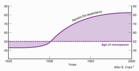

Cardiovascular Disease

Diseases of the heart are the leading cause of death for women in the United States, followed by cerebrovascular disease and malignant neoplasms. In 2005, 1 in 6 female deaths was from coronary heart disease compared with 1 in 30 for breast cancer deaths.349
More female deaths in 2005 were caused by cardiovascular diseases than the combined total from cancer, chronic lower respiratory disease, Alzheimer’s disease, accidents, and diabetes mellitus.

Most cardiovascular disease results from atherosclerosis in major vessels. The risk factors are the same for men and women: family history of cardiovascular disease, high blood pressure, smoking, diabetes mellitus, an abnormal cholesterol/lipoprotein profile, and obesity. However, when controlling for these risk factors, men prior to age 40 have a risk of developing coronary heart disease twice that of women. Even taking into consideration the changing lifestyle of women (e.g., employment outside the home), women still maintain their advantage in terms of risk for coronary heart disease. With increasing age, this advantage is gradually lost, and cardiovascular disease becomes the leading cause of death for both older women and older men.

Cardiovascular disease, especially atherosclerosis, is a consequence of multiple metabolic changes that interact with each other:


  • Adverse changes in the circulating lipid-lipoprotein profile.


  • Oxidation of low-density lipoprotein (LDL), producing a modified LDL that is chemotactic for circulating monocytes and inhibits macrophage motility (thus trapping macrophages in the intima), and that causes cell injury and death in the endothelium.


  • Endothelial injury and dysfunction affecting nitric oxide and prostacyclin production.


  • Macrophage migration and functions, influenced by growth factors and cytokines.


  • Proliferation and migration of smooth muscle cells, also influenced by growth factors and cytokines; these cells become the dominant cell type and the source of the connective tissue matrix in the atherosclerotic lesion, the fibrous plaque.


  • Vasoconstriction and thrombogenic events.


  • Remodeling of coronary arteries. An artery is able to respond to a developing atherosclerotic plaque by increasing its overall diameter in an attempt to maintain flow.350 The mechanism of this adaptive remodeling is not known, but the extent of this process must affect the risk of occlusion and infarction.

There is an established sequence of events leading to atherosclerosis. The process starts with endothelial dysfunction that leads to the fatty streak in arterial vessels, the precursor to clinically significant lesions. The fatty streak lesion, therefore, antedates the fibrous plaque, developing under the endothelial surface and dominated by fat-laden macrophages (the foam cells). The damaged endothelium expresses cytokines, adhesion molecules, and other inflammatory agents that are involved in the formation of atherosclerotic plaques. The formation of a plaque is initiated by the aggregation and adherence of circulating monocytes (macrophages) to a site on the arterial endothelium, stimulating an inflammatory response. When the monocytes penetrate through the endothelium and enter the intima, they become loaded with lipids and converted to foam cells. Modification of LDL, especially oxidation, is crucial in this conversion of monocytes to foam cells. The adherence of monocytes to endothelium can be induced by elevated cholesterol and LDL-cholesterol in the circulation. Most of the cholesterol that accumulates in atherosclerotic plaques is derived from circulating LDL-cholesterol. As plaques become significant in size, they are prone to instability, rupturing and creating a prothrombotic state. Matrix metalloproteinase enzymes are secreted by inflammatory cells and smooth muscle cells. These enzymes digest the proteins
in the fibrous cap of an atherosclerotic plaque, making the plaque unstable and predisposed to rupture. Estrogen induces matrix metalloproteinase production or activity, which digests the fibrous cap of a plaque exposing the underlying thrombogenic collagen, and this is believed to be the mechanism involved in the adverse thrombotic effects of estrogen in the presence of established atherosclerosis.351 In addition, 27-hydroxycholesterol, a cholesterol metabolite elevated in atherosclerotic lesions, competitively antagonizes estrogen receptor activity in cardiovascular epithelium.352


image


During the reproductive years, women are “protected” from coronary heart disease. For this reason, women lag behind men in the incidence of coronary heart disease by 10 years, and for myocardial infarction and sudden death, women have a 20-year advantage. The reasons for this are complex, but a significant contribution to this protection can be assigned to the higher high-density lipoprotein (HDL) levels in younger women, an effect of estrogen and lower levels of testosterone. Throughout adulthood, the blood HDL-cholesterol level is about 10 mg/dL higher in women, and this difference continues through the postmenopausal years. Total and LDL-cholesterol levels are lower in premenopausal women than in men, although the levels gradually increase with aging and after menopause they rise rapidly.353,354,355,356 and 357 After menopause the risk of coronary heart disease doubles for women as the atherogenic lipids at about age 60 reach levels greater than those in men. These changes can be favorably reduced by dietary modifications.358,359 Of course, these lipid changes at menopause (whether natural or surgical) can be reversed with estrogen treatment.360

Prospective studies have documented the strong association between total cholesterol and coronary heart disease in women, although coronary heart disease risk appears at higher total cholesterol levels for women than for men.361,362 Women with total cholesterol concentrations greater than 265 mg/dL have rates of coronary heart disease 3 times that of women with low levels. Even in elderly women, a high total cholesterol remains a significant predictor of heart disease but the strength of the association between the cholesterol level and cardiovascular disease decreases with aging, and by age 80 the cost and benefits may not justify cholesterol intervention.363 This is the reason for ceasing lipoprotein screening after age 75 in patients with normal lipids. However, this decision should be individualized, taking into account the vigor and health of the patient.

The strongest predictor of coronary heart disease in women is a low HDL-cholesterol,361,362,364 The average HDL-cholesterol in women is approximately 55-60 mg/dL. A decrease in HDL-cholesterol of 10 mg/dL increases coronary heart disease risk by 40-50%. In women (and men) who had normal total cholesterol and LDL-cholesterol levels, but low HDL-cholesterol levels, treatment with lovastatin reduced the risk of an acute major coronary event by approximately 37%.365 High HDL-cholesterol levels are uncommon in women with coronary heart disease, but even women with high levels do develop coronary heart disease.366 Because, the most powerful predictive value associated with HDL-cholesterol is the increased risk of coronary heart disease observed in individuals with low levels, it is appropriate to be concerned when HDL-cholesterol levels are less than 50 mg/dL. It should be emphasized that modest elevations in blood pressure markedly increase the risk associated with an elevated LDL-cholesterol or a low HDLcholesterol.

Keep in mind that low HDL-cholesterol levels are a component of the metabolic syndrome related to insulin resistance. The metabolic syndrome is partly a result of heredity, but strongly influenced by obesity and physical inactivity. In the U.S., the overall estimated prevalence of the metabolic syndrome is 24%, higher in women (40% by age 60) and increasing with age.367 The prevalence increases with increasing body weight, from about 5% in normal weight individuals to 60% in obese men and women, and the prevalence is highest in Mexican-Americans and lowest in blacks.368


The diagnosis of the metabolic syndrome in an individual requires that three abnormal findings are present out of the five following clinical characteristics:369


Hypertension—130/85 or higher.


Triglyceride levels—150 mg/dL or higher.


HDL-cholesterol levels—less than 50 mg/dL.


Abdominal obesity—greater than 35 inches waist circumference.


Fasting glucose—100 mg/dL or higher.


An increasing prevalence of metabolic syndrome during the perimenopausal and menopausal transition is correlated with increasing androgen dominance as estrogen secretion declines.370,371 Adiposity of the trunk is a risk factor for coronary heart disease in women and is associated with a relatively androgenic hormonal state, as well as hypertension, and disorders of lipid and carbohydrate metabolism.372 Central fat distribution in women is positively correlated with increases in total cholesterol, triglycerides, and LDL-cholesterol and negatively correlated with HDL-cholesterol.373 The atherogenic lipid profile associated with abdominal adiposity is at least partly mediated through an interplay with insulin and estrogen.374 It is worth noting that there is a strong correlation between the magnitude of the worsening in cardiovascular risk factors (lipid and lipoprotein changes, blood pressure, and insulin levels) and the amount of weight gained during the menopausal transition.375 Attention to weight gain during middle age is one of the most important components of good preventive health care. However, weight gain at menopause is not an effect of hormonal changes; it reflects diet, exercise, and aging.375

Current recommendations regarding the optimal cholesterol/lipoprotein profile are more aggressive, urging more intensive treatment aimed at lowering LDL-cholesterol levels; in the presence of coronary heart disease, the goal is to lower LDL-cholesterol to less than 100 mg/dL.376 Cholesterol-lowering drugs, specifically the statin family, have been repeatedly demonstrated in clinical trials to have a marked reduction in the risk of clinical cardiovascular events in both men and women.377,378

Triglycerides are also an important risk factor for coronary heart disease in women, but are most commonly encountered in individuals with the metabolic syndrome.376 If the triglyceride level is greater than 400 mg/dL and the HDL-cholesterol is less than 50 mg/dL, the risk of heart disease is substantially increased. Patients with an elevated triglyceride level and a positive family history for heart disease most likely have an autosomaldominant disorder classified as familial combined hyperlipidemia. This disorder accounts for most myocardial infarctions in women less than 40 years old. Triglyceride levels of 150-200 mg/dL are considered borderline elevated. Triglyceride levels can be elevated because of obesity, smoking, and lack of exercise. Weight loss alone can return elevated triglyceride levels to normal.

Observational studies and clinical trials indicate that the major determinants of blood lipid levels are the same for both sexes. A diet high in saturated fatty acids and dietary cholesterol unfavorably increases blood lipids. Excess caloric intake and obesity decrease HDL-cholesterol and increase total cholesterol, LDL-cholesterol, and triglycerides. Smoking decreases HDL-cholesterol (and also produces lower estrogen levels and an earlier menopause). Genetic defects of receptor-mediated cholesterol uptake account for only a small percentage of hyperlipidemia in men and women. There is also evidence that men and women who had impaired fetal growth have increased levels of cholesterol and LDL-cholesterol in middle age.379 The speculation is that impaired liver growth in utero produces a permanent adverse change in cholesterol and lipoprotein metabolism. Reduced fetal growth also leads in adulthood to insulin resistance and lower HDL-cholesterol levels, most severe in those who become obese.380


The Optimal Cholesterol/Lipoprotein Profile


Total cholesterol


Less than 200 mg/dL


HDL-cholesterol


Greater than 50 mg/dL


LDL-cholesterol


Less than 100 mg/dL


Triglycerides


Less than 150 mg/dL


The Role of Estrogen Exposure

By a large margin, the leading cause of death among women continues to be coronary heart disease. Coronary atherosclerosis is a lifelong process that varies in its slope of development according to the presence or absence of risk factors. The landmark Pathobiological Determinants of Atherosclerosis in Youth (PDAY) study documented the presence of fatty streaks in adolescents and an increasing prevalence with increasing age.381 The PDAY study further established that abnormal lipid profiles early in life are a major factor in determining the extent and age of onset of atherosclerosis.382 It is important for clinicians caring for women to appreciate the importance of premenopausal atherosclerosis and to understand that appropriate medical interventions can reduce the risk of later clinical events. Because atherosclerosis begins early in life, it is logical to conclude that the postmenopausal risk of coronary clinical events is influenced by the degree of coronary artery atherosclerosis already present at the time of menopause.

Women with premature ovarian failure are at increased risk for cardiovascular disease.383 In other words, there is an inverse relationship between the risk of cardiovascular disease and the age of menopause.384 Endothelial function in women with premature ovarian failure is impaired, as measured by dilation of the brachial artery in response to blood flow, a response known to be mediated by estrogen-modulated endothelial nitric oxide.385,386 This association between endothelial dysfunction and hypoestrogenemia is reinforced by the observation that endothelial dysfunction in women with premature ovarian failure was improved by hormone therapy.385

An important contribution to the gender difference in cardiovascular disease prevalence and age of onset is the favorable effect of estrogen on important endothelial events. Vasodilatory and antithrombotic activities can be attributed to endothelial production of nitric oxide and prostacyclin, a process favorably influenced by estrogen. Hypercholesterolemia adversely affects this important endothelial process, and estrogen protects this important endothelial function in the presence of hypercholesterolemia.387 Estrogen inhibits the oxidation of LDL, and also protects against the toxic effects of oxidized LDL on the endothelium. Women in the SWAN study who complained of hot flushing had more evidence of subclinical cardiovascular disease, such as aortic calcification, compared to women without hot flushes.388

A Chinese comparison study concluded that Chinese men and women with angiographically-determined coronary artery disease differ in the sex steroid environment presented to the heart by the circulation, compared with age-matched healthy individuals.389 Straightforward reasoning has led investigators to connect the different prevalence of coronary artery disease in men and women to the obvious differences in circulating sex steroids determined by the testicles and ovaries. The newly appreciated importance of estrogen in the premenopausal years has added strength to this connection. For many years, it has been generally believed that higher estrogen exposure in women protects against coronary artery disease, and the difference in coronary artery disease prevalence between men and women diminishes after menopause because of the loss of estrogen. At the heart of the matter is the gonadal difference between men and women.

Acute coronary events in premenopausal women occur more frequently when estrogen levels are the lowest during the menstrual cycle.390 In the national SWAN study, cardiovascular risk factors were more favorable in women with higher levels of estrogen and less favorable in women with longer menstrual cycles.391 Even amenorrheic athletes in good physical condition have demonstrated endothelial dysfunction, a condition that responded favorably to estrogen-containing oral contraceptives.392,393


In the WISE (Women’s Ischemia Syndrome Evaluation) Study, a study of premenopausal women undergoing coronary angiography for suspected myocardial infarction, coronary artery disease was more prevalent in those women who had low estrogen levels because of hypothalamic suppression.394 These findings are similar to the pioneering studies in monkeys that demonstrated acceleration of atherosclerosis in animals with low estrogen because of stress-induced hypothalamic suppression, an effect that could be prevented by oral contraceptive treatment.395,396 and 397 Postmenopausal women studied with coronary angiography in the WISE study who had used oral contraceptives in the past had less coronary artery disease.398 In addition, premenopausal women with coronary artery disease have lower circulating levels of estrogen compared with normal women.399

Depression is a recognized risk factor for heart disease, but its contribution to premenopausal atherosclerosis is just beginning to be appreciated. Premenopausal monkeys that exhibit depressive behavior (induced by their lower social rank in a colony of animals) develop a more adverse lipid profile and an increasing degree of atherosclerosis when fed atherogenic diets.400 Premenopausal women with a history of recurrent depression and without known coronary disease are more likely to have coronary and aortic calcification, a marker for early atherosclerosis.401 The SWAN study found more aortic calcification in black women with depressive symptoms, although an association between coronary calcification and depression in black or white women could not be detected.402

Thus hypoestrogenemia in the premenopausal years, whatever the cause, can increase the progression of atherosclerosis. This would include suppressed ovarian function associated with stress, depression, or athletic activity. The progressively deleterious effects of hypoestrogenemia include endothelial dysfunction, lower levels of HDL-cholesterol, an increase in central obesity, and possibly strengthening of depression. In monkeys and in women, lipid effects account for only 25-30% of the atheroprotective effects of estrogen.403,404

A vast literature has documented multiple mechanisms favorably influenced by estrogen that would inhibit the development of atherosclerosis.405 Clinicians tend to view testosterone as an estrogen opponent, and this is supported, for example, by studies such as lipid responses to testosterone that move in the opposite directions to those of estrogen.406 Both beneficial and detrimental vascular actions of testosterone have been documented with in vitro and animal studies. Clinical studies in women have in general supported an association between hyperandrogenism and an increase in risk for cardiovascular disease. The lipid and lipoprotein profile in androgenized women with polycystic ovaries (who are also exposed to relatively lower estrogen levels over time) is similar to the male pattern with higher levels of cholesterol, triglycerides, and LDL-cholesterol and lower levels of HDL-cholesterol, and this abnormal pattern is independent of body weight.407,408,409,410 and 411 An adverse lipid and lipoprotein profile is a distinguishing feature of these patients even when body mass index, insulin, and age are controlled in case-control studies.412 Subclinical atherosclerosis can be demonstrated by carotid ultrasonography to be prevalent in premenopausal women with a history of anovulation and polycystic ovaries.413 In women undergoing coronary angiography, the prevalence of polycystic ovaries is increased, and women with polycystic ovaries have more extensive coronary atherosclerosis.414 In the Nurses’ Health Study, women with very irregular cycles compared to women with regular cycles had an adjusted increased risk of coronary heart disease.415 Thus anovulatory women with polycystic ovaries develop risk factors for atherosclerosis and ultimately clinical disease comparable with that found in older, very overweight, postmenopausal women.

Only gold members can continue reading. Log In or Register to continue

Stay updated, free articles. Join our Telegram channel

Jul 5, 2016 | Posted by in GYNECOLOGY | Comments Off on Menopause and the Perimenopausal Transition

Full access? Get Clinical Tree

Get Clinical Tree app for offline access