Fig. 12.1
Large, multiple leiomyomas
Fibroids
The most common solid pelvic tumors, fibroids affect approximately 20–25 % of women of reproductive age [15, 16]. They are result of benign transformation and proliferation of a single smooth muscle cell. The growth and development of myomas are influenced by many factors, some of which are not well defined. Increased estrogen stimulation, alone or synergistically with growth hormone or human placental lactogen, appears to be the major growth regulator. In contrast, progesterone appears to inhibit the growth of fibroids, although some evidence indicates that under certain circumstances it can promote their growth [17].
The severity of symptoms associated with uterine leiomyomas depends on the number of tumors, their size, and location. They may cause abdominal pressure, urinary frequency, constipation or alter blood flow to the uterus and endometrium, resulting in menorrhagia. Uterine leiomyomas are seldom the only cause of infertility, but data from several studies demonstrate a link between fibroids, fetal wastage, and premature delivery. The indications for treatment of fibroids are summarized in Table 12.1.
Table 12.1
Indications for myomectomy
Menometrorrhagia and anemia |
Pelvic pain and pressure |
Enlarging leiomyoma (greater than 12 weeks), inability to evaluate the adnexa and possibility of neoplasia |
Associated fetal wastage and infertility |
Obstructed ureter |
It is important to remember that uterine leiomyosarcomas are extremely difficult to differentiate from benign fibroids preoperatively. Screening techniques are limited, with endometrial biopsies being only 38–62 % accurate at its best [18]. Clinical factors, such as advancing patient age, enlarged uteri in a postmenopausal patient and a newly diagnosed uterine mass in the climaterium, should raise suspicion for malignancy [19]. Undiagnosed neoplasias render intra-abdominal morcellation extremely detrimental to the patient due to the risk of spreading carcinomatosis, and favor laparoscopic-assisted myomectomy with extraabdominal morcellation as the preferred approach.
Preoperative Evaluation
In women with menorrhagia, the hematocrit is used to assess the degree of anemia. Patients with large broad ligament fibroids may require an intravenous pyelogram to search for ureteral obstruction. Periodic pelvic and ultrasound examinations help monitor the growth rate of asymptomatic leiomyomas. Factors such as the size, number, and location of leiomyomas will influence the decision to perform a myomectomy by laparotomy or laparoscopy, or even hysterectomy. Submucous fibroids can be detected by ultrasound, hysterosalpingography, or hysteroscopy. Small intramural myomas may be palpated during laparotomy and missed at laparoscopy. Therefore, a vaginal ultrasound should be performed preoperatively [20, 21].
Laparoscopically Assisted Myomectomy (LAM), Laparoscopic Myomectomy (LM), and Myomectomy by Laparotomy
Patients with uterine myoma who desire future fertility present a challenge to most physicians attempting a laparoscopic approach. The risk of a future uterine rupture is a major concern following any operation involving the myometrium [22]. The difficulties of adequately closing all layers laparoscopically and using electrocoagulation for hemostasis may contribute to the risk of uterine rupture [23, 24].
Uteroperitoneal fistulas may follow laparoscopic myomectomy because meticulous laparoscopic approximation of all layers may be difficult. The use of electrocoagulation for hemostasis inside the uterine defect may also increase the risk of uteroperitoneal fistula formation. Postoperative adhesions increase when sutures are placed in the serosal layer [24, 25]. A single uterine incision for removal of multiple leiomyomas and subserosal approximation of the uterine defect is advised.
A combination of laparoscopy and mini-laparotomy may reduce some of these problems. The simpler procedure and reduced operative time will enable more gynecologists to apply this technique. The uterine closure also is improved when a mini- laparotomy is used for conventional suturing in two or three layers, thereby decreasing the possibility of uterine dehiscence, fistulas, and adhesions. The laparoscopic portion of the procedure allows the diagnosis and treatment of associated endometriosis or adhesions.
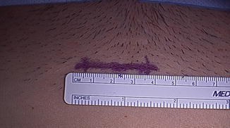
As a safe alternative to laparoscopic myomectomy (LM), laparoscopic-assisted myomectomy by mini-laparotomy (LAM) is a less technically difficult procedure and may require less time to complete. A decrease in operative time results from removing the myomas from the abdomen through a mini-laparotomy incision (see Fig. 12.2). Further, the risk of uterine rupture is lowered by suturing the uterine defect in layers and avoiding excessive electrocoagulation.


Fig. 12.2
Mini-laparotomy incision
The decision to proceed with LAM usually is made in the operating room after the diagnostic laparoscopy and treatment of associated pathology are completed. The criteria for LAM are myoma greater than 5 cm or numerous myomas requiring extensive morcellation, deep intramural myoma, and removal that requires uterine repair with sutures in multiple layers.
To objectively compare the three techniques mentioned, charts from 143 patients who had either myomectomy by laparotomy (22; 15.3 %), LM (64; 44.7 %), or LAM (57; 39.8 %) were evaluated [22]. The 22 myomectomies by laparotomy were performed before the development of the LAM technique. The data are summarized (Table 12.2). The leiomyoma weight was greater in the LAM than LM group (P < .05). LAM replaced myomectomy by laparotomy, patient selection criteria were comparable, and the myoma weights of these two groups were similar.
Table 12.2
Comparison of hysterectomy, abdominal myomectomy, and laparoscopic myomectomy for the management of symptomatic leiomyomas
Hysterectomy | Abd myomectomy | LM | |
|---|---|---|---|
Degree of difficulty | Low | Moderate | High |
Patient age | >45 years | Childbearing | >45 years |
Recurrence (%) | None | 10–15 | 10–15 |
Blood loss | <500 mL | Occasional >500 mL | <500 mL |
Postop adhesion formation | Minimal | >30 % | >30 % |
Postop hospitalization (days) | 3–4 | 3–4 | 1–2 |
Type of myoma | All types | Intramural, subserosal, pedunculated | Subserosal, pedunculated |
Uterine rupture | None | 1 % | Risk of uteroperitoneal fistula |
The mean estimated blood loss of the LAM and laparotomy groups was not different. In contrast, blood loss among the LM patients was significantly lower and may be attributed to the smaller leiomyomas. Previous studies [25, 26] have underscored the need to decrease the operative time of LM. Although subserosal myomas less than 5 cm can be managed easily laparoscopically, larger and intramural lesions require prolonged morcellation and laparoscopic suturing of the uterine defect. The largest reported myomas removed by laparoscopy were 15–16 cm [25, 26] and one group reported that 10 cm was their limit [27]. Both laparoscopic morcellation and myometrial suturing may be difficult and can prolong operations. Laparoscopic morcellation has also the risk of spreading an undiagnosed malignancy. LAM, with conventional morcellation and suturing through the mini-laparotomy incision, reduces the duration of the operation and the need for extensive laparoscopic experience. Similar mean operating times for LM and LAM techniques were observed despite larger myomas and their intramural positions, adjunctive laparoscopy, and the smaller incisions of the LAM group.
Hospitalization was longer for the patients who underwent myomectomy by laparotomy (P < .05) compared to both LAM and LM groups. When comparing the hospitalization time of the LAM and LM patients, the LAM group was longer (P(LM) = .014). This may be explained by the initial reluctance of some physicians to discharge LAM patients on the day of surgery or on the first postoperative day. After the initial 10–15 cases, all women underwent LAM on an outpatient basis. In fact, by removing the initial 15 cases from the LAM group, the mean hospital stay drops to 1.06 days, a time period not statistically different from the LM group.
The comparison of postoperative recovery time is important between the LAM and LM groups. Here, despite the differences in size and location of myoma, the recovery time can be compared because of the different incisions. The time elapsed before patients resumed work or regular activity is similar (P > .05). Introducing a 4-cm incision in the LAM group only slightly prolonged (P > .05) the subjectively perceived time for the women to achieve 100 % recovery.
Four major objectives of LAM are: minimizing blood loss, preventing postoperative adhesions, maintaining uterine wall integrity, and avoiding unprotected intracorporeal morcellation and its related complications.
Minimizing Blood Loss
Significant intraoperative blood loss can occur during the excision of subserosal and intramural leiomyomas. Depending on the tumor size and location, preoperative auto logous blood donation is suggested. Patients are counseled regarding the consequences of intraoperative and postoperative bleeding and the possible need for a laparotomy. For anemic patients, preoperative treatment with gonadotropin-releasing hormone (GnRH) may enable restoration of a normal hematocrit, decrease the size of the myoma [28] and reduce the need for transfusion [29] Intraoperatively, the use of dilute vasopressin helps to minimize blood loss. Vertical uterine incisions bleed less than transverse incisions [15] and pneumoperitoneum seems to decrease intraoperative bleeding.
Preventing Postoperative Adhesions
Although myomectomy is performed to preserve fertility, postoperative adhesion formation often jeopardizes this goal. Several procedures can minimize postoperative adhesions. Single, vertical, anterior, and midline uterine incisions are least to cause adhesions [30]. Although sutures predispose patients to adhesions, they are often necessary to close the uterine defect. While there are several adhesion barriers available or currently under development, none have proven effective [31, 32].
Stay updated, free articles. Join our Telegram channel

Full access? Get Clinical Tree


