Epidemiological case definition (classic clinical criteria)a
Fever persisting at least 5 daysb
Presence of at least 4 principal features:
1. Changes in extremities
(a) Acute: Erythema of palms, soles; edema of hands, feet
(b) Subacute: Periungual peeling of fingers, toes in weeks 2 and 3
2. Polymorphous exanthem
3. Bilateral bulbar conjunctival injection without exudate
4. Changes in lips and oral cavity: Erythema, lip cracking, strawberry tongue, diffuse injection of oral and pharyngeal mucosae
5. Cervical lymphadenopathy (>1.5-cm diameter), usually
Exclusion of other diseases with similar findings
Cutaneous manifestations are striking and should alert the clinician to the possibility of disease; they are present in 90 % of the patients [2]. A maculopapular, nonspecific exanthema may be seen accompanying the fever during the acute phase (Fig. 46.1). It can affect the trunk and sometimes involves more than 90 % of the skin (erythroderma). In other cases, it can be very subtle and affect only the perineal area or the skin folds. The changes in extremities include the erythema and edema, which can be sometimes painful. Desquamation of the tips of the fingers usually occurs later in the disease [12]. Mucous membrane manifestations include nonpurulent, bilateral conjunctivitis and hemorrhagic enanthem, with strawberry tongue with prominent papillae. A bright erythema of the lips with cracks is highly characteristic (Fig. 46.2).
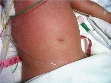
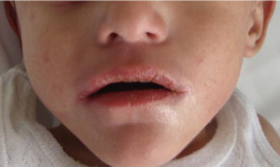
Fig. 46.1
Morbilliform exanthema in Kawasaki disease (KD) (courtesy Dr. Carola Durán)
Fig. 46.2
Lip edema and cracking in KD (courtesy Dr. Carola Durán)
At least one lymph node should be enlarged to more than 1.5 cm in diameter. Enlarged lymph nodes can be conspicuous in some cases. Heart involvement is a major risk and includes in the acute phase the presence of myocarditis or pericarditis and in later stages the formation of coronary aneurisms. Other clinical findings of KD can be seen in Table 46.2.
Cardiovascular findings |
Congestive heart failure, myocarditis, pericarditis, valvular regurgitation |
Coronary artery abnormalities |
Aneurysms of medium-size non-coronary arteries |
Raynaud’s phenomenon |
Peripheral gangrene |
Musculoskeletal system |
Arthritis, arthralgia |
Gastrointestinal tract |
Diarrhea, vomiting, abdominal pain |
Hepatic dysfunction |
Hydrops of gallbladder |
Central nervous system |
Extreme irritability |
Aseptic meningitis |
Sensorineural hearing loss |
Genitourinary system |
Urethritis/meatitis |
Other findings |
Erythema, induration at Bacille Calmette–Guérin (BCG) inoculation site |
Anterior uveitis (mild) |
Desquamating rash in groin |
Laboratory findings in acute KD |
Leukocytosis with neutrophilia and immature forms |
Elevated erythrocyte sedimentation rate (ESR) |
Elevated C-reactive protein |
Anemia |
Abnormal plasma lipids |
Hypoalbuminemia |
Hyponatremia |
Thrombocytosis after week 1 |
Sterile pyuria |
Elevated serum transaminases |
Elevated serum gamma glutamyl transpeptidase |
Pleocytosis of cerebrospinal fluid |
Leukocytosis in synovial fluid |
The diagnostic criteria of KD have low sensitivity and specificity. Therefore, patients with suspected KD but who do not meet the criteria and whose diagnosis is made upon heart involvement on echocardiography are included under the umbrella term ‘incomplete KD’ [12].
Complications and Prognosis
KD is a multisystem vasculitis, mainly involving small arteries. Coronary arteries are often affected. It is known that in the acute phase (9–10 days after fever onset) about 30–50 % of children may have a transient coronary dilatation. If left untreated, about 15–25 % of the children will develop coronary artery aneurysms (CAAs), which can lead to myocardial infarction, sudden death, and ischemic heart disease. Myocarditis and pericarditis can also be present in the acute phase, whereas CAAs appear in the convalescence phase. The occurrence of coronary artery lesion (CAL) is associated with many factors in children with KD. Age of less than 1 year or greater than 8 years, male sex, incomplete KD, delayed IVIG treatment after onset, no response to intravenous immunoglobulin treatment, and prolonged fever duration have all been identified as risk factors for the development of CAL [13].
Stay updated, free articles. Join our Telegram channel

Full access? Get Clinical Tree


