Stages of pre-embryonic development during the first week
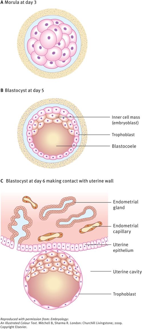
The morula soon shows signs of further differentiation. Cavities appear within the centre of the sphere of cells, forming a blastocyst, the cavity itself being the blastocoele (Figure 11.2B and C). Once this stage has been reached, the outer layer of the blastocyst soon thins to single-cell thickness to become the trophoblast, enclosing the enlarging fluid-filled blastocyst cavity. The central group of cells moves to one pole of the blastocyst (the embryonic pole) to form the inner cell mass from which the whole embryo itself will form. The trophoblast contributes to the fetal component of the placenta.
The process of morula and blastocyst formation occurs while the sphere of dividing cells is in transit along the uterine tube (Figure 11.1). The first mitotic division of cleavage will be completed by the time the two-cell stage embryo reaches the middle of the tube, at about 30 hours post-fertilisation. By 3 days the morula of 12–16 cells will have reached the junction of the uterine tube and the uterus. By 4–5 days the fully formed blastocyst reaches the uterine lumen in preparation for implantation, which occurs a day later (Figure 11.2).
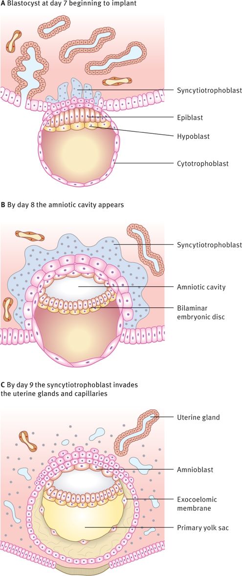
The second week – implantation and formation of bilaminar embryonic disc
At this stage the embryo is partly implanted in the endometrium. The implantation process initiates the decidual reaction or decidualisation in the uterine stroma, the cells of which contribute the maternal component of the placenta. The trophoblast begins to differentiate: its inner part becomes a single layer of cells, hence its name the cytotrophoblast (Figure 11.3A). The outer layer is more extensive and is the invasive layer. It is a syncytium and, at this stage, although it has invaded the endometrium, it has not invaded endometrial blood vessels. It is known as the syncytiotrophoblast. By this stage, the inner cell mass of the blastocyst has differentiated into two layers: the epiblast and the hypoblast (Figure 11.3A). These two layers are in contact and form a bilaminar embryonic disc (Figure 11.3B). Within the epiblast a cavity develops, the amniotic cavity, which fills with amniotic fluid. Some epiblast cells become specialised as amnioblasts and they secrete the amniotic fluid. The exocoelomic membrane is derived from the hypoblast and lines the cavity that appears beneath the endoderm, the primary yolk sac (Figure 11.3C). The fluid contained in this sac is the source of nutrition for the embryo before the placenta is fully formed and functional.
Implantation of blastocyst
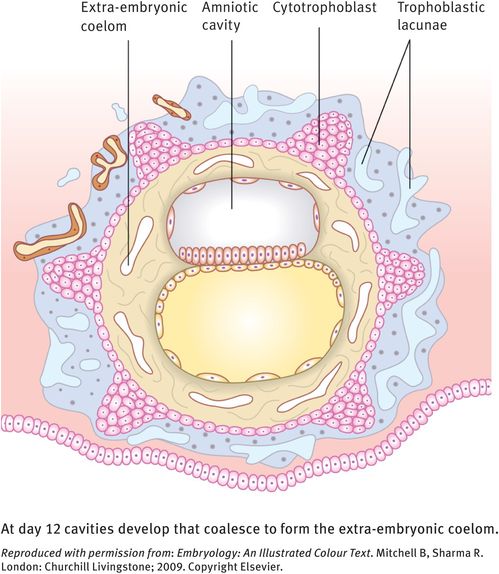
By 12 days there have been significant changes, particularly in the trophoblast. Small clefts, called lacunae, appear in the syncytiotrophoblast. These lacunae communicate with the maternal endometrial sinusoids, thereby deriving nutritional support for the developing embryo (Figure 11.4). Concurrent with this is the further development of the cytotrophoblast, which is thickest at the embryonic pole of the conceptus. Clefts appear between the exocoelomic membrane and the cytotrophoblast (Figure 11.4). These clefts merge to form the extra-embryonic coelom, which almost completely surrounds the embryo.
Implanted blastocyst
By day 13 the lacunae have enlarged substantially. The cytotrophoblast has begun to form primary chorionic villi, which are finger-like protrusions into the lacunae. The embryo proper consists of two layers, the epiblast and the hypoblast, still closely applied to each other. The two cavities continue to enlarge, with the amniotic cavity above the epiblast and the yolk sac below the hypoblast, now known as the secondary yolk sac because of the presence of the chorionic cavity. The embryo is connected to the cytotrophoblast by a connecting stalk of extra-embryonic mesoderm. This stalk is the forerunner of the umbilical cord. By this stage the uterine epithelium has reformed, thus completely engulfing the conceptus. The largest development of trophoblastic lacunae is on the deepest surface of the conceptus. By the end of the second week, the syncytiotrophoblast produces the hormone human chorionic gonadotrophin (hCG), which maintains the corpus luteum in the ovary, which in turn sustains the thickness of the endometrium. The hormone is secreted in the urine and thus its presence is an early indicator of pregnancy.
The third week – further development of the embryo and formation of trilaminar disc
The embryo develops further by forming three germ layers, the process known as gastrulation. Two layers have already formed: the epiblast and the hypoblast. These two closely apposed layers take up the form of two elliptical plates and together are termed the bilaminar embryonic disc. From this point the epiblast becomes known as the ectoderm and the hypoblast as the endoderm.
The ectoderm gives rise to the third layer that comes to lie between the two original germ layers, the intra-embryonic mesoderm. As a useful generalisation, the ectoderm (the outer skin) forms the covering of the body (the epidermis) as well as the nervous system; the endoderm (the inner skin) forms the lining of the gastrointestinal and respiratory systems; and the mesoderm (the middle skin) forms the skeletal, connective and muscle tissues of the body. The main tissue derivatives of germ layers are shown in Figure 11.5.


