Fig. 35.1
Fixed drug eruption to ibuprofen
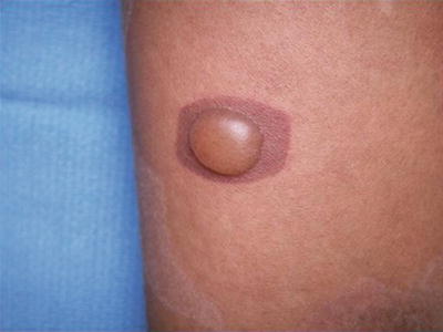
Non-pigmenting FDE (NPFDE) has only rarely been reported in children. NPFDE has similar features of FDE without the residual pigmentation [8].
Treatment
Avoid offending agent
Drug challenge may cause relapse
Identification and avoidance of the triggering agents assists in prevention of future lesions. Drug challenge tests can provoke relapses [8].
Prognosis
If the triggering agent is identified and avoided, the lesions can resolve. In skin of color, the postinflammatory hyperpigmentation may be particularly noticeable and may be persistent.
Ongoing Research
Ongoing studies to identify the causative agents associated with FDE will be useful in diagnosing this entity.
Conclusion
FDE is a unique category of drug eruptions with pigmentary changes as a prominent feature.
Photosensitive Drug Eruption
Introduction
Photosensitive drug eruptions occur with the combination of a sensitizing drug and ultraviolet (UV) exposure.
Epidemiology
Caused by a photosensitizing agent
Photoprotection may not be a usual practice in patients with skin of color
Often caused by antibiotics, thiazide diuretics, isotretinoin, and voriconazole
Photosensitive drug eruptions may be less common in skin of color because of the relative photoprotection provided by the melanin especially in darker skin types. However, when photosensitive drug eruptions do occur, they present unique challenges for the practitioner educating parents and patients with skin of color. Many patients (especially those with darker skin types) may not have been accustomed during their lives to practicing photoprotection. In general studies not particularly related to drug photosensitivity, parental use of photoprotective measures was directly related to children’s sun protection practices [9]. The most common photosensitizing drugs are antibiotics (especially tetracycline family), isotretinoin, thiazide diuretics, amiodarone [10], NSAIDs, antidepressants, psoralens, and voriconazole [11]. Some photosensitivity can also be seen as a side effect of topical retinoid use.
Clinical Presentation
Erythema
Papules
Photodistributed pattern
Photosensitive drug eruptions have erythema and papules in a photodistributed pattern. In richly pigmented skin, the erythema can be more difficult to appreciate (Fig. 35.3).
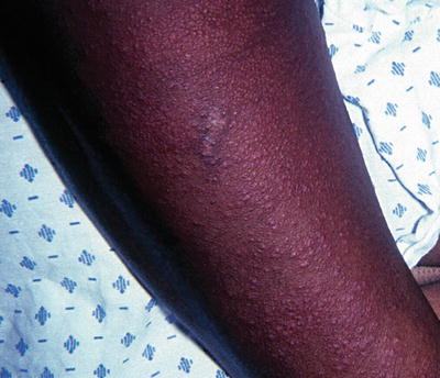
Fig. 35.3
Papules of the extensor arm in a photosensitive eruption from a thiazide diuretic
Treatment
Withdraw offending agent
Photoprotective measures
Topical and oral steroids may be useful
Withdrawal of the offending drug is the initial step for treatment. Sun avoidance and/or use of sunscreen are recommended especially if the drug is felt to be essential and is continued. Topical or oral corticosteroids can provide additional relief.
Prognosis
Once the offending drug is withdrawn and UV exposure is avoided, the eruption typically improves.
Ongoing Research
Determination of factors which predispose individuals to photosensitive drug eruptions is essential for minimizing these reactions.
Conclusion
Avoidance of UV exposure is important for a child with a photosensitive drug eruption.
Erythema Multiforme Minor
Introduction
Erythema multiforme minor (EM) is an immune-mediated condition which can occur in response to infectious or pharmacologic inciting agents. The minor designation denotes no mucous membrane involvement.
Epidemiology
Same incidence in skin of color
Associated with herpes simplex virus and Mycoplasma pneumoniae
The most common associated infectious agent in children is herpes simplex virus, although causation from Mycoplasma pneumonia infection has been reported [12]. The most common pharmacologic agents associated with EM minor include nonsteroidal anti-inflammatory drugs (NSAIDs), anticonvulsants, and antibiotics. The incidence of EM in skin of color is the same as the general population.
Clinical Presentation
“Dusky” center
Target lesions
Lesions on extensor extremities
The “target” lesion is the most well-known lesion of EM. It consists of a “dusky” center (from epidermal necrosis) with an annular edematous surround (Fig. 35.4). EM lesions are most often located on the extensor extremities, but can occur on any area of the body. The “multiforme” designation of the term refers to changing appearances of the lesions (not to any movement of the lesions).
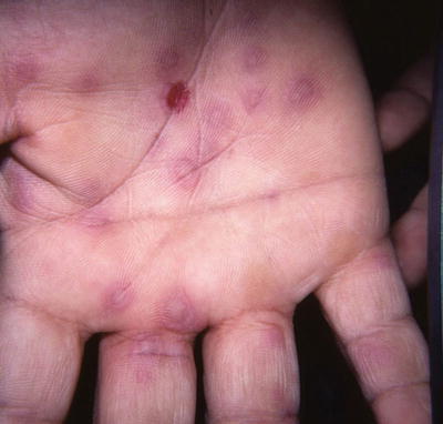
Fig. 35.4
Target lesions of erythema multiforme minor on the palms
Treatment
Anti-viral medication for herpes virus-associated lesions
Discontinue medication
Topical corticosteroids
Prognosis
EM minor has a good prognosis; however, in skin of color, postinflammatory dyspigmentation may occur.
Ongoing Research
Continued vigilance for associations of erythema multiforme minor with new medications as they are released is warranted.
Conclusion
Erythema multiforme minor is a hypersensitivity reaction which can be triggered by infectious agents or drugs.
Dress Syndrome
Introduction
DRESS syndrome, aka drug reaction with eosinophilia and systemic symptoms, is a drug reaction with characteristic features. Other terms which have been applied are: (a) Drug-induced hypersensitivity syndrome and (b) Phenytoin hypersensitivity syndrome.
Epidemiology
Increased in African Americans
Associated with mutations in drug detoxification genes
Associated with anticonvulsants, antibiotics, NSAID’s among others
Phenytoin had been the medication most often associated with DRESS; however, other medications have since been identified as causative agents. Those medications include anticonvulsants, antidepressants, antihypertensives, antimicrobials, antivirals, biologics, and nonsteroidal anti-inflammatory agents (NSAIDs) [13].
Stay updated, free articles. Join our Telegram channel

Full access? Get Clinical Tree


