Disorders of the Knee
John A. Herring
KNEE INJURIES
Knee injuries present frequently in virtually all age groups within a pediatric practice. The age and mechanism of injury are guidelines to a correct diagnosis. A swollen, painful knee in an infant should raise the suspicion of child abuse or infection. In the young child, significant ligament or meniscal injuries are rare, but epiphyseal separations and fractures are more frequent. In the adolescent, internal injuries to menisci and ligaments are common and most often result from sports activities, with or without contact. Patellar dislocations are also more common in adolescents.
The examiner should note bruising, swelling about the knee, the presence or absence of an effusion, and the ability to walk or bear weight. Most significant injuries are accompanied by an effusion or hemarthrosis, and the swollen knee is difficult to examine due to pain and limited motion. After the initial swelling has resolved, specific findings of an internal derangement can be elicited. The torn anterior cruciate is indicated by a positive anterior drawer sign in which the tibia can be anteriorly subluxated with the knee flexed.1 Meniscal tears often produce a clicking or grinding when the knee is fully flexed and extended with medial or lateral stress. Medial collateral and lateral collateral ligament injuries allow the joint to open either medially or laterally with stress in a 20° flexed position.
Radiographs should be evaluated and will show swelling only in the case of meniscal or ligamentous injury. Widening of the distal femoral growth plate suggests a separation injury of the growth plate (Salter 1). Elevation of the tibial spine indicates an injury in which the anterior cruciate pulls a fragment of bone up from the tibial articular surface.
Initial treatment usually consists of splinting with compression using a prefabricated knee immobilizer and ACE bandage.2 Aspiration of the knee is not necessary. Reevaluation at 2 weeks allows for a better physical exam for significant injury. Minor injuries will usually be recovering by then, and significant injuries will show persistent physical findings. A magnetic resonance imaging (MRI) examination will help to define internal injuries that will require orthopedic consultation.
DISCOID MENISCUS
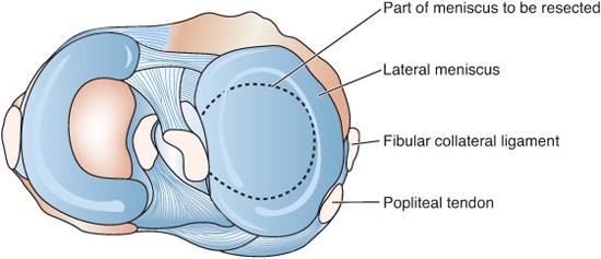
A discoid meniscus is an abnormally shaped lateral meniscus found in children of all ages (Fig. 214-1).3,4 It may slip in and out of the joint causing the patient to feel something pop over the lateral aspect of the knee joint. This may be somewhat painful. The examiner can feel something pop along the lateral joint line as the knee is flexed and extended. Over time, this may become more painful and the “popping” may begin to limit function.
An anteroposterior radiograph of the knee may show widening of the lateral joint space, and an MRI provides a definitive diagnosis. Arthroscopic reconstruction or removal of the meniscus is the treatment of choice for those with significant symptoms.5
PATELLO-FEMORAL PAIN SYNDROME
Adolescents with vague knee pain are commonly seen in a primary care office.6,7 The complaint is pain around the patella, aggravated by activities, especially physical education. The knee is often painful when the person sits in a car or theater where it is difficult to extend the knee. Straightening the knee gives immediate relief. This has been called chondromalacia patella, yet there is little evidence of damage to the cartilage of the patella. It has also been called the malalignment syndrome, and many patients have excessive femoral anteversion with patellae that face toward one another as the patient walks.
Radiographs will show no abnormalities, and the treatment is aimed at symptom relief. Nonsteroidal anti-inflammatory drugs (NSAIDs) are helpful. A well-planned physiotherapy program that emphasizes strengthening of the quadriceps and hamstrings as well as the hip abductors and external rotators will help most patients recover.

Stay updated, free articles. Join our Telegram channel

Full access? Get Clinical Tree


