Disorders of the Dermis and Subcutaneous Tissue
Anita N. Haggstrom
GRANULOMA ANNULARE
Granuloma annulare is a common benign inflammatory disorder of uncertain pathogenesis that is classically characterized by asymptomatic, flesh-colored to pink or violaceous, nonscaly annular plaques typically located over the dorsa of the hands and feet. (Fig. 359-1) The lesions of classic granuloma annulare are round to oval and are often misdiagnosed as tinea corporis (eFig. 359.1  ). However, unlike tinea infections, the lesions have a smooth, sometimes firm border that lack epidermal scale. Lesions of subcutaneous granuloma annulare appear as asymptomatic to slightly tender, flesh-colored nodules that can occur anywhere, although they are often on the lower legs and scalp. The differential diagnosis of granuloma annulare includes tinea corporis, necrobiosis lipoidica, rheumatoid nodules, and cutaneous sarcoidosis. Adults who develop this disorder may be more likely to have diabetes, but this association is not seen in children.
  Treatment is generally unsatisfactory. Topical steroids or intralesional steroids can limit progression, but results are not dramatic.
  Fortunately, granuloma annulare is an indolent process that eventually stabilizes and resolves without scarring over several years.
NECROBIOSIS LIPOIDICA DIABETICORUM
Necrobiosis lipoidica diabeticorum (NLD) is an uncommon inflammatory dermatosis characterized by well-demarcated, yellowish-pink, shiny, atrophic plaques often with prominent central telangiectasia. It is typically, but not exclusively, seen in diabetic patients. Only 0.3% of diabetics develop NLD and its occurrence is independent of glycemic control. Although it is more common in adult females, children with type I and type II diabetes rarely develop NLD. Despite its relationship to diabetes, the pathogenesis of NLD is uncertain. Lesions most commonly occur on the pretibial region; however, the upper extremities, trunk, and face can be affected. Lesions are typically asymptomatic but can develop ulcerations in response to trauma. Diagnostic evaluation should include a workup for diabetes. In general, treatment is challenging. Varied success has been reported with topical steroids, topical immunomodulators, antiplatelet agents such as aspirin and dipyridamole, antitumor necrosis factor therapies, and ultraviolet light.
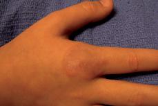
FIGURE 359-1. Dorsal hand with annulare granuloma. Note the annular pink plaque with smooth raised borders and no scale.
KELOIDS
Keloid scars represent an exuberant response to exogenous (ie, surgery, trauma, piercings) or endogenous (ie, acne, varicella) insults to the skin. Keloids are red-brown papules and nodules that extend beyond the point of initial injury, often resulting in disfiguring, bulky lesions that may be asymptomatic, pruritic, or painful. Keloids are more common in people with darker skin, although they can be seen in any skin type.3 Certain anatomic locations are considered at higher risk for keloid formation, including the skin of the ears, shoulder, and upper chest. Therapy is challenging because recurrence after surgical debulking is common (45–100%).6 A series of intralesional steroid injections is commonly used to flatten the lesions. Very exophytic lesions may benefit from surgical excision followed by adjunctive therapy such as a series of intralesional steroid injections, application of topical imiquimod, compression, or radiation to prevent recurrence.7 Silicon gel sheeting, cryotherapy, and intralesional interferon can also be used as adjunctive therapy. Carbon dioxide laser and pulsed dye laser therapy may be beneficial in some cases.8 Prevention of further keloids is crucial. Inflammatory acne should be appropriately treated, and elective procedures such as piercing should be avoided.
STRIAE (STRETCH MARKS)
Striae, or “stretch marks,” are atrophic, flesh-colored to red-violet, linear plaques seen in the setting of obesity, pregnancy, puberty, anorexia nervosa, cortisol excess (Cushing syndrome), and genetic alterations of connective tissue, including Marfan and Ehlers-Danlos syndromes. Iatrogenic causes of striae include systemic and topical steroid therapy. Initially, the lesions appear inflammatory with a pink-red color. Over several months to years, they become more subtle with textural changes reflecting atrophy. Striae are typically located on the abdomen, thighs, and breasts in girls, and on the outer aspect of the thighs and lumbosacral region in boys. Therapy for striae is suboptimal and may be more beneficial if lesions are treated in early inflammatory stages. Topical retinoids used over several months may result in modest improvement. Pulsed dye laser can be used to lighten the color. Most striae improve over time, and reassurance is important.
PANNICULITIS
Panniculitis is inflammation in the subcutaneous fat and is a feature of a variety of diverse disorders. Lesions of panniculitis are typically tender, red subcutaneous nodules and plaques.
 ERYTHEMA NODOSUM
Erythema nodosum is the most common panniculitis in children. It presents as tender, red, warm nodules or plaques without overlying scale, which are typically located on the extensor surfaces of the lower legs, although lesions can rarely be seen on the upper extremities. The onset of skin lesions may coincide with symptoms of fever, malaise, and arthralgias. Erythema nodosum may develop in response to infection, medication, and systemic inflammatory disorders. β-Hemolytic streptococcal infections are the most common causative factor in children.9 Epstein-Barr virus, tuberculosis, and other infections, such as Yersinia, have also been implicated. Erythema nodosum can also be associated with inflammatory disorders, including inflammatory bowel disease, sarcoidosis, and Behçet disease.10 Lesions of erythema nodosum are self-limited and typically resolve over several weeks with erythema evolving into a bruiselike appearance before resolving completely. Treatment is targeted at pain associated with the lesions. Rest, nonsteroidal anti-inflammatories, supersaturated potassium iodide, and systemic steroids can improve symptoms.
 COLD PANNICULITIS
The skin of infants and young children is sensitive to cold temperatures. Cold panniculitis is a form of circumscribed panniculitis caused by cold injury to the subcutaneous tissues. Classically, infants and children present with reddish-blue, ill-defined, warm plaques on one or both cheeks 2 to 3 days after eating a popsicle or ice cube.  Avoidance of hypothermia is curative, and lesions resolve without further sequelae.
 SUBCUTANEOUS FAT NECROSIS OF THE NEWBORN
Subcutaneous fat necrosis of the newborn (SCFN) is a distinctive, self-limited condition of healthy neonates characterized by nodular, erythematous, indurated plaques most commonly on the back. Children with SCFN may develop metabolic abnormalities, including hypoglycemia, thrombocytopenia, hypertriglyceridemia, anemia, and hypercalcemia. Hypercalcemia can be severe.  Treatment is not required for SCFN unless metabolic abnormalities are present. Careful and frequent monitoring for hypercalcemia is necessary for the first 6 months of life.14
REFERENCES
See references on DVD.
Stay updated, free articles. Join our Telegram channel

Full access? Get Clinical Tree


