Figure 36.1
How would you interpret this result?
A. Dilutional anaemia
B. Folate deficiency anaemia
C. Iron deficiency anaemia
D. Normal full blood count
E. Vitamin B12 deficiency
Comment: The patient is not anaemic despite the clinical history. All parameters with the full blood count are normal.
When looking at a full blood count result, make sure to examine all the parameters given.
Other questions may surround electrolyte disturbance.
SBA
A 70-year-old woman who is taking bendroflumethazide for hypertension presents to the GP with muscle weakness, muscle cramps and constipation.
The GP arranges to check serum urea and electrolytes.
Which electrolyte disturbance is most likely to be present?
A. Hypercalcaemia
B. Hyperkalaemia
C. Hypernatraemia
D. Hypocalcaemia
E. Hypokalaemia
Comment: The symptoms are all features of hypokalaemia. The patient is taking a thiazide diuretic, a side effect of which is hypokalaemia.
It is important to have an understanding of all varieties of electrolyte disturbance, their causes and clinical manifestations (Table 36.1).
| Electrolyte | Function | Distribution | Disturbance and causes |
|---|---|---|---|
| Sodium (Na) | Key role in fluid balance Contributes half the osmolarity of the extracellular fluid | Predominantly in extracellular fluid Regulated by antidiuretic hormone, aldosterone and atrial natriuretic peptide | Hyponatraemia: caused by insufficient intake (e.g. inadequate sodium in intravenous fluids),excessive water, diuretic therapy or hypoadrenalism Hypernatraemia: caused by excessive salt intake, excessive water depletion or hyperaldosteronism |
| Potassium (K) | Maintenance of intracellular fluid volume Regulation of pH Establishes resting membrane potential of cells | Predominantly in intracellular fluid Serum level regulated by aldosterone | Hypokalaemia: caused by dietary insufficiency, inadequate intravenous therapy, insulin therapy, beta agonists, vomiting, diarrhoea Hyperkalaemia: caused by excessive intravenous administration, blood transfusion, Addison’s disease, potassium-sparing diuretics |
| Calcium (Ca) | Role in excitable cells, neurotransmitter release and blood clotting | Predominantly in bone Mainly extracellular Regulated by parathyroid hormone | Hypocalcaemia: caused by hypoparathyroidism, inadequate vitamin D intake and renal disease Hypercalcaemia: caused by malignancy or hyperparathyroidism |
| Chloride (CL) | Balances anions in all fluid compartments | Diffuses easily between extracellular fluid and intracellular fluid Levels linked to sodium concentration | Hypochloraemia: found in pyloric stenosis and respiratory alkalosis Hyperchloraemia: caused by excessive intravenous saline administration or severe dehydration |
| Bicarbonate (HCO3) | Major buffer in plasma Helps maintain balance of anions and cations in all fluid compartments | Predominantly on extracellular fluid although small amounts also found in intracellular fluid Serum level controlled by kidneys | Deficit leads to metabolic acidosis; caused by use of carbonic anhydrase inhibitors, diarrhoea, fistulae Excess leads to metabolic alkalosis; caused by excessive bicarbonate administration, chronic vomiting, diuretic use |
Fluid Balance
Fluid balance is the state where the required amount of bodily water is present and correctly distributed amongst bodily compartments.
Total body water is 70% of lean body weight. Of this:
One third is extracellular
25% as plasma
75% as interstitial fluid
and two thirds is intracellular.
Normal fluid intake is 2.5–3.0 litres per day.
Fluid loss has three main components:
- Urine
1500 ml
- Insensible loss (sweat, lungs)
850 ml
- Faeces
100 ml
Urine Output
There are often questions about urine output after surgery, or after delivery.
For definitions of urine output, see Table 36.2.
| Urine output | Numerical definition | Comments |
|---|---|---|
| Anuria | <100 ml urine produced in 24 hours | – |
| Oliguria | <400 ml urine produced per day, but more than 100 ml/day | Many causes including drugs, dehydration, endocrine disturbance, abnormal renal function |
| Normal urine output | 0.5–1.0 ml/kg/h (In infants 2 ml/kg/h) | Dependent upon age and renal function |
| Polyuria | >3 litres urine production per day | Many potential causes including diuretics, increased fluid intake, diabetes mellitus, diabetes insipidus, Addison’s disease |
Microbiology
Regarding swabs, different organisms inhabit different sites, or different types of epithelium. It is therefore important to have the correct swab to identify the correct organism (Table 36.3).
| Organism | Epithelium | Swab |
|---|---|---|
| Trichomonas vaginalis | Vaginal mucosa | High vaginal swab |
| Candida albicans | Vaginal mucosa | High vaginal swab |
| Neisseria gonorrhoeae | Columnar epithelium | Endocervical swab, urethral and rectal swabs |
| Chlamydia trachomatis | Columnar epithelium | Endocervical and urethral swabs |
| Bacterial vaginosis (Coliforms) | Vaginal mucosa | High vaginal swab |
Similarly, there may be results from the culture and sensitivity of a mid-stream specimen of urine where you are asked to make an interpretation.
Module 3: IT, clinical governance and research
There have been questions surrounding accuracy of tests in every paper since SBA questions were introduced in 2012. These questions are simple for examiners to write, and any mathematics will always be straightforward. If your calculation becomes very complex, then it is almost certainly wrong!
Make sure you have a firm understanding of the 2 × 2 table of diagnostic test and condition:
Sensitivity: the percentage of cases with the condition that are correctly identified by the test (TP/TP + FN) × 100.
| Condition | |||
|---|---|---|---|
| Present | Absent | ||
| Diagnostic test | Positive | True positives (TP) | False positives (FP) |
| Negative | False negatives (FN) | True negatives (TN) | |
Specificity: the percentage of cases without the condition that have a negative test (TN/TN + FP) × 100.
Positive predictive value: the chance of having the condition with a positive test (TP/TP + FP).
Negative predictive value: the chance of not having the condition with a negative test (TN/TN+ FN).
SBA
A specialty trainee undertakes a study of heavy menstrual bleeding. She surveys 200 women attending a gynaecology clinic and notes a history of passing clots vaginally during menstruation. She then takes a full blood count to look for the presence of anaemia. From the study, 110 women give a history of passing clots, and of these, 40 are shown to be anaemic. Sixty women are found to be anaemic in total.
What is the specificity of a history of passing clots for detecting the presence of anaemia?
1. 20%
2. 40%
3. 50%
4. 60%
5. 70%
Comment: The first thing to do here is to construct a 2 × 2 table:
There are 110 women with a history of clots, and 40 are anaemic:
| Anaemia | |||
|---|---|---|---|
| Present | Absent | ||
| History of passing clots | Positive | ||
| Negative | |||
There are 60 women that are anaemic in total, and the total number of women studied was 200:
| Anaemia | |||
|---|---|---|---|
| Present | Absent | ||
| History of passing clots | Positive | 40 | 70 |
| Negative | |||
Specificity = TN/(TN + FP) = 70/(70 + 70) × 100 = 50%, so the answer is C.
| Anaemia | ||||
|---|---|---|---|---|
| Present | Absent | Total | ||
| History of passing clots | Positive | 40 | 70 | 110 |
| Negative | 20 | 70 | 90 | |
| Total | 60 | 140 | 200 | |
The questions may look complex, but actually the mathematics tends to be very straightforward if the 2 × 2 table is used correctly.
Module 5: core surgical skills
Questions in this area will tend to surround measurement of variables in relation to preoperative assessment, most commonly electrocardiogram and spirometry.
A 12-lead electrocardiogram may be supplied, with supplementary clinical information. For example:
SBA
A 65-year-old woman with a history of hypertension (treated with lisinopril) and hyperthyroidism (treated with carbimazole) presents to her GP with episodes of palpitations and fainting.
An ECG is arranged with the following result (Figure 36.2).
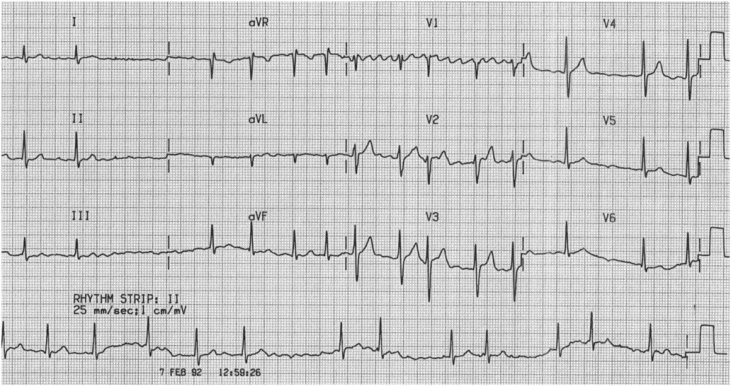 Figure 36.2
Stay updated, free articles. Join our Telegram channel

Full access? Get Clinical Tree


