Fig. 42.1
Annular, erythematous, scaly plaques in the face and trunk of a neonate with NLE
Periorbital erythema, referred to as “raccoon eye” or “owl eye,” is a very common characteristic [6]. At times, the lesions may be urticarial, desquamative, ulcerative, or crusted [6, 14]. Bullous lesions may be seen with a particular predilection for the soles of the feet [14]. In some infants, solar exposure seems to precipitate the eruption [15]. These lesions typically last for weeks or up to 6 months and then resolve spontaneously consequent to the disappearance of maternal antibodies in the neonatal circulation [12]. Dyspigmentation is frequent but it may resolve spontaneously. Atrophic lesions and, rarely, atrophic scars may develop [6, 13]. Telangiectasia is often prominent and is the sole cutaneous manifestation reported in some patients. The atrophic telangiectatic changes are most evident near the temples and scalp and do not necessarily occur in the same sites as the erythematous lesions [12]. The latter site may occasionally be associated with a permanent alopecia. Telangiectasia, scarring, and atrophic changes are expected to persist.
The cardiac manifestations include conduction abnormalities (first-, second-, and third-degree heart block) and cardiomyopathy [1, 2, 16]. Third-degree heart block, once established, is usually irreversible [4]. Congenital heart block may present as bradycardia noted in utero or during physical examination at birth. Conduction disturbances may also present as irregular heartbeat and prolongation of the QT interval. Congenital heart block may be associated with endocardial fibroelastosis and cardiomyopathy [1]. In some cases, myocarditis and pericarditis can develop which may lead to bradycardia. Heart failure is a well-recognized complication during the neonatal period.
Hepatobiliary involvement may present with elevation of liver enzymes (such as aspartate aminotransferase and alanine aminotransferase) and/or conjugated hyperbilirubinemia occurring a few weeks or months after birth and resolving thereafter. Some infants may have mild hepatomegaly and, less commonly, splenomegaly [1]. The hepatomegaly and splenomegaly are usually transient. Cholestatic hepatitis and hepatic failure may also rarely occur [17].
Hematologic disturbances (e.g., hemolytic anemia, thrombocytopenia, and neutropenia) may occur in the first 2 weeks of life. Infants with hematological involvement are usually asymptomatic [1]. Autoantibodies, mainly anti-Ro, bind directly to the neutrophil and cause neutropenia. Thrombocytopenia may manifest as petechiae. Hematologic symptoms usually appear at around the second week of life and disappear by the end of the second month. Lymphopenia is not a characteristic hematologic abnormality of NLE [12].
Other abnormalities such as hydrocephalus and macrocephaly may occur [18]. Aseptic meningitis and myelopathy have rarely been reported [6]. Pneumonitis may manifest as tachypnea and/or tachycardia [1].
Pathogenesis
Anti-52/60-kDa Ro/SSA and anti-48-kDa La/SSB antibodies are associated with heart block
Anti-50-kDa La/SSB antibodies are associated with cutaneous disease
Anti-ENA/U1RNP antibodies are usually associated with atypical cutaneous features and thrombocytopenia
NLE is caused by the transplacental passage of maternal autoantibodies [2, 3]. Approximately 98 % of the affected infants have maternal transfer of autoantibodies against Ro/SSA, La/SSB, and, less commonly, ENA/U1-RNP.
The antibodies associated with heart block and with cutaneous disease are believed to be different; antibodies against the 52/60-kDa Ro/SSA and 48-kDa La/SSB ribonucleoproteins are associated with heart block, whereas antibodies against the 50-kDa La/SSB ribonucleoprotein are associated with cutaneous disease [11]. Ultraviolet radiation and estrogens increase Ro antigen expression on the surface of the keratinocytes [1].
The 52-kDa Ro/SSA (Ro52) ribonucleoprotein is an antigenic target strongly linked with the autoimmune response in mothers whose children have NLE, congenital heart block, and other conduction abnormalities [1]. Anti-Ro52/SSA autoantibodies antagonize the serotonin-induced L-type calcium channel activation on human fetal atrial cells and trigger an inflammatory response, leading ultimately to fibrosis and scarring of the atrioventricular node, sinus node, and His bundle [6].
Anti-ENA/U1RNP autoantibodies are usually associated with atypical cutaneous lesions without cardiac or systemic abnormalities in a small number of NLE cases and may play a role in the pathogenesis of thrombocytopenia [6].
In addition to its presence in the skin and heart, the Ro antigen is also found in the liver, bowel, lungs, brain, and blood cells—the tissues that are most often affected by NLE [1]. However, only some neonates exposed to these autoantibodies develop complications. Therefore, other factors such as titers of maternal antibodies, genetic predisposition, and environmental factors such as viral infection may be involved [19].
Diagnosis and Differential Diagnoses
Diagnosis depends on clinical features on the infant and the demonstration of NLE-associated antibodies in maternal serum
Skin biopsy is useful when the diagnosis is in doubt
Several differential diagnoses include seborrheic dermatitis, epidermolysis bullosa, atopic dermatitis, and tinea corporis among others
The diagnosis is usually established based on the clinical features and the demonstration of NLE-associated antibodies in the serum of the mother or the affected infant. Screening of infants with NLE for the presence of these antibodies is strongly recommended [2, 4, 6].
Skin biopsy is useful in patients with NLE when the diagnosis is in doubt. Histologic examination shows interface dermatitis, keratinocyte damage, moderate hyperkeratosis, follicular plugging, and vacuolar degeneration in the basal cell layer. Epidermal atrophy may be found. Inflammatory infiltrate may be intense with bulla formation histologically. An immunofluorescent examination reveals a granular IgG deposition at the dermoepidermal junction; IgM and C3 deposition may also be evident [12].
Laboratory investigations may reveal pancytopenia, thrombocytopenia, leukopenia, or elevated transaminase levels [20].
Differential diagnosis of NLE includes seborrheic dermatitis, atopic dermatitis, neonatal acne, epidermolysis bullosa, tinea corporis, psoriasis, granuloma annulare, erythema multiforme, Langerhans cell histiocytosis, congenital rubella, congenital syphilis, Bloom syndrome, and Rothmund–Thomson syndrome [20].
Treatment
Photoprotection and topical steroids for cutaneous lesions
Evaluation of pacemaker need for cardiac lesions
Systemic steroids or immunosuppressive agents for severe systemic involvement
Photoprotection and low-potency topical corticosteroids are the mainstay of therapy for cutaneous lesions of NLE. Parents of the affected children must be thoroughly counseled regarding avoidance of sunlight, proper use of sunscreen, and protective clothing [21].
Sometimes, though there is resolution of active skin lesions, pigmentary changes, atrophy, and telangiectasia may persist over the face and other exposed body parts. In such cases, cosmetic camouflage helps these children in social interaction. Persistent telangiectasia may be managed with vascular laser [21].
Systemic corticosteroids or immunosuppressives are not recommended to treat cutaneous lesions of NLE. In rare cases, if systemic therapy is indicated, hydroxychloroquine may be used [21].
Patients with NLE with cardiac involvement require regular monitoring to assess cardiac function and the need for a pacemaker. A pacemaker is often necessary for those who are unable to compensate for a slow heart rate. Serial echocardiography to monitor for a prolonged PR interval should also be arranged. If the cardiac involvement is severe, activity may have to be restricted in the young child [21].
Infants with severe hepatic and hematological involvement may require treatment with systemic corticosteroids, intravenous immunoglobulin, and/or immunosuppressive agents [6].
Children with NLE need continued follow-up, especially before adolescence and if the mother herself has an autoimmune disease [22]. Although the child may not be at increased risk of developing SLE, the development of some form of autoimmune disease in early childhood may be of concern. As many mothers are unaware of their diagnosis at the time of the child with NLEs birth, mothers must be advised to seek work up and clinical management of their systemic disease, as well as guidance regarding future pregnancies.
Prognosis
The morbidity and mortality of NLE of childhood depend on the organ systems affected [2, 3]. Children with NLE have an excellent long-term outcome when only skin lesions are present [16, 23]. The cutaneous lesions usually disappear by 6 months of age, coincident with the clearance of maternal antibodies from the child’s circulation [2]. Involvement of the skin may, rarely, lead to scar formation. Although children with cutaneous disease may be more prone to develop SLE or autoimmunity later in life, this is mainly due to their genetic predisposition, not that they had NLE. Their unaffected siblings are also at risk for development of SLE or autoimmunity. While the cutaneous lesions of NLE are themselves benign, cutaneous NLE is associated with a 6–10-fold risk for a subsequent child with cardiac NLE [2, 16, 23].
NLE with cardiac involvement is associated with a 20–30 % mortality rate in the neonatal period [2, 16]. Mortality is particularly high in cases of congenital heart block with concurrent cardiomyopathy [7, 12]. Death most often results from congestive heart failure caused by congenital heart block. Approximately 57–66 % of patients with congenital heart block eventually require a pacemaker [2, 16, 23]. Those with pacemakers are at risk of developing dilated cardiomyopathy in their lives [1]. However, many children with congenital heart block may be relatively asymptomatic until adolescence, when they begin to exercise. At that time, they may develop syncope and require a pacemaker implantation. Prospective clinical trials on the use of antenatal fluorinated steroids in women with anti-SSA/Ro and/or anti-SSB/La antibodies and fetuses with heart block identified in utero are required before definitive recommendations can be made. A number of anecdotal cases support the use of dexamethasone for treatment of hydrops and possibly incomplete block [2].
Most patients with NLE affecting liver or blood have transient disease that spontaneously resolves within 4–6 months. In some cases, cholestatic hepatitis and liver failure may occur, which is associated with a poor prognosis [1]. Anemia, thrombocytopenia, and neutropenia are self-limited. However, if severe thrombocytopenia is present, internal bleeding can lead to a poor prognosis [1].
Ongoing Research
Currently, the main focus of the investigation in NLE lies on the search for reliable markers that predict the specific involvement of fetal organs and for a safe and effective treatment to prevent definitive lesions [24].
Conclusion
NLE refers to a clinical spectrum of cutaneous, cardiac, and systemic abnormalities observed in newborn infants whose mothers have autoantibodies against Ro/SSA and La/SSB. The condition may be associated with serious sequelae. Neonates with NLE should be managed at a tertiary care center, and multidisciplinary team involvement may be indicated.
Cutaneous Lupus Erythematosus
Introduction
Lupus erythematosus is a complex autoimmune disease with variable clinical features
Cutaneous lupus erythematosus (CLE) can be classified as acute, subacute, and chronic
All forms of CLE may progress to SLE
Lupus erythematosus (LE) is a complex autoimmune disease that generates a chronic, persistent multiorgan inflammatory process with variable clinical features and course. CLE can be classified as acute, subacute, which includes neonatal lupus, and chronic [25, 26]. Acute CLE may produce mid-facial or disseminated lesions and will be discussed in another chapter as it is a common feature of SLE. SCLE was first described in 1979 by Sontheimer et al. [27] as a distinct subset of cutaneous lupus clinically characterized by psoriasiform and/or annular lesions. Chronic cutaneous lupus erythematosus (CCLE) is typically characterized by discoid lesions, but other varieties can be observed [26]. All forms of CLE may progress to SLE [25].
Epidemiology
SCLE is extremely rare in childhood and female predominance is noted among the reported cases
Less than 3 % of patients develop discoid lupus erythematosus (DLE) before the age of 10 years
Female predominance in DLE patients might depend on the age of onset
SCLE is extremely rare in childhood though a few cases have been reported in literature. Among the described cases, there is a frank female predominance (2.5:1) and the mean age at diagnosis is 6.6 ± 3.7 years [28–34].
DLE is uncommon in childhood (with around 80 published cases) [35–46] and exceptional in infancy [40, 46]. Less than 3 % of patients with DLE develop the disease before the age 10 years [40]. Some authors note the absence of female predominance [40, 46] whereas others have noted a female predominance, more marked in patients less than 10 years (5:1) than in older than 10 years (1.5:1) of age [41].
Clinical Features
SCLE is characterized by annular or psoriasiform photodistributed lesions
DLE is the most common form of CCLE and may present with a linear arrangement
Lupus tumidus, lupus panniculitis, and chilblain lupus may not exhibit epidermal involvement
Subacute Cutaneous Lupus Erythematosus
This type of cutaneous lupus is typically photosensitive, with lesions confined to sun-exposed skin. Despite this fact, mid-facial skin is usually spared, while the sides of the face, V of the neck, and extensor aspects of the upper extremities are commonly involved. SCLE lesions may have an annular configuration, with raised red borders and central clearing, or a papulosquamous presentation (either eczematous or psoriasiform in appearance). Lesions often heal without scarring but with long-lasting hypopigmentation or even vitiligo-like leukoderma and telangiectases [28–34]. Nail dystrophy affecting almost all the nails has been described by Rai and Balachandran [30]. Although the classic presentation is in the form of annular or papulosquamous lesions, poikilodermatous [47], erythrodermic [48], and bullous SCLE [49] have also been reported in adults.
Patients with SCLE can complain of mild systemic symptoms (the most prominent of which are musculoskeletal complaints); however, approximately one half of them fulfill the criteria for SLE as per the American Rheumatism Association criteria, although it is usually a milder form of the disease [30].
Chronic Cutaneous Lupus Erythematosus
The term CCLE encompasses several variants of cutaneous lupus: DLE, LE tumidus, lupus panniculitis, and chilblain lupus, DLE being the most common form among these [26].
Discoid Lupus Erythematosus
DLE lesions are found most often on the face, scalp, and ears, but may be present in a widespread distribution [50]. Occasionally, DLE lesions may appear on mucosal surfaces, including the lips, nasal mucosa, conjunctivae, and genital mucosa [51].
DLE consists of erythematous plaques with atrophy, scale, alopecia, follicular plugging, and telangiectasias (Fig. 42.2). The lesions have the potential for scarring and dyspigmentation, typically with hypopigmentation in the center and hyperpigmentation at the periphery, but sometimes with vitiligo-like depigmentation [2, 35–46]. Prevalence of photosensitivity in children with DLE is thought by some authors to be lower than in adults [46]. However, Mokhtar et al. [52] and Cherif et al. [40] in two Tunisian studies reported a prevalence of photosensitivity as high as 81 %.
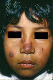
Fig. 42.2
Localized DLE lesions on the face of a Mexican Mestizo patient
Several authors have described linear chronic DLE as a rare manifestation of lupus in which the erythematous, atrophic, dyschromic lesions are located along the lines of Blaschko, affecting more commonly the face and neck. Unlike typical DLE, the linear variant onset occurs frequently in childhood [53–56].
Lupus Tumidus
LE tumidus represents an uncommon but distinct subtype of CCLE. The lesions are characterized by erythematous, indurated, urticarial, nonscarring plaques in sun-exposed areas. Unlike other variants of CCLE, there is no epidermal involvement; therefore they lack associated scale or follicular plugging (Fig. 42.3). The lesions tend to resolve without scarring or atrophy [50, 57].
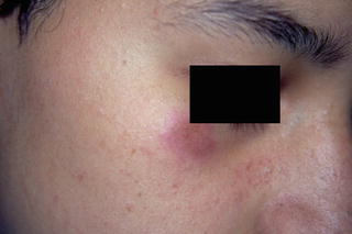
Fig. 42.3
LE tumidus—erythematous, indurated, urticarial plaque with no scale or follicular plugging
Lupus Panniculitis/Lupus Profundus
Intense inflammation in the subcutaneous tissue leads to indurated plaques, which may have overlying hyperpigmentation in patients of color, that can evolve to depressed, disfiguring areas. The lesions of lupus panniculitis occur predominantly on the face, upper arms, upper trunk, breasts, buttocks, and thighs (Fig. 42.4). Some patients have discoid lesions overlying the panniculitis and it is sometimes referred to as lupus profundus [40, 42, 58].
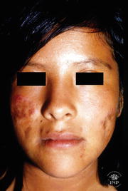
Fig. 42.4
Lupus profundus—deep infiltration with overlying discoid lesions and residual atrophy on the face of a teenager
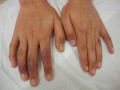
Chilblain Lupus Erythematosus
Chilblain lupus erythematosus (CHLE) is a rare, chronic form of CLE. It consists of red or dusky purple papules and plaques on the toes, fingers, and sometimes the nose, elbows, knees, and lower legs (Fig. 42.5). The lesions are triggered or exacerbated by cold, particularly in moist cold climates [59, 60].