(1)
Hospital del Niño Jesús, Tucumán, Argentina
Abstract
Becker’s nevus is characterized by the presence of a light or dark brown patch or plaque with a sharply outlined but irregular border that resolves into smaller pigmented spots, arranged in a checkerboard pattern. In male patients, the lesion may show increased hairiness after puberty. Becker’s nevus is a fairly common condition, but often overlooked or misdiagnosed.
“Becker’s nevus syndrome” represents one of the epidermal nevus syndromes and denotes the simultaneous occurrence of Becker’s nevus and unilateral breast hypoplasia or other cutaneous, muscular, or skeletal defects. All of these anomalies tend to show a regional correspondence to the nevus and are mostly ipsilateral.
Keywords
Becker’s nevusBecker’s nevus syndromeBecker’s pigmented hamartomaMosaicismIntroduction
Becker’s nevus is an organoid nevus reflecting mosaicism
It is characterized by a hyperpigmented, often hairy macule with a sharp outline but irregular borders
Becker’s nevus is arranged in a checkerboard pattern, more often located in the upper half of the thorax
S.W. Becker first described Becker’s nevus (BN) in 1949 as “concurrent melanosis and hypertrichosis in the distribution of nevus unius lateris”; it was subsequently named after him [1].
Other names given to this nevus are Becker’s melanosis, pigmented hairy epidermal nevus, and Becker’s pigmented hamartoma. Because melanocyte counts are not increased in this lesion, these names should be discouraged in order to avoid further confusion with a melanocytic lesion.
BN is an organoid nevus, reflecting mosaicism, that manifests as one or more lightly to deeply hyperpigmented, usually hairy patches or plaques, with a sharp demarcation from normal skin, but irregular borders. Lesions are arranged in a checkerboard pattern, more often located on the upper half of the thorax, shoulder, or arms. It can be seen in any part of the body. The association of this nevus with unilateral breast hypoplasia, muscle, skeletal, and/or skin anomalies has been named Becker’s nevus syndrome. All of these anomalies tend to show a regional correspondence to the nevus and are mostly ipsilateral [2–4].
Epidemiology/Demographics
The prevalence of BN is not known, but a study involving 19,302 men (17–26 years old) showed that it is close to 0.52 % [5]. Hair density may be variable and may even be absent. In 5 % of the cases, BN is itchy, and in 25 % there is a history of severe sunburn prior to the lesion. There is a strong association with smooth muscle hamartoma [3–5].
A retrospective study was performed by Patrizi et al. in Italy, covering a 10-year period, to better define the clinical characteristics of the BN in childhood; they analyzed 118 cases and found that the BN was more frequent than other studies suggested and had similar predilection sites to those of adults but that hypertrichosis was rarely seen in the condition [1]. Contrary to expectations, the peak incidence of BN was observed not at puberty but at birth. No sex predilection was found in their study, in agreement with data reported by Happle and Koopman [6].
Most authors believe that isolated BN occurs more frequently in men than in women, with a 2:1 ratio. Happle and Koopman [6], however, stated that the true sex ratio may in fact be 1:1, because BN tends to be less conspicuous in women [4–7].
This nevus can occur in all races. It usually appears around puberty and in 75 % of instances it has appeared before the age of 15 years. Although in its classic form it is considered to be an acquired disorder, the occurrence of congenital Becker’s nevus has been reported [7–10]. Familial cases can also occur [7, 9–13].
Clinical Presentation
BN is a congenital lesion noticed in childhood
Is an organoid epidermal nevus characterized by light to brown patches in a checkerboard distribution
Is an androgen dependent lesion, showing increase in hairness and color at puberty
Histologically there are no nevus cells so there is no increased risk of malignant transformation
BN is usually first noticed in childhood, after sun exposure, as a grayish, light, or dark brown pigmentation on the chest, back, or upper arm, although it can be seen in any body site. It spreads in an irregular fashion until it reaches an area of 10–15 cm in diameter. The outline is sharply demarcated, irregular, and often surrounded by islands of blotchy pigmentation [3, 14] (Figs. 28.1, 28.2, and 28.3).


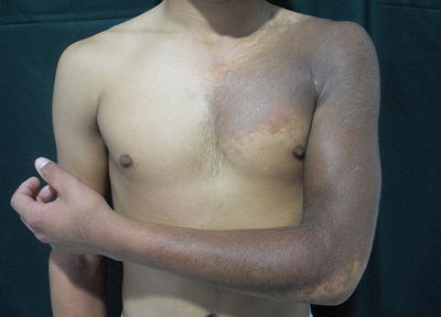
Fig. 28.1
Dark pigmented, hairy patch in a checkerboard distribution. Note thoracic asymmetry

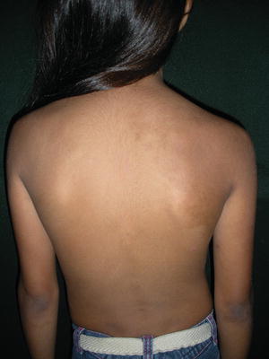
Fig. 28.2
Female patient. Note light hyperpigmented macule over the right shoulder and scapula. Note scoliosis

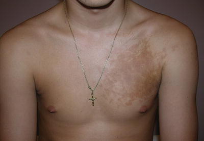
Fig. 28.3
Sharply demarcated, irregular brown macule surrounded by islands of blotchy pigmentation
Within the group of epidermal nevi, it can be classified as an organoid epidermal nevus, arranged in a checkerboard pattern. It can be present as a single or as multiple lesions. The lesion represents a form of mosaicism; therefore, it is a congenital lesion with growth of the lesions near puberty because it is an androgen dependent nevus [3]. Near puberty the lesion becomes thicker and darker and in most of them dark, coarse hairs appear in the region of, but not necessarily coinciding with, the pigmented area [14] (Fig. 28.1).
Histopathologic features reveal epidermal thickening, elongation of the rete ridges, and increased pigmentation in the basal layer. There is no increase in number of melanocytes, and since there are no nevus cells, malignant transformation does not generally occur. Hair structures and smooth muscle fibers are usually increased in number [7, 14, 15].
The entity known as smooth muscle hamartoma could be into the clinicial as well as histopathological spectrum of BN.
Treatment
BN is a benign condition; therefore, treatment is merely for cosmetic reasons. The most effective treatment is not been well defined. Traditional surgical excision usually is unsuccessful and may result in unacceptable scars. Lasers have been used for improving the pigmentary component as well as for reducing the associated hypertrichosis, with variable results 4, 7.
Trelles et al. [16] demonstrate the superiority of Erbium:YAG laser when compared to Q-switched Neodymium:YAG laser, showing complete clearance in 54 % of the patients. Moreno-Arias et al using intense pulsated light showed not very satisfactory results, with clearance of less than 25 % of the lesions [17]. Choi et al. showed a fair to excellent clinical response in 11 patients treated with long-pulsed alexandrite laser with mild to moderate side effects in some patients, consisting of hypopigmentation, skin texture change, and scar [18].
Lapidoth et al. reported the use of low fluence with high-repetition-rate diode laser hair removal as a safe and effective method for the management of hypertrichosis in Becker’s nevus. No adverse events were reported [19].
Glaich et al. used a fractionated laser showing good response, without side effects [20]. In a study by Meester et al., an ablative 10,600-nm fractional laser therapy was moderately effective in patients with BN [21]; postinflammatory hyperpigmentation and relatively negative patient-reported outcomes still preclude ablative fractional laser therapy from being the standard therapy [22].
Stay updated, free articles. Join our Telegram channel

Full access? Get Clinical Tree


