- Gene structure
- Commonly used investigations
- Genetic variation
- Multifactorial inheritance
- Approach to the dysmorphic neonate
- Prevention of congenital abnormalities
Introduction
Human genetics is a rapidly expanding field in almost all fields of medicine. The list of genetic loci and mutations that may cause genetic disease is ever expanding and are catalogued in the Online Mendelian Inheritance in Man website (http://www.ncbi.nlm.nih.gov/entrez/query.fcgi?db=OMIM). It is now possible to sequence an individual’s DNA, thereby identifying risk factors for the development of future disease. There is an increasing recognition of polygenic diseases, where a number of genes interact to increase the risk of disease. A detailed discussion of all the causes and types of mutations that may occur is beyond the scope of this book. For further information the reader should refer to specific texts on the molecular basis of genetic disease.
Gene Structure
Every cell in the body normally contains 2 sets of 23 chromosomes (the haploid number). Therefore there are 46 chromosomes (the diploid number) in every cell, 2 are sex chromosomes (either XX in females or XY in males) and 44 are autosomes. A chromosome contains a double strand of DNA tightly wound together. During meiosis the chromosomes separate and align themselves around the centre of the cell, and half migrate into each daughter cell. Fertilization requires fusion of the maternal oocyte (containing 23 chromosomes) and the paternal sperm (containing 23 chromosomes). Mitochondria also have their own distinct genome. Mitochondria are passed only through the ova and therefore all mitochondrial DNA comes from the mother.
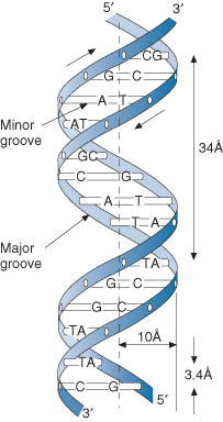
DNA consists of two strands of very long sequences of nucleotides (see Fig. 8.1). A nucleotide consists of a five-carbon ringed sugar, a triphosphate (attached to the 5 carbon), a hydroxyl group (attached to the 3 carbon), a hydrogen group (attached to the 2 carbon) and one of four bases (attached to the 1 carbon): adenine (A), cytosine (C), guanine (G) and thymidine (T). The other strand of DNA (also called the antisense strand) contains the complementary bases, wherever there is an A the opposite strand has a T (and vice versa), and wherever there is a G the opposite strand has a C (and vice versa). In RNA, thymidine is replaced by uracil (U). A series of 3 nucleotides makes a codon (e.g. UAA, AUG, UGA) and each is a specific instruction for either stopping transcription or insertion of a specific amino acid. In total there are 64 possible codons for the 20 amino acids.
The total amount of chromosomal DNA codes for about 3 billion base pairs. However, only 1.5% contain codes for genes (of which there are approximately 23,000). This coding DNA is subsequently translated into proteins. The remainder is RNA genes, sections involved in gene regulation, and other non-coding sections which have poorly understood functional properties but do not translate into proteins.
Commonly Used Investigations
Genetic testing can now be undertaken prior to conception (e.g. test parents for gene), in vitro (e.g. test embryo preimplantation), during gestation (e.g. perform chorionic villus sampling or amniocentesis) or after birth. For any genetic investigation, a specimen containing genetic material is required. Various tissues can be sampled or cultured, depending on the timing and urgency of the diagnostic test being undertaken. Commonly used tissues are listed in Box 8.1
- Blood: lymphocytes (collected in a heparinized tube), ideally from a patient who has not recently ingested alcohol, antibiotics or drugs of addiction
- Bone marrow
- Embryo
- Skin (fibroblast culture): useful up to 2 days after death
- Desquamated cells in amniotic fluid
- Chorionic villus sample
Chromosome Analysis
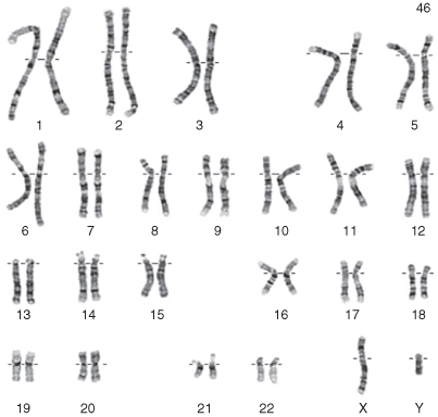
A Giemsa (G) stain gives the overall appearance of the karyotype. G-banding of the chromosomes gives detailed information about more subtle abnormalities. Chromosomes are grouped according to banding characteristics, size of chromosomes and centromere position. The larger chromosomes are designated by the lowest numbers, and each chromosome is divided by the centromere into a short arm (p) and a long arm (q) (see Fig. 8.2). Karyotype analysis should be available in 24–48 h but banding can take up to 14 days.
Figure 8.2 Normal chromosome pattern and number after Giemsa staining. This is an example of a male karyotype. The hashed horizontal line is at the centromere and divides the chromosome into short (p) and long (q) arms.

Polymerase Chain Reaction
Although it is not itself a genetic test, the development of the polymerase chain reaction (PCR) technique has proved incredibly useful for providing genetic material for other investigations. This technique allows for rapid amplification of small fragments of DNA. It is extremely sensitive, to the point that DNA from a single cell can be amplified a million times in only a couple of hours. This technique is relatively inexpensive and is performed in most laboratories.
Fluorescence In-Situ Hybridization
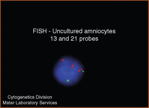
The fluorescence in-situ hybridization (FISH) technique allows more careful examination of chromosomes for very small abnormalities of which the DNA sequence is already known. The DNA is initially denatured and then a specific sequence of DNA with a fluorescent marker is added and the culture is incubated to allow hybridization. The DNA is then washed removing any unbound sequences. If the marker has bound to its complementary sequence it will be detectable on fluorescence microscopy examination. It can be used for rapid diagnosis of conditions such as trisomy 21, trisomy 13 and trisomy 18 as well as others such as velocardiofacial syndrome. See Fig. 8.3 for an example.
Figure 8.3 Image showing FISH probes for chromosome 21 (red) and chromosome 13 (green). There are three red signals (abnormal) and two green signals (normal); this patient therefore has trisomy 21.

Microarrays
Microarray testing is a method of analysing whether or not a number of specific genes are being expressed in cells. A microarray works by measuring the amount of sample mRNA molecules bound to a specific template of the gene. By using an array of hundreds or thousands of gene templates, the amount of mRNA bound to each site on the array can be measured. This process generates a profile of gene expression in the cell and can accomplish many genetic tests in parallel.
Indications for Investigations
While it is now much easier to perform many of the above investigations, there are still a number of important issues that need to be considered before they are ordered. Some of these include: purpose of test (e.g. diagnosis versus screening), tissue sample required to get the result, length of time before results will be available, cost of the investigation and ethical implications for child/siblings/parent/relatives. Consultation with a clinical geneticist may be required. Some possible indications for genetic testing are listed in Box 8.2.
- Recurrent unexplained first-trimester pregnancy loss
- High risk of genetic abnormality on antenatal testing (e.g. increased risk of chromosomal disorder on triple test or abnormalities on ultrasound)
- Fresh stillbirth with physical abnormalities
- Infant with previous chromosomal disorder
- Known genetic disorder in family
- Neonate with dysmorphic features
Genetic Variation
Changes in the genome account for our individuality but can also impact on the normal function of cells and organs. There are three basic categories for genetic variation:
- structural changes or changes in large segments of DNA that affect more than one gene
- changes in the DNA sequence
- multifactorial changes.
Stay updated, free articles. Join our Telegram channel

Full access? Get Clinical Tree


