Fig. 17.1
(a) Placement of the Allis clamps and catheterization of the urethral meatus. (b) A U-shaped incision in the vulva, mobilization of the tissues and placement of the first suture. (c) Closing of the first layer with placed of sutures between the inner skin margins. (d) Closing of the second layer and completion of the operation [2, 3]
Closing of the inner skin margins followers. The knots are placed inside the created neovagina to avoid early decomposition, which could lead to wound opening.
A layer of sutures is followed to approximate the subcutaneous fat and the perineal muscles. Finally, the external skin is closed (Fig. 17.1d). For the closing of both the skin layers (Fig. 17.1b, c), interrupted absorbable 2-0 sutures are used, starting posteriorly and proceeding anteriorly.
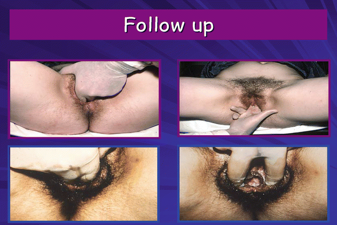
The criterion for the success of the operation is the creation of a neovagina up to 10–12 cm in depth and 4–5 cm in width. The functional dimensions of the neovagina are measured using sonovaginography [1]. A clinical re-examination in 4 weeks and 6 months and then on a yearly basis is recommended (Fig. 17.2). Following our procedure, no significant postoperative complications were reported and all patients have had a satisfactory sexual intercourse. A mean hospital stay up to 6 days is required to prevent postoperative complications such as dehiscence and to maximize patient’s compliance. Finally, there is no need for postoperative vaginal dilatations, which usually reduce the psychological impact on the patient [3, 6].


Fig. 17.2
Postoperatively results of Creatsas vaginoplasty
Comparative Advantages of Vulvo-perineoplasty
The McIndoe’s vaginoplasty was a common used vaginoplasty among other available operative techniques. However, several complications were reported such as the risk of injuries of the neighboring organs. Also, graft shrinkage, due to the development of granulomatous tissue, caused neovaginal stenosis. The aesthetic outcome should be taken into consideration.
The Vecchetti’s operation and its laparoscopic version are frequently performed in several European centers over the last years, with low perioperative morbidity and a short recovery period. Potential important complications may occur, namely passing the cutting needle from the abdominal wall to the retrohymenal fossa. Frequent follow-up evaluations to adjust the device tension and the use of dilators after the removal of the apparatus, are also required.
The sigmoidal colpoplasty is considered to be a major, complicated intraperitoneal operation that carries intraoperative risks and complications. Satisfactory anatomical and functional results have been reported by the use of pelvic peritoneum from the pouch of Douglas [7, 8].
Stay updated, free articles. Join our Telegram channel

Full access? Get Clinical Tree


