Case Note
A 35-year-old woman presented with premature menopause and previous failure of oocyte donation; our pregnancy rate for this procedure is 80%. She had undergone, at another clinic, operative hysteroscopy intervention during which 2 polyps were removed. However, this last intervention was followed by inflammatory complications. We decided to perform virtual ultrasonographic hysteroscopy as a more patient-friendly procedure.
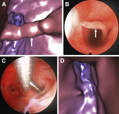
This intervention was performed by transvaginal ultrasonographic examination using “fly-through” technology (Fly Thru, Toshiba American Medical Systems, Tustin CA) enabling computerized 4-dimensional virtual reconstruction of the uterine cavity, without the need for physically entering the uterus. Her uterine cavity was slightly distended by injecting 10 mL of saline through a narrow soft balloon catheter fixed to the cervical canal. Immediately thereafter, images of the cavity were taken ( Video 1 ). The whole procedure took <10 minutes. As compared to conventional saline-infusion sonography, fly-through technology makes it possible to minimize the volume of injected saline without compromising image quality. Image analysis showed a beamlike adhesion crossing the uterine cavity ( Figure , A, and Video 1 ). Conventional hysteroscopy was thus performed 2 days later, under general anesthesia. The uterine adhesion was visualized ( Figure , B) and removed ( Figure , C). One month later, virtual ultrasonographic hysteroscopy was repeated to evaluate the result of the previous hysteroscopic surgery. The images showed complete disappearance of the intracavital adhesion in its previous location ( Figure , D, and Video 2 ). The patient thus prepared for transfer of her cryopreserved embryos, resulting from the previous oocyte donation attempt. After conventional uterine preparation protocol, 2 embryos were transferred, resulting in an ongoing singleton pregnancy.


