Use of Monitoring Devices
Kenneth Yen
Marc H. Gorelick
Introduction
Monitoring is the process of observing the clinical condition of a patient, typically by repeated measurement of vital signs. Although such monitoring may be done by human observers, the task is frequently performed by automated devices. In this section, methods and equipment for several of the most commonly monitored parameters in pediatric emergency medicine—heart rate and rhythm, blood pressure, and temperature—are discussed. Capnography is a commonly used type of monitoring but will not be covered here because it is the subject of Chapter 76. Pulse oximetry, which is also the subject of another chapter (Chapter 75), is briefly discussed below.
Pulse Oximetry
The use of pulse oximetry, a noninvasive transcutaneous method of measuring the oxygen saturation of blood, has expanded rapidly since its introduction into clinical practice in the early 1980s.
Physiology
Noninvasive arterial oxygen saturation measurement is obtained by directing red and infrared light through a pulsating vascular bed. The pulsating arterioles in the path of the light beam cause a change in the amount of light detected by a photodiode. Different hemoglobin species (oxygenation) have different light absorption spectra (1). The oximeter measures within the pulse wave form the ratio of transmitted red to infrared light and uses it to determine the oxygen saturation of the blood. The nonpulsatile signal is removed electronically for the purpose of calculation, and therefore skin, bone, and other nonpulsating substances do not interfere with the calculation of arterial saturation.
Procedure
The pulse oximeter consists usually of a small and portable monitor and a sensing unit that clips or tapes onto the patient’s finger. Toes and ear lobes are also often used. Along with the arterial oxygen saturation (SaO2), the meter also typically provides a tracing of the pulse rate. The strength of the identified pulse rate can also help determine if the oximeter reading is accurate.
Indications for pulse oximetry include screening for the presence or development of hypoxemia, as well as continuous monitoring to measure response to therapy. It is commonly used in the assessment of children with acute respiratory illness such as pneumonia, asthma, and upper airway obstruction. It is also recommended for monitoring patients undergoing conscious sedation (2). The anatomy, physiology, and technology underlying pulse oximetry are detailed in Chapter 75, which also contains a discussion of proper technique and interpretation of findings.
Several things have to be kept in mind. Measurements in hypothermic patients and in patients with poor perfusion (e.g., decompensated shock) are often inaccurate. The reading can be falsely high in patients with carbon monoxide poisoning or in a chronic smoker who has elevated blood carbon monoxide levels. Other things that may affect pulse oximetry readings include extremely bright lights, dense fingernail polish, and patient movement.
Electrocardiographic Monitoring
Physiology
Electrocardiograph (ECG) monitors measure and display heart rate and rhythm by detecting the electrical activity of cardiac muscle. The device consists of a sensing component, the electrode, which when placed on the skin surface can detect changes in electrical potential (i.e., depolarization and repolarization) originating in the heart. Two electrodes comprised in a lead are required to detect cardiac activity by measuring the difference in electrical potential between two sites. Standard limb leads are placed on the left arm, right arm, and left leg. A right leg electrode serves as ground. Each lead detects a different view of the heart, and all the leads together give a complete picture of cardiac activity. A 12-lead ECG involves both limb and chest leads placed in a standard configuration and allows a look at the activity of a specific lead. Routine monitoring in the emergency department (ED) usually involves only three electrodes, one placed at the base of the heart, one at the apex, and a reference electrode on the upper left chest wall to reduce electrical interference (3).
Electrical potentials detected by leads require amplification to detect the low-voltage signal. The amplifier also has a filtration system to reduce electrical “noise.” Cardiac activity can be displayed continuously on an oscilloscope and/or recorded on ECG graph paper as a plot of voltage versus time. Most monitors also have the capacity to freeze and record a tracing in the last few seconds before an abnormality activates the monitor alarm (4). Monitor alarms, both auditory and visual, indicate equipment problems, including loose or defective electrodes, heart rate parameters that have exceeded the programmed limits, and abnormal cardiac rhythms.
Procedure
Intended electrode sites are cleansed with alcohol. Conductive gel is used at each area of contact between the electrode surface and skin to stabilize the electrode. Most disposable electrodes are pre-gelled. The electrode and lead wire should be firmly applied to dry skin. As previously noted, for routine continuous monitoring, three thoracic sites (at the base and apex of the heart and on the upper left chest) are usually chosen (Fig. 5.1).
Potential sources of difficulty can be patient- or equipment-related. Any voluntary movement, involuntary shivering, seizure activity, or interference by nearby electronic devices can produce artifacts. Good contact must occur between the skin and the electrode to reduce artifacts and produce an accurate tracing. The machine must be standardized so that a given voltage activity produces a consistent deflection, and both patient and machine should be properly grounded.
As with all monitoring devices, the ECG must not replace frequent clinical assessment of the patient. The results of ECG monitoring can be normal in a patient with organic heart disease or abnormal in a healthy individual. As with any laboratory test, ECG findings must be interpreted in conjunction with the clinical status of the patient.
Blood Pressure
Monitoring of blood pressure in the ED is used as a screen to (a) assess the level of cardiovascular stability for patient triage, (b) follow clinical response or adverse reaction to therapy, and (c) follow clinical course in a potentially unstable condition. In the majority of cases, this can be done noninvasively with a blood pressure cuff and stethoscope or with an automated oscillometric device such as the Dinamap. Invasive blood pressure monitoring is reserved for those critically ill patients requiring intensive care unit admission, multiple repeated measurements of arterial blood gases, and continuous monitoring.
Physiology
Noninvasive blood pressure monitoring involves the use of a compression cuff applied to the limb and inflated with air to a pressure above systolic blood pressure, thereby terminating distal flow. Blood pressure is determined by detecting the pressure at which blood flow resumes as the cuff is deflated. Auscultatory methods include using a stethoscope to detect Korotkoff sounds, which are the result of turbulent blood flow as cuff pressure falls below arterial systolic pressure (see also Chapter 4). The first sound auscultated approximates systolic blood pressure. Diastolic blood pressure is the pressure at which muffling of sounds occurs. If auscultation is difficult because of noise level or a low-flow state, the arterial pulse can be palpated as the cuff is deflated or an
ultrasonic flow detector can detect when flow resumes. These two latter methods cannot be used to determine diastolic pressure.
ultrasonic flow detector can detect when flow resumes. These two latter methods cannot be used to determine diastolic pressure.
An alternative method of blood pressure detection is the oscillometric method utilized in equipment such as the Dinamap machines. This method is based on the principle that pulsatile blood flow through a blood vessel produces arterial wall oscillations that are transmitted to a blood pressure cuff (5). The machine operates with a pressure transducer and a microcomputer, both of which sense cuff pressure, initiate cuff inflation and deflation, control for motion artifacts, and record points of the changing amplitude of oscillations. At systolic blood pressure, a rapid increase occurs in the amplitude of oscillations. Diastolic pressure is the point at which a sudden decrease occurs in oscillations. The mean arterial pressure is the point at which the amplitude of oscillations reaches a maximum. These three independently measured values are typically displayed on the monitor at the completion of each inflation-deflation cycle.
Procedure
Errors in blood pressure measurement can be cuff- or patient-related (6). The most common error is incorrect cuff size—in particular, a cuff that is too small—which falsely elevates the blood pressure (see Figs. 4.2 and 4.3). The cuff should cover two thirds of the upper arm length, fully encircling the arm with a snug fit. In obese children, a cuff width 20% greater than arm diameter will provide a more accurate reading. Rapid deflation rate is another source of measurement error, creating falsely low blood pressure measurements. Cuff deflation rate should be no more than 2 to 3 mm Hg per second. Before cuff application and between determinations, ensure that all residual air is eliminated.
Patient-related sources of error include having a crying or anxious child and placing the extremity above or below heart level during measurement.
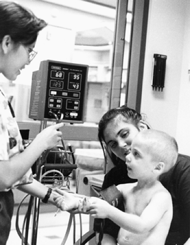
Use of Dinamap machine and other similar oscillometric blood pressure–monitoring equipment begins with placing the proper size cuff on the selected limb, which in most cases is the upper arm. Other sites, including the forearm and ankle, are more comfortable for prolonged periods of monitoring in the conscious patient but are less helpful when dealing with the patient with shock or peripheral vasoconstriction. Before the cuff is applied, all residual air must be squeezed out; the cuff is then put on snugly but not so tightly as to cause venous congestion. If residual air remains in the cuff or the cuff is not snug, blood pressure readings will be falsely elevated. The cuff and the patient’s heart must be at the same level to avoid the effect of hydrostatic pressure on the reading, which is falsely high when the cuff is below the heart and falsely low when the cuff is above the heart. The conscious, cooperative patient must be told to remain quiet and avoid movement (Fig. 5.2; see also Fig. 4.4).
Problems with noninvasive blood pressure monitoring occur with shock and peripheral vasoconstriction. Palpation and auscultatory methods are insensitive in low-flow states. The Dinamap is more useful during shock because it measures pressure oscillations instead of sounds. In severe cases, only mean arterial pressure may be measurable, if even that, and invasive blood pressure monitoring may be warranted. Accurate noninvasive monitoring is also compromised in patients with frequent irregular heart rhythms; patients with significant tachycardia; and patients who are moving excessively (shivering) or on rapid cycling mechanical ventilators, when large variability in systolic blood pressures exists.
 Figure 5.2 The child is relaxed and comfortable, with arm at heart level, for blood pressure monitoring.
Stay updated, free articles. Join our Telegram channel
Full access? Get Clinical Tree
 Get Clinical Tree app for offline access

|
