Case Notes
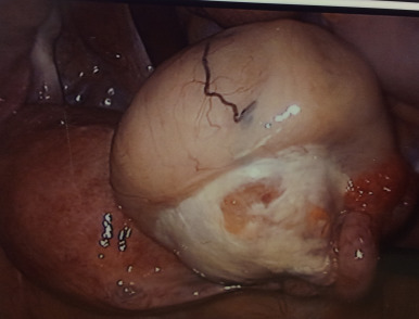
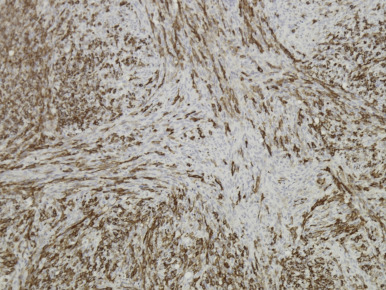
A 45-year-old, para 1 woman with abdominal pain was referred to our outpatient clinic. On physical examination, a fixed soft mass was palpated between left adnexa and pelvic sidewall. Transvaginal ultrasound revealed a left ovarian mass measuring 4.3 × 3.0 cm, with blood flow. Cancer antigen-125 was 70.5 U/mL. During laparoscopic left salpingo-oophorectomy, the tumor was found to be homogeneous and gray-yellowish with a yolky appearance ( Figure 1 ). On gross cut, it was firm with lobulation, without hemorrhage or necrosis. The pathological biopsy showed a thecoma of the ovary composed of interlacing spindle thecoma cells with long fascicles in a storiform and whorl arrangement ( Figure 2 ) and the fallopian tube demonstrated no abnormality.
Stay updated, free articles. Join our Telegram channel

Full access? Get Clinical Tree


