Chapter 26 The newborn infant
TRANSITION TO EXTRA-UTERINE LIFE
Following birth the neonate undergoes several physiological changes so that it can adapt to extra-uterine life. Two main changes affect respiration and cardiovascular function.
First breaths
The several control systems that regulate breathing are already present in fetal life. Most of them operate mainly in fetal active states, such as REM (rapid eye movement) sleep. In quiet sleep, long periods without breathing occur. At birth there is a need to exchange oxygen and carbon dioxide. Breathing is augmented by an elevated carbon dioxide. Non-specific stimuli, such as pain, cold and light also lead to stimulation of breathing. In most infants the first breathing efforts occur within 30 seconds of birth and are forceful enough to overcome the high resistance of the liquid in the airways and to inflate the lungs. In immature and abnormal infants the breathing efforts may be reduced.
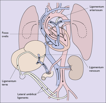
Changes in the pattern of blood circulation
The second change is the redirection of the blood flow following the cessation of the high blood flow through the umbilical arteries that perfused the placental villi, and a lowering of the large volume of blood returning through the umbilical vein and the vena cava. The venous pressure in the vena cava falls and the ductus venosus closes. The lungs expand with the first breath and the pulmonary vascular resistance falls abruptly. The infant’s systemic blood pressure rises slightly at the same time, which results in a temporary reversal in the direction of the blood flowing through the ductus arteriosus. As the infant breathes, the oxygen tension in the blood rises and the muscular walls of the ductus contract, the passage of blood through it ceasing. At the same time, the pressure in the right atrium falls. There is a simultaneous increase in the blood flow through the lungs. This blood enters and increases the pressure in the left atrium. Because of the changes in pressure between the two atria, the foramen ovale closes.
With the closure of the ductus venosus, the foramen ovale and the ductus arteriosus, the adult pattern of circulation of the blood is established (Fig. 26.1; compare with Fig. 4.1, p. 29).
Changes in liver function
After the cardiovascular changes have taken place the infant’s liver perfusion is increased. The liver can efficiently convert glucose into glycogen and vice versa, but some of its enzymatic functions are immature. The most important is its limited ability to conjugate bilirubin. In consequence, physiological jaundice may occur in the first week of life.
Most of the glycogen stored in the liver is laid down in the last 8 weeks of intrauterine life. Glucose from glycogen and the metabolism of ketone bodies contribute significantly to the energy needs of the first few days of life. Preterm and growth restricted babies have smaller liver glycogen stores and may develop hypoglycaemia. After a few days the neonate obtains energy from food and by oxidizing fats stored in adipose tissue.
Temperature control
At birth, vasoconstriction of the skin vessels preserves body heat, but temperature maintenance is made difficult because of the newborn infant’s relatively large surface area. The body temperature of newborn infants may fall by up to 1.5 °C immediately after the birth, especially if wet skin is exposed allowing rapid heat loss. A mature infant will have laid down fat in brown adipose tissue and can utilize this for heat production without shivering. Preterm babies have less brown fat and may become hypothermic.
Because of the instability of temperature control, neonates should be wrapped properly in cold environments, but in hot environments too much wrapping should be avoided.
PERINATAL HYPOXIA
Any condition occurring during late pregnancy or labour that reduces the oxygen available to the fetus will predispose it to cardiorespiratory and neurological depression at birth. These conditions have been mentioned when discussing the at-risk fetus in pregnancy and labour (see p. 152).
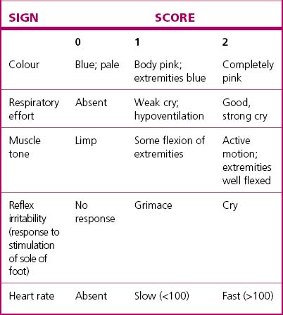
The severity of cardiorespiratory and neurological depression around the time of birth can be assessed by the Apgar scoring system (Table 26.1) or by the pH and/or blood lactate of umbilical artery blood. Both are useful in managing the immediate problem but are relatively poor indicators of long-term outcome.
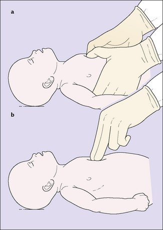
The basic resuscitation of the newborn is described on page 77 (see Fig. 8.15). The most important step in resuscitation is to initiate ventilation with intermittent positive-pressure respiration; current evidence suggests that ventilation using air (21% oxygen) should be the initial step for term babies, with oxygen being added only if hypoxia persists despite adequate ventilation. Initial ventilation should be with a mask and inflating device; failure to initiate spontaneous breathing within 2–5 minutes may require endotracheal intubation – but only if a person skilled in this technique is present. If the infant remains bradycardic (HR <60) despite adequate ventilation, the circulation should be supported by external cardiac massage (Fig. 26.2) and, possibly, endotracheal 1: 10 000 adrenaline 0.3–1.0 mL/kg. Once the umbilical vein is cannulated a further 0.1–0.3 mL/kg dose of adrenaline is given, and if there is no response further doses of adrenaline 0.1– 0.3 mL/kg can be given at 3–5-minute intervals.

Fig. 26.2 Closed cardiac massage. Intermittent pressure is applied over the middle third of the sternum (which is pressed down about half an inch) with two fingers or thumbs. The heart is compressed between the rib cage and the vertebral column, and blood is expelled into the great vessel. For clarity, the endotracheal tube has been omitted in the drawing.
A 5-minute Apgar of less than 6 should be followed by measurements of arterial blood gases and pH, and treatment given to adjust the findings if necessary.
Infants who have required advanced resuscitation or have prolonged cardiorespiratory depression should be closely observed in a special care nursery until it is clear that they have recovered or need continuing specialist care.
BIRTH INJURIES
With increasingly good obstetric care during childbirth, birth injuries are becoming less common and less severe. Only those injuries most likely to occur will be discussed. In all cases, the injury must be explained to the parents and the treatment and prognosis discussed.
Cranial injuries (Fig. 26.3)
Caput succedaneum
Caput succedaneum is caused by pressure on the fetal scalp when the head is being pushed deeply into the pelvis and the venous return from the scalp is impeded. The oedematous swelling and bruising is subcutaneous and can cover a large area of scalp. Treatment is not required; the swelling of the caput disappears rapidly and the bruising within a few days.
Cephalhaematoma
Cephalhaematomas may occur after a spontaneous vaginal delivery or following trauma from the obstetric forceps or the ventouse. The periosteum is sheared from the underlying parietal bone and subperiosteal haemorrhage occurs. The swelling is limited to the outline of the bone; initially it is tense, subsequently it is fluctuant and the rim may calcify. The haematoma remains for 2 weeks to 3 months and is slowly absorbed.
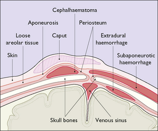
Subaponeurotic (subgaleal) haemorrhage
Subaponeurotic haemorrhage, while rare, occurs most commonly after a traumatic vacuum extraction (see p. 195–197) and may result in significant blood loss from the infant’s circulation. Occasionally the bleeding is very rapid and life-threatening and requires urgent blood transfusion. Frequent observation for diffuse head swelling following vacuum extraction is mandatory.

Stay updated, free articles. Join our Telegram channel

Full access? Get Clinical Tree

