Ovulation and the menstrual cycle

Chapter 2 Ovulation and the menstrual cycle


The endocrinological changes that take place to transform a female child into an adolescent who menstruates and ovulates and is capable of conceiving a child, begin several years before puberty, but the most marked changes occur in the 2 years before the girl’s first menstrual period (menarche).


Disturbances occurring during the menstrual cycle are discussed in Chapters 28 and 29.


MENARCHE


The underlying major endocrinological change is that the hypothalamus begins to secrete releasing hormones. These lead to the release into the circulation of adrenal androgens and pituitary human growth hormone (hGH). It is hGH that causes the growth spurt which begins 3–4 years before the menarche, and which is maximal in the first 2 years (Fig. 2.1). The physical growth slows down as the first menstruation (menarche) approaches. This is because increasing quantities of oestrogen are secreted by the ovaries and feed back negatively, reducing hGH secretion. Shortly after the secretion of hGH starts, the hypothalamus begins to release gonadotrophin-releasing hormone (GnRH) in an episodic pulsed manner. At first, the pulses are greater in amplitude during sleep, but after 2 years they occur by day and night at about 2-hour intervals. GnRH induces the release of follicle-stimulating hormone (FSH) and luteinizing hormone (LH) from the pituitary gland, which in turn bind to receptors in the ovaries and induce the secretion and release of oestrogen and progesterone into the circulation. The quantity of FSH and LH increases as the girl matures.


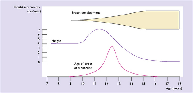
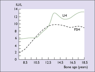
Until the age of 8 years, only small quantities of oestrogen are secreted (and less of progesterone). After that age oestrogen secretion begins to rise, slowly at first, but after the age of about 11 years the rise is quite rapid. The FSH levels reach a plateau when the girl is aged about 13. LH levels rise more slowly until 1 year before menarche, at which time a rapid rise occurs (Fig. 2.2). By this time, the GnRH pulses occur every 90 minutes. These hormonal changes persist until after the age of 40, when changes presaging the menopause begin (see Ch. 42).


It is thought that the rapid rise of LH induces the onset of menarche, but other factors are also involved. These include an increase in the fat : lean ratio of body composition, which in turn is related to good nutrition and the absence of debilitating diseases.


Between the early 1800s and the mid 1900s the average age at menarche fell from 15–17 years to 13–13.5 years. There has been little change over the past 30 years the mean age being around 12.5 years with the exception that obese girls tend to enter puberty earlier than those of normal weight. The initial reduction is believed to be due to better childhood nutrition. It is hypothesized that the greater amount of body fat in girls today permits the greater aromatization of androgens to oestrogens. Rapidly rising levels of oestrogens feed back positively to the hypothalamus and pituitary gland, leading to the LH surge that precedes the menarche.


The menarche may be delayed in women who are of low body weight, such as ballet dancers, women who have anorexia nervosa, or those who are compulsive exercisers.


EFFECTS OF OESTROGEN AND PROGESTERONE ON BODY TISSUES


More than 20 oestrogens have been isolated, the three considered the most important being oestrone, oestradiol and oestriol. Oestrone is a relatively weak oestrogen and interconverts with 17-β oestradiol, which is the most active and the predominant oestrogen in the reproductive years. Oestradiol is rapidly transported in the blood to tissues that have oestrogen-binding receptors. In the blood, 60% is bound to albumin, 37% to sex hormone-binding globulin, and 3% is free. The tissues of the genital tract and the lobular elements of the breasts have the highest concentration of cells containing specific oestrogen-binding receptors, and consequently are most affected by circulating oestradiol.


Once attached to the specific binding sites oestradiol is transferred to the cell’s nucleus, where it activates genes, leading to RNA synthesis. This process is regulated to some extent by progesterone, which blocks the formation of new receptors and induces intracellular enzyme production; these enzymes also regulate oestrogen metabolism. Following nuclear gene activation, oestradiol is rapidly converted to the relatively inactive oestriol, which is transported to the liver where it is conjugated with glucuronic acid. The conjugate is then excreted, mostly in the urine. This leaves the cell receptors free to bind more oestradiol.


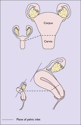
Oestradiol stimulates the growth of the vulva and the vagina after the menarche, the hormone causing proliferation of both the epithelial and the muscular layers. This oestrogen also stimulates the formation of more blood vessels, which supply the organs. The uterus is particularly stimulated by oestradiol, which increases its vascularity. Oestradiol causes endometrial proliferation, stimulating the growth of the glands and stroma as well as the growth of the muscular layers of the uterus, so that the uterus grows from its prepubertal size to its adult size in the perimenarchal years (Fig. 2.3). The great increase in circulating oestrogen in pregnancy causes the rapid growth of the uterus, and the lack of this hormone after the menopause leads to uterine atrophy.


Stay updated, free articles. Join our Telegram channel

Jun 15, 2016 | Posted by in OBSTETRICS | Comments Off on Ovulation and the menstrual cycle

Full access? Get Clinical Tree

Get Clinical Tree app for offline access