The Challenged Child


The Challenged Child


Cheryl E. Goble


This chapter is designed to assist the doctor of chiropractic in the care and management of the challenged child. In today’s society, children who are physically or mentally challenged are accepted and therefore seem to be more prevalent. The health care provider must be prepared to aid in the treatment of these children. One of the most significant necessities of this assistance is to participate as part of the child’s health care team. Services from therapists, teachers, optometrists, nutritionists, psychologists, medical physicians, and social workers are many times essential to the total well-being of the patient; therefore, the doctor of chiropractic must be cooperative with these providers.

This chapter will review the clinical history, chiropractic evaluation, and chiropractic treatment of the challenged child. The author will address the chiropractic diagnosis, management, and prognosis of four specific disorders: cerebral palsy, Down’s syndrome, attention deficit hyperactivity disorder, and fetal alcohol syndrome.


HISTORY

The clinical history of the child is of utmost importance; adequate time must be made available with the child and parents or caretaker of the child to properly evaluate the child’s history and current presentation. The environment of the office should be one of comfort and accessibility. It is suggested the child have appropriate toys, such as puzzles, puppets, or drawing tools, to occupy his or her time during the evaluation period.

The first element of the assessment is the child’s birth. Information should be compiled regarding the type of delivery. Complications of pregnancy or birth trauma are more frequent in cases of the handicapped child than in the histories of other children (1). Because a number of childhood disabilities are caused by abnormalities of pregnancy and labor, a history of gestation and delivery should be obtained (2). Prenatal and intranatal complications arouse the suspicion of central nervous system (CNS) damage (3).

The child’s development, including physical, mental, and emotional abilities, should be assessed. Accomplishment of skills in gross and fine motor function, along with social skills, are significant in the child’s overall developmental representation. Oftentimes the parent or caretaker is unable to adequately detail the progress of these developmental milestones. Table 27.1 provides guidelines regarding normal skills (4). The parent or caretaker can provide information regarding the child’s academic progress and social behavior, as well as the child’s ability to adapt in a normal everyday lifestyle. Many times, the child’s capabilities and limitations can be observed in the office during the evaluation period.

During review of the general health of the child, a chronological order of symptoms, signs, and treatment should be analyzed. The category of general health should include immunizations, medications, diagnostic testing, injuries, surgical procedures, medical treatment, and hospitalizations (3). Social behavior can be easily observed, and with adequate inquiry one can determine the activity capabilities, temperament, sleeping habits, eating patterns, discipline problems, and interactive skills of the child. The doctor can gain a vast amount of knowledge about the child by having the child, parent, or caretaker give a description of a “typical day” for the child. As with all patients, the clinical history provides a wealth of cumulative information that the doctor can use to determine an accurate diagnosis, treatment plan, and prognosis for the patient.


EXAMINATION

As with any patient, cooperation is essential to obtain accurate findings. The patient should be in a comfortable and appropriate position for examination.
A flexible approach to the child is necessary; initial evaluation should begin with general observation of the child and his or her capabilities. Throughout the evaluation, the doctor should be aware of the developmental stage of the child (5) (Table 27.1). A significant amount of information can be collated about the neuromuscular system, alertness, behavior, and parent-child interaction by observing the child in spontaneous activities (6). The neurologic examination begins with observation; special attention is given to the child’s gait, motor function, and postural deviances. The doctor must take advantage of opportunities provided by the patient (4). Should the patient be confined to a wheelchair, the doctor must make adaptations to properly evaluate the child in this position (Figs. 27-1 and 27-2). If the child is able to sit on his or her own, a cervical chair is an appropriate tool to be used for the evaluation.


TABLE 27.1 Screening Protocol for Developmental Delay


Age


Appropriate Skills


3 mo


Gross motor: Supports weight on forearms


Fine motor: Opens hands spontaneously


Social Skills: Smiles appropriately


Language: Coos, laughs


6 mo


Gross motor: Sits


Fine motor: Momentarily transfers objects


Social Skills: Shows like and dislike


Language: Babbles


9 mo


Gross motor: Pulls to stand


Fine motor: Grasps (pincer)


Social skills: Plays patty cake and peek-a-boo


Language: Imitates sound


12 mo


Gross motor: Walks with one hand held


Fine motor: Releases object on command


Social skills: Comes when called


Language: Speaks one or two meaningful words


18 mo


Gross motor: Walks upstairs with assistance


Fine motor: Feeds from a spoon


Social skills: Mimic actions of others


Language: Speaks at least six words


24 mo


Gross motor: Runs


Fine motor: Builds a tower of six blocks


Social skills: Plays with others


Language: Speaks two- to three-word sentences.


One component of the chiropractic evaluation is the use of a handheld temperature differential instrument (7,8,9). If the patient is able to sit, the instrument can be used in a normal glide pattern. If unable to sit, the child may be more comfortable in a prone position for this portion of the examination.


FIGURE 27-1 Assessment of upper cervical mobility in Y-axis rotation in a patient with cerebral palsy.


FIGURE 27-2 Motion palpation of the lower cervical spine in a patient with cerebral palsy.


When evaluating the child for evidence of vertebral subluxation, motion and static palpation of the spine and paraspinal areas for tenderness, edematous areas, and hypertonicity or hypotonicity of musculature reveals potential levels of vertebral subluxation (10). Intersegmental motion palpation of the spine from occiput to the sacrum (including sacroiliac articulations) provides useful clinical information regarding the biomechanical function or lack of normal function of the joint (10).

To appropriately evaluate the child, plain film radiography is a fundamental way of determining if chiropractic treatment will be appropriate (11). Spinal alignment derived from a specific radiographic marking system can add to the analysis of the child regarding the application of a specific chiropractic adjustment. To appropriately evaluate the child, full-spine radiographs taken anterior to posterior in a single exposure and a lateral projection taken in two exposures are recommended (12), provided the patient can be adequately positioned. For certain disorders covered in this chapter, additional views may be recommended to properly assess the biomechanics of the spine.

In summary, the chiropractic evaluation and examination of the challenged child requires patience on the part of all parties involved. Many times, the examination may require more than one office visit, depending on the child’s physical status, emotional and mental capabilities, and the child’s level of cooperation during this important portion of the child’s care.


COMMON DISORDERS

The following section is a collation of the most commonly seen challenged-child disorders in this author’s practice. A chiropractic approach to each disorder is presented as well as information regarding prognosis, where applicable.


Cerebral Palsy

In the United States, approximately 300,000 individuals manifest this central motor deficit. This syndrome is described as a nonprogressive motor disorder resulting from gestational or perinatal CNS damage and characterized by an impairment of voluntary movement (13). In this category of neurologic defect there is evidence of a major disturbance of motor function. Numerous causes of cerebral palsy have been investigated; these include physical trauma to the brain at birth (4) and complications of pregnancy, which include placental complications; abnormalities or presentation (face, brow, and transverse); breech delivery; prolapse of the umbilical cord; and fetal growth retardation, although most term growth-retarded infants develop normally (1,14). For low birth weight infants born by breech delivery, the cerebral palsy rate reached almost 5% in a study performed at the National Institute of Neurological and Communicative Disorders (1). Risk factors include child abuse, prematurity, encephalopathy or seizures during the perinatal period, interventricular hemorrhage during the perinatal period, in-utero infections, postnatal meningitis or encephalitis, and hypoxic ischemia (15).

Intrapartum fetal asphyxia has been implicated as a cause of cerebral palsy, although most cases of cerebral palsy occur in persons without evidence of birth asphyxia or other intrapartum events (16,17,18,19). Based on formalized review processes, the U.S. Preventive Services Task Force has not recommended that routine electronic fetal monitoring be used for low-risk women in labor (20). The Task Force also concluded that there was insufficient evidence to recommend for or against intrapartum electronic fetal monitoring for high-risk pregnant women.

Clinical Signs and Symptoms Cerebral palsy can be grouped into four main categories: spastic, athetoid, ataxic, and mixed forms. Spastic syndromes are most common, representing approximately 70% of the cases (13). The spastic form of cerebral palsy is characterized by stiff, awkward movements of the affected limbs, muscular hypertonicity, increased deep tendon reflexes, “scissors gait,” and toe-walking of the lower extremities. There may be stiffness and incoordination of the hands and arms, evident in spastic quadriplegia. This form accounts for 30% of cerebral palsy cases, and these children are generally markedly retarded and nonambulatory (21). Children with spastic hemiplegia or paraplegia frequently have normal intelligence and a good prognosis for social independence (13). Movements of the extremities are slow, writhing, and involuntary. Emotional tension seems to cause an increase in the motions, which disappear during sleep. Ataxic cerebral palsy is commonly associated with sclerotic lesions of the cerebellum (21). These patients have difficulty with rapidity of fine movement, weakness, incoordination, and a wide-based gait. The fourth category of cerebral palsy is the mixed form. These are classified most often as spasticity and athetosis and, less often, ataxia and athetosis (13).

Radiologic Evaluation In addition to the full spine AP and lateral views, a flexion view of the cervical region is beneficial to determine the potential vertebral subluxation. It is often difficult to radiograph these children, and technological assistance is of utmost importance. Because of the patient’s condition, multiple levels of malalignment will usually be present. It is of utmost importance to evaluate the mobility of any motion segment that is determined to need an
adjustment, and all adjustments should be administered in ways that promote improved posture of the spine at both the segmental and global levels.

A subluxation is often detected in the upper cervical region in the patient with cerebral palsy, either as an effect of the disorder or preceding it, but most commonly the antero-superior (AS) condyle (22). The integrity of the position of the occiput in relationship to the atlas is noted on the neutral lateral, flexion, and AP cervical views. When the condyle slips anterior and superior, the space between the posterior arch of the atlas and the occiput is diminished. The space will remain closed even when the cervical spine is in flexion (23).

Abnormalities of the brain, identifiable with computed tomography and/or magnetic resonance imaging, include cysts, cerebral atrophy, calcification, tumors, malformation, and stroke (15).

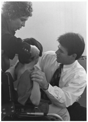
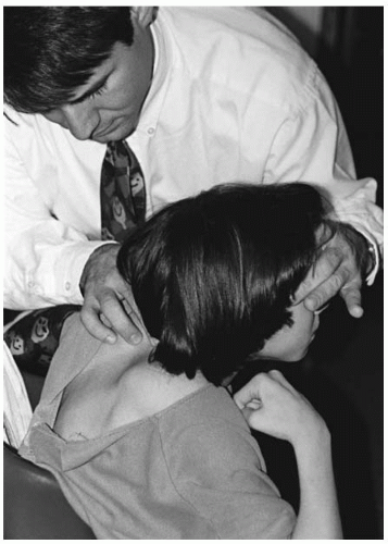
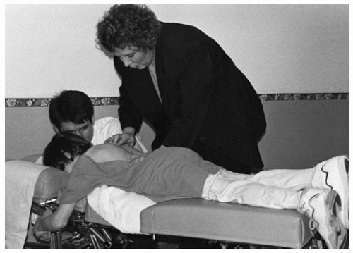
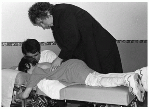
Subluxation Considerations The vertebral subluxation complex can be determined by visual inspection, motion and static palpation, and instrumentation. The correlation of these findings, along with radiographic studies, are optimal for the location and correction of the vertebral subluxation. Special attention should be directed to the upper cervical region; many times the child will present with a “starward gaze” in their head presentation. This could be indicative of an AS condyle misalignment. Careful analysis of this region and the entire spine (including the cranium and pelvis) can produce the specific level of the vertebral subluxation. When correlating the objective findings with the radiological findings, a specific listing can be determined, thus assisting in a specific correction (Figs. 27-3,27-4,27-5,27-6).

After analyzing the child with cerebral palsy and determining the area of subluxation, a specific adjustment is necessary. The adjusting thrust should be gentle and specific because of the spasticity in many of these children. The thrust should be high velocity and low amplitude.

Cranial and pelvic structures should be checked carefully for subluxation and addressed accordingly. The removal of neurological stresses that may be present can promote a more normal functioning of the nervous system, resulting in improved general resistance.

Prognosis The child with cerebral palsy has many special needs. When caring for these children, special time should be appropriated for not only evaluation and adjusting, but for communication with the children’s caretakers. In this author’s experience, many parents have noted a more positive attitude, relaxation, improved sleep patterns, better coordination, and stronger resistance to stresses. These observations should be evaluated systematically for evidence of efficacy.


FIGURE 27-3 Adjustment of an AS condyle subluxation in a patient with cerebral palsy.

Spinal contractures will increase during growth, resulting in scoliosis. This factor will require careful scrutiny by the attending chiropractor. The general course for most patients is functional improvement with time (15).

General follow-up by the appropriate health care provider is important for patients with cerebral palsy.
Specific areas to be addressed during the child’s growth are the development of contractures, epilepsy, learning disabilities, strabismus, hearing loss, and mental retardation (15).


FIGURE 27-4 Demonstration of a vertebral count in the prone position.


Get Clinical Tree app for offline access