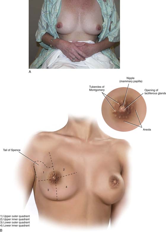
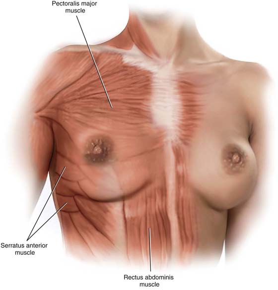
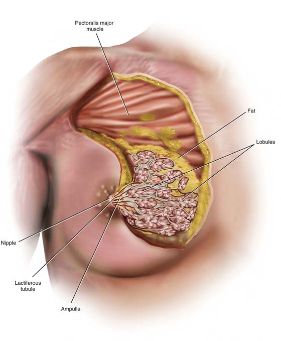
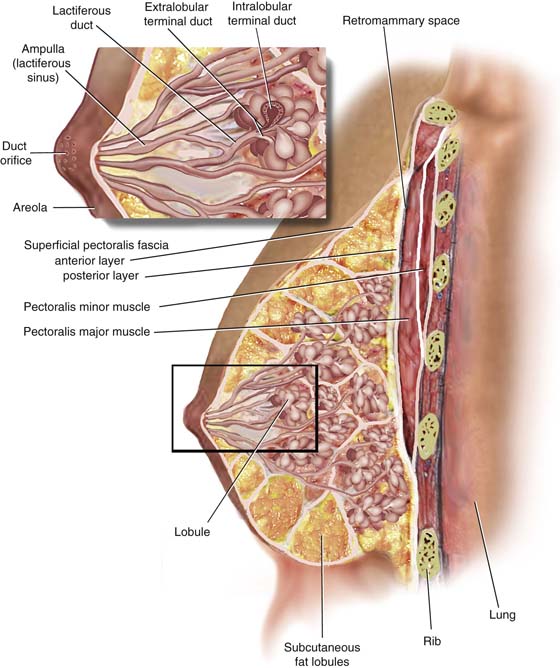
CHAPTER 105 Donna L. Stahl The breasts are modified sweat glands and specifically function as modified apocrine glands. They represent significant gender-identifying structures, which society and the individual connote as phenotypically female. Mature adult breasts occupy a prominent position on the anterior chest wall between the second and sixth ribs (Fig. 105–1A). The “average” breast measures 10 to 12 cm in diameter and is 5 to 7 cm in thickness. Across the chest wall, the breast extends from the margin of the sternum to the midaxillary line. A portion of breast tissue projects into the axilla. This entity is known as the tail of Spence (Fig. 105–1B). Anatomically, the entire breast lies between the superficial and deep layers of the superficial pectoral fascia. The latter is contiguous with Scarpa’s fascia of the anterior abdominal wall. Thus, the breast is roughly hemispherical in shape and sits on top of the deep pectoral fascia, which in turn encompasses the pectoralis major muscle (Fig. 105–2). A significant factor in the maintenance of the structure and shape of the breasts is attributed to fibrous bands that course between the deep and superficial layers of fascia. These dense Cooper’s ligaments form the so-called suspensory ligaments of the breast, which are particularly prominent at its lower portion (inframammary fold). The tissues lying between the deep and superficial layers of the superficial pectoral fascia consist mainly of fat but additionally of breast parenchyma and connective tissue (stroma). The relative amount of natural fat content contributes to the bounce of the breast with movement. As the volume of fat increases, so does the motion of the breasts, but only to the point where the quantity of fat produces such a large mass that it results in drooping, pendulous breasts. Scarring secondary to artificial augmentation of the breasts also diminishes the natural movement of the breasts on the chest wall (Fig. 105–3). The breast parenchyma is divided into 15 to 20 segments or glandular units or lobes, which are radial in arrangement and converge to a series of ducts on the nipple. Approximately 5 to 10 major collecting ducts open at the nipple. Each duct drains a segment or lobe of the breast. Each lobe contains 20 to 40 lobules. Each lobule in turn consists of between 10 and 100 alveoli (Figs. 105–3 and 105–4). Mounted on each breast is a more deeply pigmented circular area measuring an inch or more in diameter—the areola. The nipple caps the center of the areola. The dermis of the areola contains longitudinal and circular smooth muscle, which creates a wrinkled appearance when the muscles contract (see Fig. 105–1B, inset). Several elevated openings may be observed on the periphery of the areola. These are the termini of the ducts of Montgomery’s glands. The latter are modified sebaceous glands whose secretions keep the areolae lubricated and supple. During pregnancy, these same glands may secrete a milklike substance (see Fig. 105–1B, inset). The functioning unit of the breast is the lobular unit. This consists of small glands lined by cuboidal and myoepithelial cells surrounded by a vascular stroma. Small interlobular terminal ducts lead to extralobular terminal ducts, which in turn lead to larger collecting ducts and then lead to still larger lactiferous ducts that drain entire lobes. Before draining into the nipple, these ducts dilate to form a lactiferous ampulla or sinus. Contraction of the myoepithelial cells as well as the smooth muscle of the areola aids in emptying the lactiferous sinuses of milk (see Fig. 105–4, inset). Breast volume changes during the menstrual cycle. The breast parenchyma responds to stimulation with estrogen and progesterone. Similarly, water content within the fat (edema) parallels that seen within the endometrium, with peaks between days 22 and 25. The least amount of hormonal alteration occurs during the 4 to 5 days after the onset of menstrual flow. Finally, although most breasts appear roughly symmetrical in size (volume), differences between the left and right breasts are common (Fig. 105–5). FIGURE 105–1 A. The mammary glands occupy a prominent location on the female chest wall. Phenotypically, they connote female gender to the individual. B. The breasts occupy space on the anterior chest wall between the second and sixth ribs. For purposes of description, the breasts are divided into four quadrants—two upper and two lower. The sculpted lower edge of the hemispheric breast is formed by the inframammary ridge. A portion of the breast tissue in the upper outer quadrant extends into the axilla (tail of Spence). The inset shows details of the areola, which encompasses the nipple, the tubercles of Montgomery, and the pigmented skin highlighting this area. FIGURE 105–2 The breasts are contained within the superficial pectoral fascia and lie on the deep fascial layer that encloses the pectoralis major muscle. The breast encroaches inferiorly on the deep fascia of the serratus anterior, external oblique, and rectus abdominis muscles. The superficial pectoral fascia is subdivided into superficial and deep layers. The deep superficial layer is distinctly separated from the deep pectoral fascia by loose fat. FIGURE 105–3 The breast consists of fat and breast tissue. The latter is made up of lobules, ducts, and fibrous connective tissue. The apocrine glandular tissues or lobules secrete milk, which is collected and transported via a series of ducts to the nipple. The structural architecture of the breast is maintained by fibrous bands traveling between the deep superficial fascia and the dermal component of the breast’s skin. These connective tissue bands or Cooper’s ligaments largely account for the breast’s spherical shape. FIGURE 105–4
The Breast
 Karen S. Columbus
  Michael S. Baggish
Anatomy of the Female Breast
![]()
Stay updated, free articles. Join our Telegram channel

Full access? Get Clinical Tree


