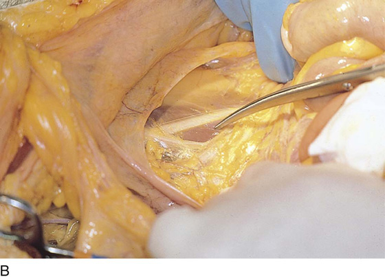
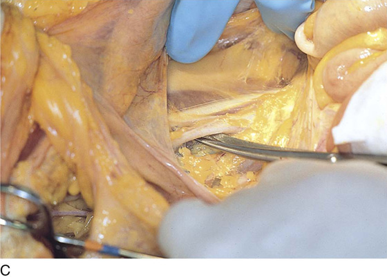
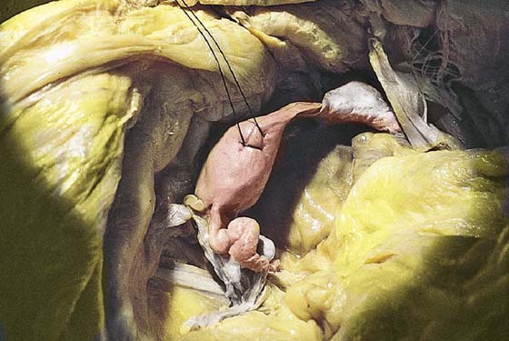
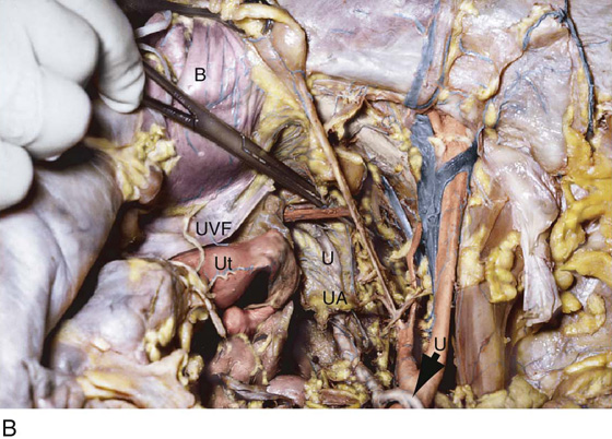
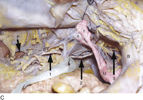
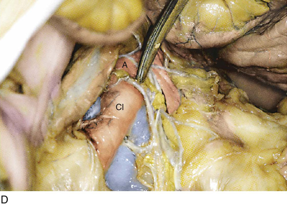
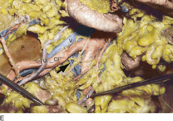
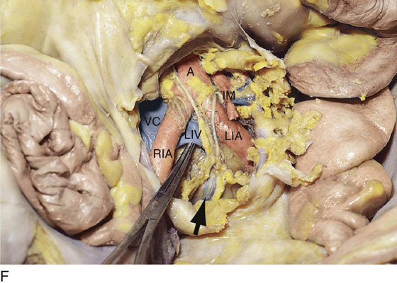
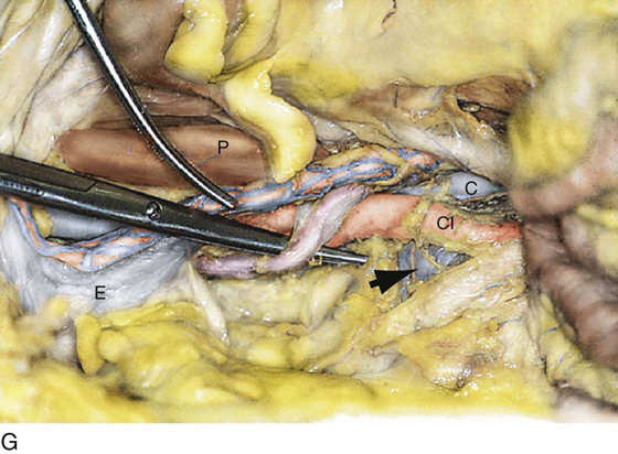
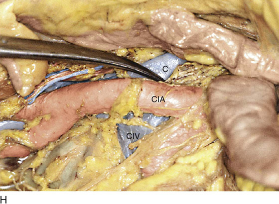
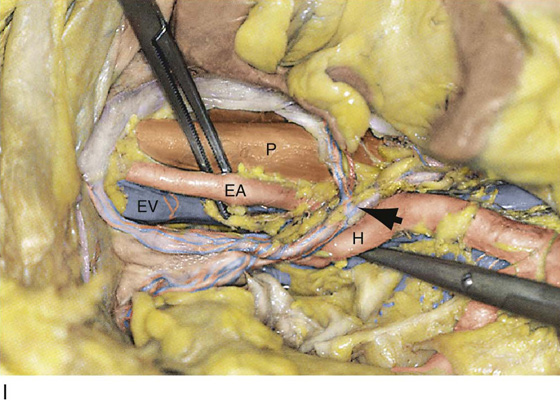
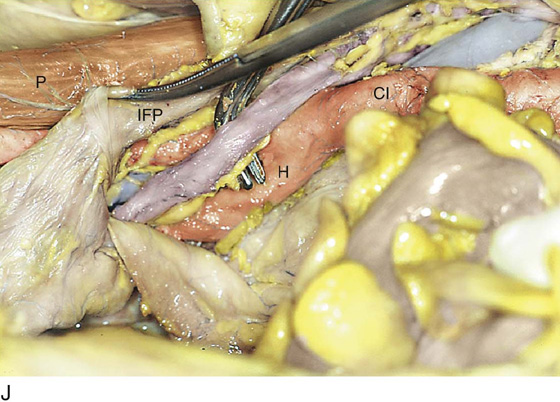
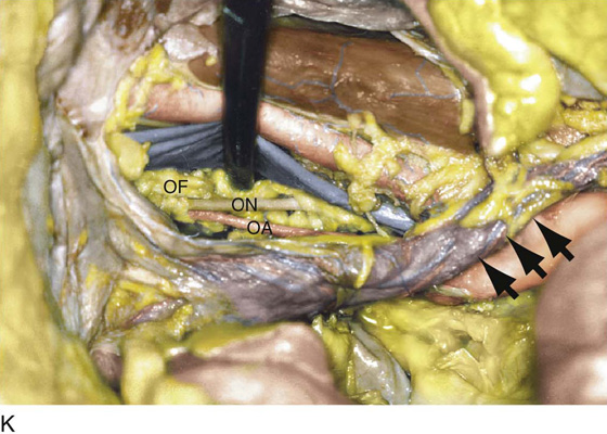
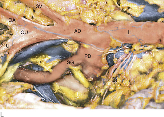
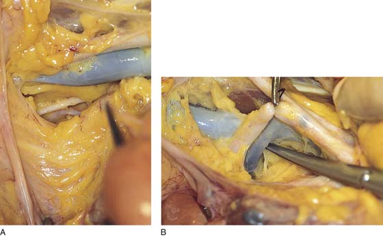
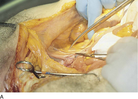
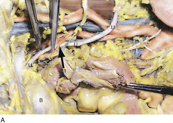
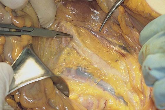
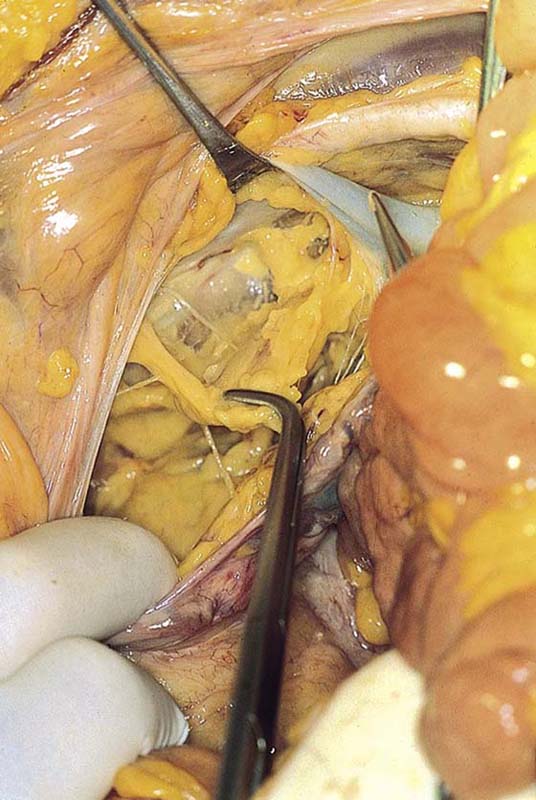
CHAPTER 12 Helmut F. Schellhas An understanding of the anatomy of the retroperitoneal space is required to initiate lymphadenectomy and exposure of the great vessels (Fig. 12–1A–C). Although the course of the ureter was covered earlier in this section, additional anatomic dissection views are needed to help the reader understand the operative steps necessary to perform radical hysterectomy (Fig. 12–2). In truth, radical hysterectomy and lymphadenectomy are exercises in anatomic dissection. It is therefore imperative to have precise knowledge of the retroperitoneum, particularly the relationships of the ureter to the great vessels and of the vessels to one another (Fig. 12–3A–L). The structures most vulnerable to injury and most difficult to repair are the veins accompanying the large pelvic arteries (Fig. 12–4). The precise location of these veins and their careful retraction are crucial to the safe performance of the surgery (Fig. 12–5A, B). All of these anatomic relationships come together during the dissection of the obturator fossa (Fig. 12–6). FIGURE 12–1 A. The retroperitoneum is entered by grasping the round ligament and cutting the peritoneum at the top of the broad ligament in a linear and foot-to-head direction. B. The scissors point to the belly of the psoas major muscle, which is lateral to the external iliac artery. C. The external iliac artery has now been exposed and lies above the spread tips of the scissors. FIGURE 12–2 Orientation view of the uterus in situ with a ligature placed into the fundus for traction. FIGURE 12–3 A. The entire course of the right ureter is shown here. U, ureter; 1, uterine artery; B, bladder (view from left side). B. The entire course of the right ureter viewed from overhead. Ut, uterus; U, ureter; B, bladder; UVF, uterovesical fold; UA, feeding vessel to ureter. The clamp points to the uterine artery. C. The entire course of the left ureter is shown. U, uterus; arrows 1, 2, and 3 point to the ureter; IFP, infundibulopelvic ligament; O, ovary; t, tube; B, bladder; P, psoas major muscle; V, uterine vessels. D. The scissors point to the aorta (A) and fibers of the hypogastric nerve plexus. CI, common iliac artery. E. The large vessels within the retroperitoneal space are well visualized in this photograph. The iliac veins and vena cava are also shown. A, aorta; M, inferior mesenteric artery; CI, common iliac artery; U, ureter; P, psoas major muscle. F. The presacral space is exposed. The arrow points to the middle sacral vessels. The clamp elevates the left internal iliac vein (LIV). A, aorta; RIA, right common iliac artery; LIA, left common iliac artery; IM, inferior mesenteric artery; VC, vena cava. G. The right ureter is elevated by the scissors. The clamp points to the ovarian vessels. The arrow points to the left common iliac vein. CI, common iliac artery (right); C, inferior vena cava; E, external iliac vein; P, psoas major muscle. H. Detail at presacral space. CIA, common iliac artery (right); CIV, common iliac vein (left); C, vena cava. I. The arrow points to the right ureter and the infundibulopelvic ligament. H, hypogastric artery; EV, external iliac vein; EA, external iliac artery; P, psoas major muscle. J. The ureter above the right-angle clamp has been separated from the ovarian vessels (infundibulopelvic ligament [IFP]). P, psoas major muscle; H, hypogastric artery; CI, common iliac artery. K. The arrows point to the right ureter. The clamp elevates the right external iliac vein to expose the obturator fossa (OF), the obturator nerve (ON), and the obturator artery (OA). L. Detailed dissection of the hypogastric artery (H) shows the anterior division (AD), the posterior division (PD), the common trunk with the obliterated umbilical and superior vesical artery (OU, SV), the obturator artery (OA), and the uterine artery (U). The superior gluteal artery (SG) branches from the posterior division (PD). FIGURE 12–4 The peritoneum has been opened over the terminus of the abdominal aorta (clamp). Both right and left common iliac arteries are clearly seen. The large vein is the left common iliac vein. FIGURE 12–5 A. The external iliac artery and vein (blue) cross above the obturator fossa. Removal of some fat and lymph nodes partially exposes the obturator nerve (white) and the obturator artery (pink). B. The anterior division of the hypogastric artery has been ligated. A clamp elevates the hypogastric artery to expose the hypogastric vein. The tip of the scissors is just beneath the junction of the hypogastric and external iliac veins. FIGURE 12–6
Radical Hysterectomy
 Michael S. Baggish
Supplemental Anatomy for Radical Hysterectomy and Pelvic Lymphadenectomy
![]()
Stay updated, free articles. Join our Telegram channel

Full access? Get Clinical Tree


